Review
. 2014 Jan:38:1-19.
doi: 10.1016/j.preteyeres.2013.08.004. Epub 2013 Oct 14.

Molecular complexes that direct rhodopsin transport to primary cilia

Affiliations
Review

Molecular complexes that direct rhodopsin transport to primary cilia

Jing Wang et al. Prog Retin Eye Res. 2014 Jan.

Abstract

Rhodopsin is a key molecular constituent of photoreceptor cells, yet understanding of how it regulates photoreceptor membrane trafficking and biogenesis of light-sensing organelles, the rod outer segments (ROS) is only beginning to emerge. Recently identified sequence of well-orchestrated molecular interactions of rhodopsin with the functional networks of Arf and Rab GTPases at multiple stages of intracellular targeting fits well into the complex framework of the biogenesis and maintenance of primary cilia, of which the ROS is one example. This review will discuss the latest progress in dissecting the molecular complexes that coordinate rhodopsin incorporation into ciliary-targeted carriers with the recruitment and activation of membrane tethering complexes and regulators of fusion with the periciliary plasma membrane. In addition to revealing the fundamental principals of ciliary membrane renewal, recent advances also provide molecular insight into the ways by which disruptions of the exquisitely orchestrated interactions lead to cilia dysfunction and result in human retinal dystrophies and syndromic diseases that affect multiple organs, including the eyes.

Keywords: ADRP; Arfs; Autosomal Dominant Retinitis Pigmentosa; BBS; BBSome; Bardet-Biedl Syndrome; CTS; Ciliary Targeting Signal; Cilium; DHA; Docosahexaenoic Acid; GAP; GC1; GEF; GTPase Activating Protein; Guanine Nucleotide Exchange Factor; Guanylyl Cyclase 1; IFT; Intraflagellar Transport; JBTS; Joubert Syndrome; MKS; MT; MTOC; Meckel Syndrome; Microtubule Organizing Center; Microtubules; NPHP; Nephronophthisis; OCT; Optical Coherence Tomography; PLA; Proximity Ligation Assay; RIS; ROS; RPGR; RTC(s); Rabs; Retinitis Pigmentosa GTPase Regulator; Rhodopsin; Rhodopsin Transport Carrier(s); Rod Inner Segment(s); Rod Outer Segment(s); SNARE; Soluble N-ethylmaleimide-sensitive Factor Attachment Protein Receptor; TGN; Trafficking; Trans-Golgi Network; a conserved complex of BBS proteins.


Figures

Figure 1
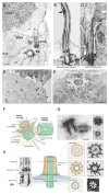
Figure 1. ROS is a primary cilium
A. Rhodopsin transport carriers (RTCs) that replenish the ROS sensory membrane are detected by EM immunocytochemistry in the rod inner segment (RIS), at the base of the cilium (C) (also called the connecting cilium) of Rana Berlandieri photoreceptors. BB, basal body, m, mitochondria. Modified from Deretic and Papermaster, 1991. Bar = 0.3 μm. B. and C. Comparison of Chlamydomonas flagellum and rod connecting cilium, respectively. Arrows point to the IFT particles underlying the ciliary membrane. Reproduced from Rosenbaum et al, 1999. D. Membranes clustered at the base of the cilium carry newly synthesized rhodopsin, detected by EM autoradiography of Xenopus photoreceptors. E. Rhodopsin transport vesicles (v) fuse with the plasma membrane at the periciliary ridge complex (PRC) of Xenopus rods. D and E Adapted from Papermaster et al, 1985. Bar = 0.5 μm. F. and G. Schematic representation (F) and the EM (G) of the basal body (centrosome). A, B, and C-tubules in each triplet correspond to the internal, middle and external one, respectively. Mother centriole is surrounded by PCM and contains distal and subdistal appendages on the B and C-tubule, respectively. Bar = 0.2 μm in (G). H. Organization of the axoneme, transition zone and the basal body of Chlamydomonas flagellum. An array of doublets arising from the distal end of the centriole fills the axoneme. CW, cartwheel, at the proximal end of the basal body. Bar = 0.25 μm. F–H Reproduced with permission from Bettencourt-Dias and Glover, 2007.
Figure 2
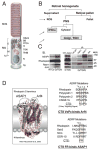
Figure 2. Rhodopsin trafficking to the primary cilium: Ciliary targeting signals and regulators
A. Schematic of a rod photoreceptor outlining rhodopsin trafficking pathways. B. Schematic of retinal subcellular fractionation that generates fractions enriched in rhodopsin transport membranes. C. Immunoprecipitation using the transport membranes as a source of protein complexes reveals regulators of rhodopsin trafficking. Reproduced from Wang et al, 2012. D. The rhodopsin cytoplasmic C-terminal VxPx and H-8 FR are its ciliary targeting signals (CTSs), that are recognized by Arf4 and ASAP1, respectively.
Figure 3
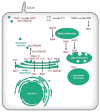
Figure 3. Pathways regulated by Arf GTPases
A. Arf proteins have distinct localizations and functions in the endoplasmic reticulum (ER)–Golgi system. Arf1 and Arf4 localize to the early cis-Golgi. Arf3 and Arf4 localize to the trans-Golgi network (TGN). In addition to the recruitment of coat proteins (coatomer complex I (COPI), GGA (Golgi-localized, γ-ear-containing, Arf-binding protein) and adaptor protein 1 (AP1)) to the Golgi, Arf1 binds to ceramide transfer (CERT) and FAPP2 to mediate the transport of ceramide and glucosylceramide lipids from the cis-Golgi to the trans-Golgi. At the ER–Golgi intermediate compartment (ERGIC), Arf1 and the GEF GBF1 act with COPII to regulate the formation of lipid droplets and for the replication of several viruses. GBF1 is also localized at the TGN where it activates Arf4 (Lowery et al, 2013). CAPS (Calcium-dependent activator protein for secretion), which is involved in regulated secretion, is recruited to the TGN by Arf4 and Arf5. At the ER, Sar1, activated by SEC12, recruits COPII to allow vesicle transport to the Golgi. B. In rod photoreceptors Arf4 binds specifically to rhodopsin in the TGN membrane and, together with FIP3, ASAP1 and Rab11, it facilitates the transport of rhodopsin in transport vesicles from the inner segment to the outer segment, which is a specialized cilium. Arf-like 3 (Arl3) has been localized to the connecting cilium, and retinitis pigmentosa 2 (RP2; also known as XRP2), an Arl3 GAP, localizes to the TGN, the basal body and the membrane adjacent to the connecting cilium. C. In primary cilia, Arl6 recruits the BBSome coat complex that facilitates the transport of membrane proteins into the cilium. Arl13 is localized to the cilium and has been implicated in intraflagellar transport. ADRP, adipose differentiation-related protein (also known as adipophilin); ATGL, adipose triglyceride lipase; PtdIns4K, phosphatidylinositol 4-kinase. Figure is reproduced with permission from Donaldson and Jackson, 2011. The figure legend is adapted from Donaldson and Jackson, 2011.
Figure 4
Figure 4. Formation of the rhodopsin ciliary targeting complex at the TGN
The schematic was generated by combining, scaling, merging and outlining the known crystal structures, as described in the text. The width of the complex containing the dimmer of the BAR, PH and GAP domains of ASAP1 is approximately 22 nm. Assembly of the complex at the TGN can be divided in two stages: The Arf4-dependent Stage I and the post-Arf4 Stage II. In Stage I, cytosolic Arf4 is activated by the Arf-GEF GBF1 and becomes membrane-associated. Activated Arf4 recognizes the VxPx CTS of rhodopsin. ASAP1 is recruited to the TGN by activated Arf4, acidic phospholipids and PIP2 (indicated by red head groups). There, it recognizes the FR CTS of rhodopsin. ASAP1 dimerizes through the BAR domain, which likely induces membrane deformation necessary for budding. It selectively binds Rab11a and FIP3, which also acts as a dimer. The CTS proofreading phase is completed through the GTP hydrolysis on Arf4 by ASAP1, assisted by FIP3. In Stage II inactivated Arf4 departs the TGN, but the rest of the complex remains associated to regulate subsequent events. This includes the recruitment of the GEF Rabin8 and the inactive, GDP-bound Rab8 by ASAP1 and Rab11a. This endows the nascent buds with the ciliary targeting information.
Figure 5
Figure 5. Rab lipid modification and the GTPase cycle
Newly synthesized Rabs bind to Rab-escort protein (REP) encoded by the choroideremia gene, which presents them to Rab geranylgeranyl transferase (RGGTase) that prenylates Rabs (1) and delivers them to membranes of donor compartments (2). Rabs are activated by Rab GEFs and recruit effectors, which regulate budding (3), movement (4) and tethering/docking (5) of the carrier vesicle. As membrane carriers fuse with the acceptor compartment membrane, Rabs are inactivated by Rab GAPs. Rab guanine-nucleotide dissociation inhibitor (Rab GDI) extracts GDP-bound Rabs into the cytosol (6) and delivers them to donor compartment membranes with the assistance of a GDI-displacement factor (GDF), also known as Prenylated Rab Acceptor 1 (PRA1). Adapted with permission from Seixas et al., 2013. Note that only Qa SNARE (syntaxin) and R-SNARE (VAMP) are presented in this schematic, whereas the Qbc SNAREs that provide two helices to the SNARE complex are omitted. See text for the details on SNARE complex assembly.
Figure 6
Figure 6. Localization of Rab11-Rabin8-Rab8 GTPase cascade in a ciliated cell
Rab GTPases involved in ciliary trafficking are conserved from yeast to mammals. In yeast, Ypt31p/Ypt32p interacts with Sec2p, a GEF for the next Rab in cascade, which is Sec4p. Sec2p activates Sec4p on the secretory vesicle, which leads to vesicle tethering and fusion with plasma membrane. Mammalian homologues Rab11, Rabin8 (a GEF for Rab8) and Rab8 participate in the ciliogensis cascade, which regulates targeting of ciliary transport carriers, including RTCs. Both Rab8 and Rabin8 are required for primary cilium formation. At the plasma membrane, Arf6 activates the small GTPase Rac1. Rab5 and Rab7 regulate early and late endocytic events, respectively. Modified with permission from Mizuno-Yamazaki and Novick, 2012.
Figure 7
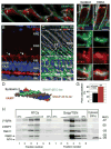
Figure 7. Interactions that regulate rhodopsin transiting from the Golgi/TGN into ciliary-targeted RTCs
A. Proximity Ligation Assay (PLA) involves dual target recognition by antibodies covalently linked to DNA primers. Circularization and ligation takes place only between the connector oligonucleotides located at <16 nm. DNA amplification is followed by hybridization with fluorescent oligos (excitation: 598 nm). B. When PLA is performed on 100 μm thick sections of Rana Berlandieri retinas using the modified method adapted for brain slices (Trifilieff et al, 2011) with antibodies to rhodopsin and an unrelated Golgi-associated protein no red fluorescence is detected, signifying an absence of protein-protein interactions. C. By contrast, rhodopsin-ASAP1 interaction sites (red dots) are detected by PLA. D. Frog retina labeled with mAb 11D5 to the VxPx motif of rhodopsin. Rhodopsin is concentrated in the ROS and the photoreceptor Golgi (G). E. The Golgi complex is localized in the photoreceptor myoid (M). The cis-Golgi is visualized by GM 130 (red) and the trans-Golgi by Rab6 (green). E, Ellipsoid. F. ASAP1 (blue) and Rab11 (red) co-localize on the nascent buds at the tips of the trans-Golgi (Rab6, green). G. and H. ASAP1-Arf4 interaction sites (red dots) are detected by PLA and retinal sections are subsequently stained with Rab6-Alexa Fluor 488 conjugate (green). Arrows indicate ASAP1-Arf4 interaction sites juxtaposed to the trans-Golgi. I. and J. Rhodopsin-Arf4, or ASAP1-Rab11 interaction sites are detected by PLA, respectively. Bar = 5 μm in D, E, I and J; 1 μm in B, C, G and H; 0.5 μm in F. (K) Red fluorescent signals are quantified, the data are expressed as a percent of total interaction sites within the RIS and presented as the means ± SEM (***, p<2.27E−6; *, p=0.01). Modified from Wang et al, 2012.
Figure 8
Figure 8. Molecular interactions and the sequence of events taking place during rhodopsin trafficking to the cilia
At the TGN, rhodopsin forms a complex with activated Arf4 and ASAP1, which recognize its VxPx and FR CTSs, respectively. This leads to the formation of the Arf4-based ciliary targeting complex, which, in addition to Arf4, contains ASAP1, Rab11 and FIP3. Following GTP hydrolysis on Arf4, the remaining components recruit Rabin8 and Rab8, which is a regulator of fusion with the plasma membrane. On RTCs, ASAP1 serves as a scaffold, or an affinity adaptor for spatially restricted activation of Rab8. Activated Rab8 permits RTCs fusion and cargo delivery across the diffusion barrier surrounding the cilium, based in part on its ability to interact with the Sec15 subunit of the exocyst, a membrane tethering complex involved in ciliary targeting. The approximate size of the targeting complex is as in Figure 4.
Figure 9
Figure 9. Interactions between the Arf4-based ciliary targeting complex and IFT20
A. – E. Rhodopsin-Rab8, -Rabin8, -IFT20, and Rabin8-IFT20, or Rab8-IFT20 interaction sites were detected by PLA on Rana Berlandieri retinas, respectively. The antibody to IFT20 was a kind gift of Dr. Greg Pazour. F. By confocal microscopy Rabin8 (green) and Rab11 (red) colocalize (yellow) on RTCs (arrow). G. Rabin8 (green) and Rab11 (red) are present on nascent buds associated with the trans-Golgi (G), revealed by Rab6 (blue). N, nucleus. H. Magnified Golgi area from G. Arrows indicate carriers containing Rabin8 (green) and Rab11 (red, yellow in the merged image). Rabin8-positive carriers (green, yellow arrowheads in G. and H.) are juxtaposed to Rab11-positive carriers (red, white arrowheads). I. Rabin8 (green) and ASAP1 (red) colocalize (yellow) on RTCs (arrow). Yellow arrowhead indicates Rabin8-positive carrier (green) juxtaposed to ASAP1-positive carrier (red, white arrowheads). J. and K. Microtubule depolymerization with nocodazole, or treatment with propranolol (Ppl), respectively, does not interfere with the interaction of Rabin8 with ASAP1. An arrow and arrowheads in K point to the same structures as in I. that are enlarged upon treatment with propranolol, as reported (Mazelova et al, 2009a). L. Rabin8 and IFT20 were detected by PLA (red dots) and retinal sections were then stained with Rab8 Alexa Fluor 488 conjugate (green). Arrowheads indicate separate structures containing Rabin8 and IFT20, or Rab8. M.–O. Rabin8 and IFT20-containing carriers (red, arrows) are found in the proximity of the trans-Golgi labeled with Rab6 Alexa Fluor 488 conjugate (green), in control, nocodazole or Ppl-treated retinas, respectively. Bar = 5 μm in A–E and M–O; 2 μm in F–L. P. A model summarizing interaction sites of IFT20 with Rabin8, as suggested by our study.
Figure 10
Figure 10. Tethering complexes at the periciliary plasma membrane
A. Three consecutive tomographic slices of the mouse photoreceptor cilium. Filaments originating from the IS (red) interact with vesicles (yellow), C-microtubule distal appendages (green), and ciliary membrane filaments (blue). B. Segmentation from A. of vesicles (yellow), IS filaments (red), distal appendages (green), and membrane tethers (blue) in the pericentriolar region. Bar = 0.1 μm. Reproduced with permission from Gilliam et al, 2012. C. Rab8-postive RTCs (red, arrows) are in close proximity and partially overlap (yellow) with Sec8-labeled structures (green) in Rana photoreceptors. Bar = 1 μm. Reproduced from Mazelova et al., 2009b. D. A structure and a model for the assembly of the exocyst, modified with permission from Munson and Novick, 2006. A quick-freeze/deep-etch EM of purified mammalian brain exocyst complexes, either unfixed or fixed with glutaraldehyde (from Hsu et al., 1998). Bar = 0.1 μm. The schematic representation of the yeast exocyst complex as an elongated helical-bundle structure packed together to form a structure similar to the fixed complex. The exocyst is linked to the carrier membrane via the interaction of Rab8 with the Sec15 subunit. Note that the size of the exocyst closely matches the size of the membrane tethers (blue) in A. and B.
Figure 11
Figure 11. SNAREs regulate fusion of RTCs with the periciliary plasma membrane
A. In Rana photoreceptors SNAP-25 (green) and Syntaxin 3 (STX3, red) co-localize in the RIS at the RTC fusion sites (arrows, yellow). B. Syntaxin 3 (red) is highly concentrated in the RIS PM (arrows), but is absent from the ROS and from the RPE. AJ, adherens junctions that form the outer limiting membrane (OLM, dotted line); retinal layers: ONL-outer nuclear, OPL-outer plexiform, INL-inner nuclear. Syntaxin 3 is also abundant in the OPL. Asterisks indicate the protruding RIS of green rods that account for ~5% of total rods. Nuclei are stained with TO-PRO-3 (blue). C. Anti-SNAP-25 (green) outlines the photoreceptor PM. The CPs, which are in continuum with the RIS PM, also contain SNAP-25. The ROS, which are visible by DIC, are completely devoid of this SNARE. SNAP-25 co-localizes with synaptophysin (SYP, red) in the OPL. D. Space-filling model of the SNARE helical bundle. E. and F. DHA treated retinas display increase in Syntaxin 3 (red) content of the PM surrounding the cilium. Upon DHA treatment Syntaxin 3 (red) and SNAP-25 (green) increasingly colocalize (yellow, merge). G. DHA significantly increases Syntaxin 3-SNAP-25 pixel co-localization (**, p=0.003). Bar = 10 μm in B and C; 5 μm in A, and in upper panels in E and F; 0.5 μm in lower panels in E and F. H. PNS isolated from one frog retina fractionated by velocity sedimentation followed by 20–39% sucrose density gradients. Membrane and cytosolic proteins were separated by SDS-PAGE. Radiolabeled rhodopsin was detected by autoradiography. VAMP7, Rab8 and Arf4 were detected by immunoblotting on a single blot, whereas Rab11 was probed on a duplicate blot. Arf4 is also detected in the cytosol (arrow on the right). A.-C. and E.-G. Reproduced from Mazelova et al., 2009b. H. Modified from Wang et al., 2012.
Figure 12
Figure 12. Comparison of the OCT and DIC image of frog retinas
A. Cross-section optical coherence tomography (OCT) image of living frog retinas averaged over 120 μm along the y-axis. B. Confocal optical section of the fixed frog retina imaged by differential interference contrast (DIC). Nuclei are stained with TO-PRO-3. Retinal layers: ONL-outer nuclear, OPL-outer plexiform, INL-inner nuclear, OPL-outer plexiform, GCL-ganglion cell layer. Note that the hyper-reflective band visualized by OCT and commonly attributed to the rod photoreceptor inner/outer segment (RIS/ROS) junction originates from the ellipsoid region of the RIS, which is detectable in the DIC image by the change in optical properties associated with the tight mitochondrial packing. Bar = 10 μm. Modified from Lu et al., 2012.

Similar articles

Cited by

References

    1. Alberts P, Galli T. The cell outgrowth secretory endosome (COSE): a specialized compartment involved in neuronal morphogenesis. Biol Cell. 2003;95:419–424. - PubMed
    1. Alberts P, Rudge R, Irinopoulou T, Danglot L, Gauthier-Rouviere C, Galli T. Cdc42 and actin control polarized expression of TI-VAMP vesicles to neuronal growth cones and their fusion with the plasma membrane. Mol Biol Cell. 2006;17:1194–1203. - PMC - PubMed
    1. Anderson RG. The three-dimensional structure of the basal body from the rhesus monkey oviduct. J Cell Biol. 1972;54:246–265. - PMC - PubMed
    1. Arshavsky VY, Burns ME. Photoreceptor signaling: supporting vision across a wide range of light intensities. J Biol Chem. 2012;287:1620–1626. - PMC - PubMed
    1. Arts HH, Doherty D, van Beersum SE, Parisi MA, Letteboer SJ, Gorden NT, Peters TA, Marker T, Voesenek K, Kartono A, Ozyurek H, Farin FM, Kroes HY, Wolfrum U, Brunner HG, Cremers FP, Glass IA, Knoers NV, Roepman R. Mutations in the gene encoding the basal body protein RPGRIP1L, a nephrocystin-4 interactor, cause Joubert syndrome. Nat Genet. 2007;39:882–888. - PubMed

Publication types

MeSH terms