

Calcium influx through L-type CaV1.2 Ca2+ channels regulates mandibular development
- PMID: 23549079
- PMCID: PMC3613930
- DOI: 10.1172/JCI66903
Calcium influx through L-type CaV1.2 Ca2+ channels regulates mandibular development
Abstract
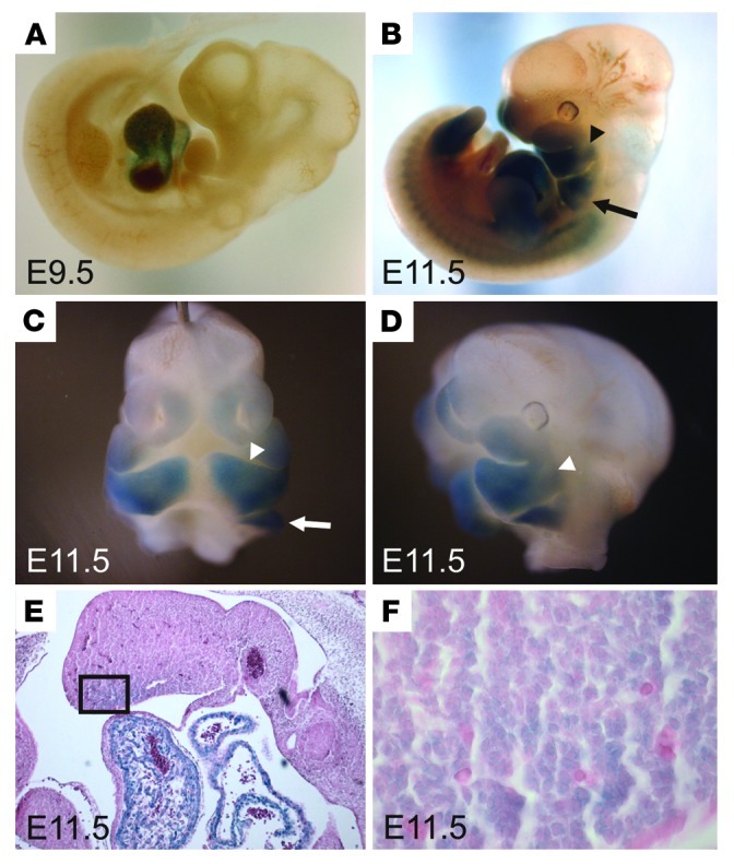
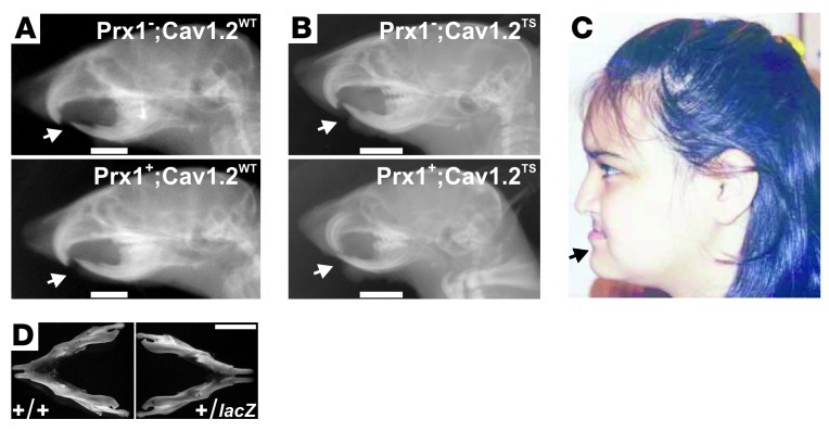
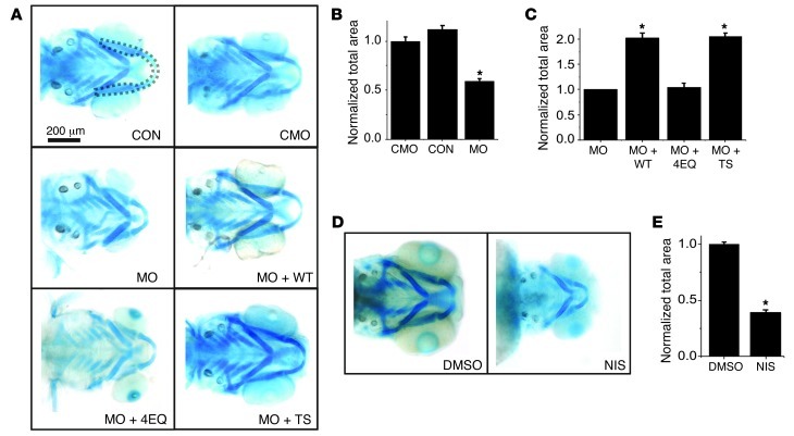
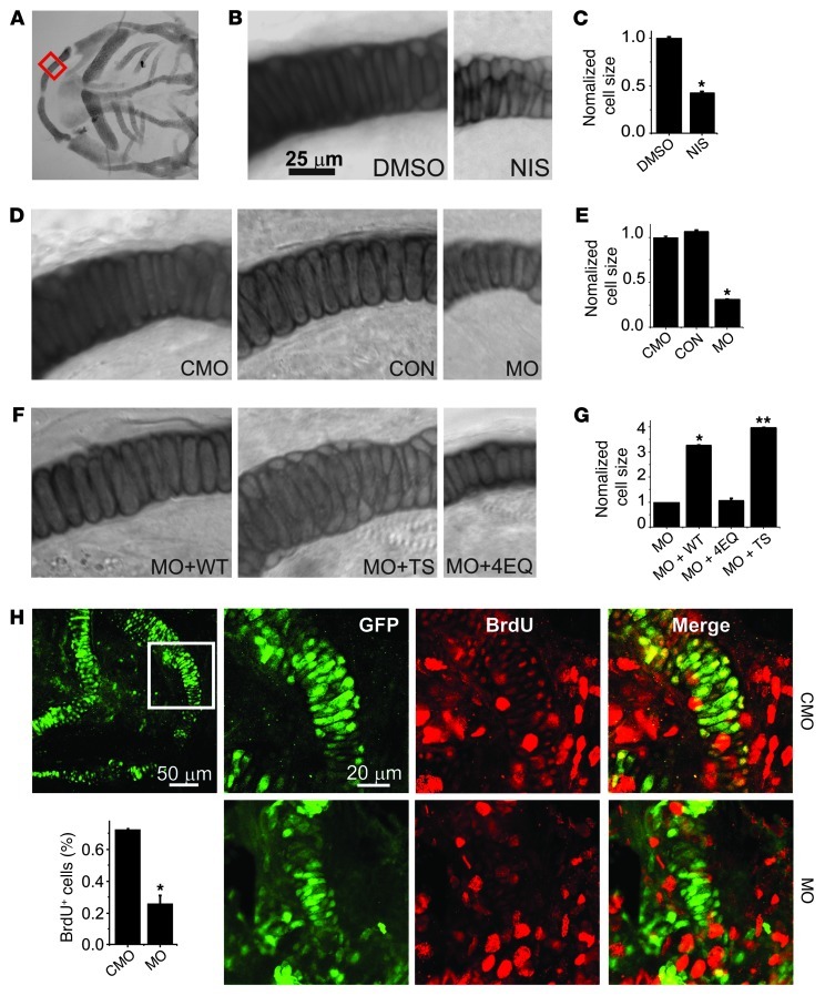
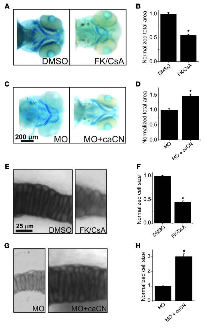
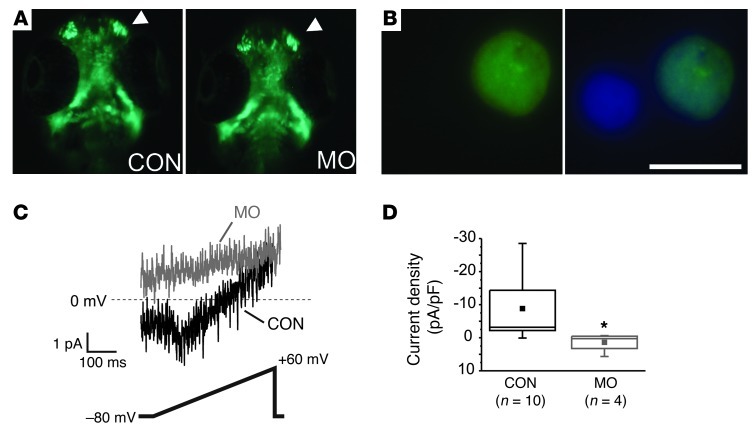
The identification of a gain-of-function mutation in CACNA1C as the cause of Timothy Syndrome (TS), a rare disorder characterized by cardiac arrhythmias and syndactyly, highlighted unexpected roles for the L-type voltage-gated Ca2+ channel CaV1.2 in nonexcitable cells. How abnormal Ca2+ influx through CaV1.2 underlies phenotypes such as the accompanying syndactyly or craniofacial abnormalities in the majority of affected individuals is not readily explained by established CaV1.2 roles. Here, we show that CaV1.2 is expressed in the first and second pharyngeal arches within the subset of cells that give rise to jaw primordia. Gain-of-function and loss-of-function studies in mouse, in concert with knockdown/rescue and pharmacological approaches in zebrafish, demonstrated that Ca2+ influx through CaV1.2 regulates jaw development. Cranial neural crest migration was unaffected by CaV1.2 knockdown, suggesting a role for CaV1.2 later in development. Focusing on the mandible, we observed that cellular hypertrophy and hyperplasia depended upon Ca2+ signals through CaV1.2, including those that activated the calcineurin signaling pathway. Together, these results provide new insights into the role of voltage-gated Ca2+ channels in nonexcitable cells during development.
Figures
Similar articles
-
Impaired chromaffin cell excitability and exocytosis in autistic Timothy syndrome TS2-neo mouse rescued by L-type calcium channel blockers.J Physiol. 2019 Mar;597(6):1705-1733. doi: 10.1113/JP277487. Epub 2019 Jan 28. J Physiol. 2019. PMID: 30629744 Free PMC article.
-
Cav1.2 channelopathies causing autism: new hallmarks on Timothy syndrome.Pflugers Arch. 2020 Jul;472(7):775-789. doi: 10.1007/s00424-020-02430-0. Epub 2020 Jul 3. Pflugers Arch. 2020. PMID: 32621084 Review.
-
Cellular mechanisms of ventricular arrhythmias in a mouse model of Timothy syndrome (long QT syndrome 8).J Mol Cell Cardiol. 2014 Jan;66:63-71. doi: 10.1016/j.yjmcc.2013.10.021. Epub 2013 Nov 9. J Mol Cell Cardiol. 2014. PMID: 24215710 Free PMC article.
-
State-dependent signaling by Cav1.2 regulates hair follicle stem cell function.Genes Dev. 2013 Jun 1;27(11):1217-22. doi: 10.1101/gad.216556.113. Genes Dev. 2013. PMID: 23752588 Free PMC article.
-
Voltage-Gated Calcium Channels in Nonexcitable Tissues.Annu Rev Physiol. 2021 Feb 10;83:183-203. doi: 10.1146/annurev-physiol-031620-091043. Epub 2020 Oct 26. Annu Rev Physiol. 2021. PMID: 33106102 Free PMC article. Review.
Cited by
-
Excitation-Contraction Coupling in the Goldfish (Carassius auratus) Intact Heart.Front Physiol. 2020 Sep 11;11:1103. doi: 10.3389/fphys.2020.01103. eCollection 2020. Front Physiol. 2020. PMID: 33041845 Free PMC article.
-
Genetic Evidence Supporting the Role of the Calcium Channel, CACNA1S, in Tooth Cusp and Root Patterning.Front Physiol. 2018 Sep 26;9:1329. doi: 10.3389/fphys.2018.01329. eCollection 2018. Front Physiol. 2018. PMID: 30319441 Free PMC article.
-
Voltage-dependent Ca2+ channels promote branching morphogenesis of salivary glands by patterning differential growth.Sci Rep. 2018 May 15;8(1):7566. doi: 10.1038/s41598-018-25957-w. Sci Rep. 2018. PMID: 29765108 Free PMC article.
-
Genetic disruption of voltage-gated calcium channels in psychiatric and neurological disorders.Prog Neurobiol. 2015 Nov;134:36-54. doi: 10.1016/j.pneurobio.2015.09.002. Epub 2015 Sep 16. Prog Neurobiol. 2015. PMID: 26386135 Free PMC article. Review.
-
The CaV1.2 L-type calcium channel regulates bone homeostasis in the middle and inner ear.Bone. 2019 Aug;125:160-168. doi: 10.1016/j.bone.2019.05.024. Epub 2019 May 20. Bone. 2019. PMID: 31121355 Free PMC article.
References
-
- Chai Y, et al. Fate of the mammalian cranial neural crest during tooth and mandibular morphogenesis. Development. 2000;127(8):1671–1679. - PubMed
Publication types
MeSH terms
Substances
Supplementary concepts
Grants and funding
LinkOut - more resources
Full Text Sources
Other Literature Sources
Molecular Biology Databases
Miscellaneous
