Review
. 2013 Oct;28(10):1923-42.
doi: 10.1007/s00467-012-2329-z. Epub 2013 Jan 20.

Hereditary causes of kidney stones and chronic kidney disease

Affiliations
Review

Hereditary causes of kidney stones and chronic kidney disease

Vidar O Edvardsson et al. Pediatr Nephrol. 2013 Oct.

Abstract

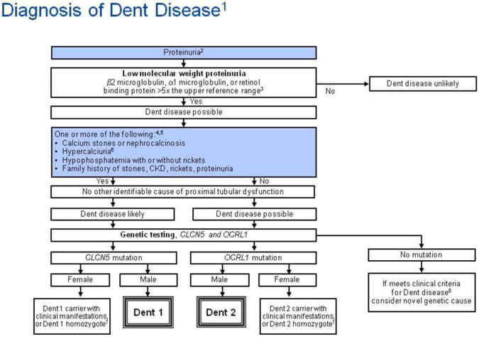
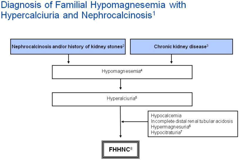
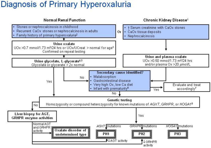
Adenine phosphoribosyltransferase (APRT) deficiency, cystinuria, Dent disease, familial hypomagnesemia with hypercalciuria and nephrocalcinosis (FHHNC), and primary hyperoxaluria (PH) are rare but important causes of severe kidney stone disease and/or chronic kidney disease in children. Recurrent kidney stone disease and nephrocalcinosis, particularly in pre-pubertal children, should alert the physician to the possibility of an inborn error of metabolism as the underlying cause. Unfortunately, the lack of recognition and knowledge of the five disorders has frequently resulted in an unacceptable delay in diagnosis and treatment, sometimes with grave consequences. A high index of suspicion coupled with early diagnosis may reduce or even prevent the serious long-term complications of these diseases. In this paper, we review the epidemiology, clinical features, diagnosis, treatment, and outcome of patients with APRT deficiency, cystinuria, Dent disease, FHHNC, and PH, with an emphasis on childhood manifestations.


Conflict of interest statement

Conflict of interest statement: Dr. Goldfarb is a consultant for Takeda.

Figures

Figure 1
Figure 1
Urinary crystals. A. Typical small and medium sized 2,8-dihydroxyadenine crystals. The medium sized cystals are brown with dark outline and central spicules. (Original magnification × 400). B. The same field viewed with polarized light microscopy. The small and medium sized crystals appear yellow in colour and produce a central Maltese cross pattern. (Original magnification × 400). C. Urinary cystine crystals. The typical 6-sided crystal is diagnostic of cystinuria. A good example can be seen on the left side of the Figure (arrow). D. Urinary calcium oxalate crystals. The typical bipyramidal calcium oxalate dihydrate crystals (arrows) and a large dumbbell calcium oxalate monohydrate crystal (asterisk) are both seen. E. Amorphous calcium phosphate crystals. Although their appearance is not as distinctive, amorphous calcium phosphate crystals should be suspected if the urine is alkaline (pH > 6.0).
Figure 2
Figure 2
Schematic overview of adenine metabolism. In adenine phosphoribosyltransferase deficiency, adenine cannot be converted to adenosine monophsophate and is instead converted by xanthine dehydrogenase to 2,8-dihydroxyadenine. Abbreviations: APRT, adenine phosphoribosyltransferase; AMP, adenosine monophsophate; HPRT, hypoxanthine-guanine phosphoribosyltransferase; IMP, inosine monophosphate; XDH, xanthine dehydrogenase.
Figure 3
Figure 3
Algorithm for diagnostic evaluation of adenine phosphoribosyltransferase (APRT) deficiency and 2,8-dihydroxyadeninuria. Annotations to Figure 3 12,8-dihydroxyadeninuria, hyperuricosuria and xanthinuria should always be considered in the differential diagnosis of radiolucent kidney stones in childhood. 2 Children with radiolucent kidney stones and chronic kidney disease (CKD) should be evaluated for adenine phosphoribosyltransferase (APRT) deficiency. 3,6Patients with APRT deficiency may present with acute kidney injury (AKI), CKD or acute allograft nephropathy, even in the absence of previous history of kidney stones. Kidney biopsy shows variable degree of tubulointerstitial injury and features consistent with crystalline nephropathy. 4Ultraviolet spectrophotometry and/or x-ray crystallography easily differentiates 2,8-dihydroxyadenine (DHA) from uric acid and xanthine. 5The pathognomonic round, brown urinary DHA crystals (Figure 1) are seen in almost all patients with the disorder. The crystals may, however, be hard to identify in patients with markedly decreased renal function due to reduced crystal clearance. 7APRT activity in red blood cell lysates ranges from 16-32 nmol/h/mg hemoglobin in healthy subjects [118]; homozygotes and compound heterozygotes have no measurable enzyme activity and in heterozygotes the activity is approximately 25%. Recent blood transfusion may falsely elevate APRT activity. 8 Genetic testing, which confirms the diagnosis when functionally significant mutations are found in both copies of the APRT gene, is not clinically indicated in patients with abolished APRT activity.
Figure 4
Figure 4
Algorithm for diagnostic evaluation of cystinuria. Annotations to Figure 4 Demonstration of aminoaciduria with excessive levels of ornithine, arginine or lysine is not required for diagnosis. All patients with stones prior to age 30 years should be screened. Sodium-cyanide-nitroprusside test can yield false positive result in heterozygotes below the age of 2 years. A positive sodium-cyanide-nitroprusside test should be confirmed with 24 hour urine collection. Homozygotes have more than 170 μmol cystine/mmol creatinine (1500 μmol cystine/g creatinine). Heterozygotes (with SLC7A9 mutations) will have between 11 and 110 μmol cystine/mmol creatinine (100-1000 μmol cystine/g creatinine), while other heterozygotes (with SLC3A1 mutations) have normal cystine excretion (≤11 μmol cystine/mmol creatinine). Adult homozygotes excrete more than 1.3 mmol cystine per day (300 mg/day); heterozygotes (with SLC7A9 mutations) will excrete more than 0.13 mmol/day (30 mg/day) while other heterozygotes (with SLC3A1 mutations) have normal cystine excretion.
Figure 5
Figure 5
Algorithm for diagnostic evaluation of Dent disease. Annotations to Figure 5 1The guideline does not address prenatal diagnosis. 2Trace or greater by dipstick; or >0.2 mg protein/mg creatinine; or >100 mg protein/day. 3Low molecular weight (LMW) proteinuria may be reduced in advanced renal insufficiency, or be present in renal failure from other causes, but remains helpful when markedly increased. 4In the presence of kidney failure, defined as a measured or estimated glomerular filtration rate (GFR) less than 15 ml/min/1.73 m2, calcium stones, nephrocalcinosis, hypercalciuria, hypophosphatemia with or without rickets, and family history of stones, CKD, rickets or proteinuria may be sufficient to consider Dent disease, even in the absence of documented LMW proteinuria, and genetic testing could be considered. 5Dent disease mutations have also been described in patients with histologic features of focal segmental glomerulosclerosis. 6Hypercalciuria is defined as a 24 hour urine calcium or random urine calcium-to-creatinine ratio of >95th percentile for age.
Random urine calcium-to-creatinine ratio (Ca/Cr) by age [119]
Age (yr)Ca/Cr ratio Upper limit of normal
(mmol/mmol)(mg/mg)
0-12.290.81
1-21.580.56
2-31.410.50
3-51.160.41
5-70.850.30
7-100.710.25
10-140.680.24
14-170.680.24
7Possibly homozygous for mutation or with inactivation of normal X-chromosome. 8Clinical criteria include low molecular weight (LMW) proteinuria plus one of the findings in Box A.
Figure 6
Figure 6
Algorithm for diagnostic evaluation of familial hypomagnesemia with hypercalciuria and nephrocalcinosis (FHHNC). Annotations to Figure 6 1The diagnosis of familial hypomagnesemia with hypercalciuria and nephrocalcinosis (FHHNC) is based on the triad of hypomagnesemia, hypercalciuria and nephrocalcinosis [5]. Tables of normal values should be consulted in the interpretation of any random urine solute-to-creatinine ratios. 2All patients have nephrocalcinosis early in the course of the disease and 30% of patients eventually develop kidney stones. 3Progressive chronic kidney disease (CKD) is apparent during childhood and adolescence, with half of patients reaching end stage renal disease (ESRD) at 20 years of age [5]. Renal histological findings are not specific and include calcium deposits, glomerular sclerosis, tubular atrophy and interstitial fibrosis consistent with a tubulointerstitial nephropathy. 4The serum magnesium concentration at presentation has been shown to range from 0.23 to 0.61 mmol/L with a median concentration of 0.40 mmol/L [76]. 5Normal limits for urinary calcium excretion is presented above in the annotations to Figure 5. 6Hypermagnesuria is defined as the random urine magnesium-to-creatinine ratio of >95th percentile for age.
Random urine magnesium-to-creatinine (Mg/Cr) ratio by age [119]
Age (yr)Mg/Cr ratioUpper limit of normal
mmo/mmolmg/mg
0-12.20.48
1-21.70.37
2-31.60.34
3-51.30.29
5-71.00.21
7-100.90.18
10-140.70.15
14-170.60.13
7Lower limits for the random citrate-to-creatinine ratio are presented below.
Random urine citrate-to-creatinine (Cit/Cr) ratio by age [120]
Age (yr)Cit/Cr ratio Lower limit of normal
mmol/mmolmg/mg
0-50.250.42
>50.150.25
8Familial hypomagnesemia with hypercalciuria and nephrocalcinosis (HHNC) is caused by mutations in the CLDN16 or CLDN19 genes. Genetic testing may be available at research institutions. Ocular abnormalities are indicative of a CLDN19 defect.
Figure 7
Figure 7
Algorithm for diagnostic evaluation of primary hyperoxaluria. Annotations to Figure 7 1Chronic kidney disease is defined as a glomerular filtration rate of less than 50 ml/min/1.73 m2, or serum creatinine that is greater than or equal to two times normal for age. 2The guideline does not address prenatal diagnosis [121, 122]. 3Urine oxalate-to-creatinine ratios in healthy children vary continuously by age. Tables of normal values should be consulted in interpretation of any random urine oxalate-to-creatinine ratio.
Random urine oxalate-to-creatinine (Ox/Cr) ratio by age [123-126]
AgeOx/Cr ratio Upper limit of normal
(mmol/mmol)(mg/mg)
<6 months0.370.29
6 months to 2 years0.260.20
>2 years to 5 years0.140.11
6 to 12 years0.080.063
Little data is available to guide interpretation of random urine oxalate-to-creatinine ratio in adolescents and adults. Upper limits of normal ratios declining to 0.04 by age 18-20 years and then remaining stable through adult age are suggested by available literature [125, 127]. In patients of all ages, confirmation of hyperoxaluria by a 24 hour urine collection with normalization of the oxalate excretion rate to 1.73 m2 body surface area, is strongly recommended. From 2 years of age through adulthood, normal urine oxalate is constant at <0.45 mmol/1.73 m2/24 hours [125]. 4Urine and plasma oxalate and urine glycolate measurements for diagnostic testing should be obtained while the patient is not receiving pyridoxine or vitamin supplements. 5 Increased urine glycolate in the presence of hyperoxaluria is suggestive, but not diagnostic of primary hyperoxaluria (PH) type 1. Increased urine L-glycerate in a hyperoxaluric patient suggests PH type 2. 6Urine oxalate-to-creatinine ratios are higher in very premature infants than in term infants, especially when they are receiving parenteral nutrition containing amino acids. The ratio falls when premature infants are receiving only glucose and electrolyte solutions [128]. 7When very high oxalate or low dietary calcium is suspected as the cause of the hyperoxaluria, the diet should be corrected and the urine oxalate remeasured for verification. 8In some cases with firm clinical diagnosis, only one mutation is found even after analysis for large rearrangements, suggesting that regulatory or deep intronic mutations may be the second, undetected mutation. In such cases, the finding of a single disease-associated mutation in the context of a typical phenotype supports the clinical diagnosis of PH.

Similar articles

Cited by

References

    1. Sas DJ. An update on the changing epidemiology and metabolic risk factors in pediatric kidney stone disease. Clin J Am Soc Nephrol. 2011;6:2062–2068. - PubMed
    1. Stamatelou KK, Francis ME, Jones CA, Nyberg LM, Curhan GC. Time trends in reported prevalence of kidney stones in the United States: 1976-1994. Kidney Int. 2003;63:1817–1823. - PubMed
    1. Beara-Lasic L, Edvardsson V, Palsson R, Lieske J, Goldfarb D, Milliner D. Genetic causes of kidney stones and kidney failure. Clin Rev Bone Miner Metab. 2011;10(6):1–18.
    1. Greenwood MC, Dillon MJ, Simmonds HA, Barratt TM, Pincott JR, Metreweli C. Renal failure due to 2,8-dihydroxyadenine urolithiasis. Eur J Pediatr. 1982;138:346–349. - PubMed
    1. Konrad M, Hou J, Weber S, Dotsch J, Kari JA, Seeman T, Kuwertz-Broking E, Peco-Antic A, Tasic V, Dittrich K, Alshaya HO, von Vigier RO, Gallati S, Goodenough DA, Schaller A. CLDN16 genotype predicts renal decline in familial hypomagnesemia with hypercalciuria and nephrocalcinosis. J Am Soc Nephrol. 2008;19:171–181. - PMC - PubMed

Publication types

MeSH terms

Substances

Supplementary concepts

LinkOut - more resources