

The genetics and neuropathology of Alzheimer's disease
- PMID: 22618995
- PMCID: PMC3708460
- DOI: 10.1007/s00401-012-0996-2
The genetics and neuropathology of Alzheimer's disease
Abstract
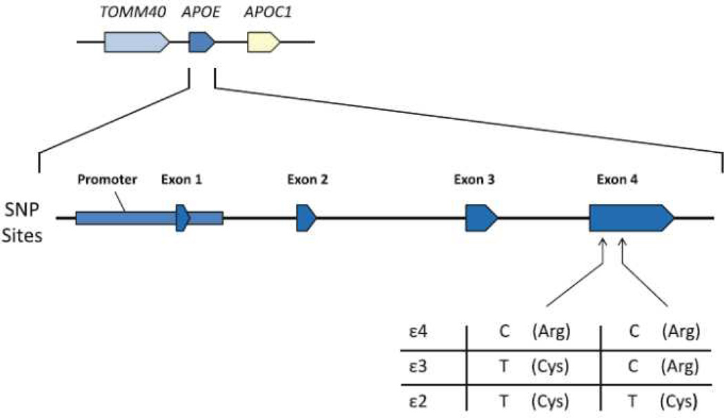
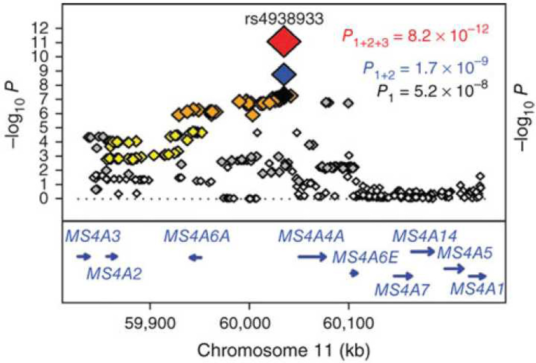
Here we review the genetic causes and risks for Alzheimer's disease (AD). Early work identified mutations in three genes that cause AD: APP, PSEN1 and PSEN2. Although mutations in these genes are rare causes of AD, their discovery had a major impact on our understanding of molecular mechanisms of AD. Early work also revealed the ε4 allele of the APOE as a strong risk factor for AD. Subsequently, SORL1 also was identified as an AD risk gene. More recently, advances in our knowledge of the human genome, made possible by technological advances and methods to analyze genomic data, permit systematic identification of genes that contribute to AD risk. This work, so far accomplished through single nucleotide polymorphism arrays, has revealed nine new genes implicated in AD risk (ABCA7, BIN1, CD33, CD2AP, CLU, CR1, EPHA1, MS4A4E/MS4A6A, and PICALM). We review the relationship between these mutations and genetic variants and the neuropathologic features of AD and related disorders. Together, these discoveries point toward a new era in neurodegenerative disease research that impacts not only AD but also related illnesses that produce cognitive and behavioral deficits.
Figures
Similar articles
-
Genomics of Alzheimer Disease: A Review.JAMA Neurol. 2016 Jul 1;73(7):867-74. doi: 10.1001/jamaneurol.2016.0301. JAMA Neurol. 2016. PMID: 27135718 Free PMC article. Review.
-
Molecular Genetics of Early- and Late-Onset Alzheimer's Disease.Curr Gene Ther. 2021;21(1):43-52. doi: 10.2174/1566523220666201123112822. Curr Gene Ther. 2021. PMID: 33231156 Review.
-
Alzheimer's disease risk genes and mechanisms of disease pathogenesis.Biol Psychiatry. 2015 Jan 1;77(1):43-51. doi: 10.1016/j.biopsych.2014.05.006. Epub 2014 May 17. Biol Psychiatry. 2015. PMID: 24951455 Free PMC article. Review.
-
Genetics of Alzheimer's disease.Adv Genet. 2014;87:245-94. doi: 10.1016/B978-0-12-800149-3.00005-6. Adv Genet. 2014. PMID: 25311924 Review.
-
The genetics of Alzheimer's disease.Clin Interv Aging. 2014 Apr 1;9:535-51. doi: 10.2147/CIA.S51571. eCollection 2014. Clin Interv Aging. 2014. PMID: 24729694 Free PMC article. Review.
Cited by
-
The Role of Clusterin Transporter in the Pathogenesis of Alzheimer's Disease at the Blood-Brain Barrier Interface: A Systematic Review.Biomolecules. 2022 Oct 10;12(10):1452. doi: 10.3390/biom12101452. Biomolecules. 2022. PMID: 36291661 Free PMC article. Review.
-
Different mechanisms of apolipoprotein E isoform-dependent modulation of prostaglandin E2 production and triggering receptor expressed on myeloid cells 2 (TREM2) expression after innate immune activation of microglia.FASEB J. 2015 May;29(5):1754-62. doi: 10.1096/fj.14-262683. Epub 2015 Jan 15. FASEB J. 2015. PMID: 25593125 Free PMC article.
-
Calpain cleavage and inactivation of the sodium calcium exchanger-3 occur downstream of Aβ in Alzheimer's disease.Aging Cell. 2014 Feb;13(1):49-59. doi: 10.1111/acel.12148. Epub 2013 Sep 18. Aging Cell. 2014. PMID: 23919677 Free PMC article.
-
Biology and genetics of prions causing neurodegeneration.Annu Rev Genet. 2013;47:601-23. doi: 10.1146/annurev-genet-110711-155524. Annu Rev Genet. 2013. PMID: 24274755 Free PMC article. Review.
-
Elevated serum pesticide levels and risk for Alzheimer disease.JAMA Neurol. 2014 Mar;71(3):284-90. doi: 10.1001/jamaneurol.2013.6030. JAMA Neurol. 2014. PMID: 24473795 Free PMC article.
References
-
- Adams RH, Klein R. Eph receptors and ephrin ligands. essential mediators of vascular development. Trends Cardiovasc Med. 2000;10:183–188. - PubMed
-
- Andersen OM, Schmidt V, Spoelgen R, Gliemann J, et al. Molecular dissection of the interaction between amyloid precursor protein and its neuronal trafficking receptor SorLA/LR11. Biochemistry. 2006;45:2618–2628. - PubMed
Publication types
MeSH terms
Substances
Grants and funding
LinkOut - more resources
Full Text Sources
Medical
Research Materials
Miscellaneous
