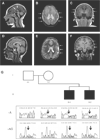
Whole-exome sequencing identifies compound heterozygous mutations in WDR62 in siblings with recurrent polymicrogyria
- PMID: 21834044
- PMCID: PMC3616765
- DOI: 10.1002/ajmg.a.34165
Whole-exome sequencing identifies compound heterozygous mutations in WDR62 in siblings with recurrent polymicrogyria
Abstract
Polymicrogyria is a disorder of neuronal development resulting in structurally abnormal cerebral hemispheres characterized by over-folding and abnormal lamination of the cerebral cortex. Polymicrogyria is frequently associated with severe neurologic deficits including intellectual disability, motor problems, and epilepsy. There are acquired and genetic causes of polymicrogyria, but most patients with a presumed genetic etiology lack a specific diagnosis. Here we report using whole-exome sequencing to identify compound heterozygous mutations in the WD repeat domain 62 (WDR62) gene as the cause of recurrent polymicrogyria in a sibling pair. Sanger sequencing confirmed that the siblings both inherited 1-bp (maternal allele) and 2-bp (paternal allele) frameshift deletions, which predict premature truncation of WDR62, a protein that has a role in early cortical development. The probands are from a non-consanguineous family of Northern European descent, suggesting that autosomal recessive PMG due to compound heterozygous mutation of WDR62 might be a relatively common cause of PMG in the population. Further studies to identify mutation frequency in the population are needed.
Copyright © 2011 Wiley-Liss, Inc.
Figures


Comment in
-
Editorial comment on "Whole exome sequencing identifies compound heterozygous mutations in WDR62 in siblings with recurrent polymicrogyria".Am J Med Genet A. 2011 Sep;155A(9):2069-70. doi: 10.1002/ajmg.a.34183. Epub 2011 Aug 10. Am J Med Genet A. 2011. PMID: 21834027 No abstract available.
Similar articles
-
Editorial comment on "Whole exome sequencing identifies compound heterozygous mutations in WDR62 in siblings with recurrent polymicrogyria".Am J Med Genet A. 2011 Sep;155A(9):2069-70. doi: 10.1002/ajmg.a.34183. Epub 2011 Aug 10. Am J Med Genet A. 2011. PMID: 21834027 No abstract available.
-
Whole-exome sequencing identifies recessive WDR62 mutations in severe brain malformations.Nature. 2010 Sep 9;467(7312):207-10. doi: 10.1038/nature09327. Epub 2010 Aug 22. Nature. 2010. PMID: 20729831 Free PMC article.
-
Characterisation of mutations of the phosphoinositide-3-kinase regulatory subunit, PIK3R2, in perisylvian polymicrogyria: a next-generation sequencing study.Lancet Neurol. 2015 Dec;14(12):1182-95. doi: 10.1016/S1474-4422(15)00278-1. Epub 2015 Oct 29. Lancet Neurol. 2015. PMID: 26520804 Free PMC article.
-
Exome sequencing identifies compound heterozygous mutations in C12orf57 in two siblings with severe intellectual disability, hypoplasia of the corpus callosum, chorioretinal coloboma, and intractable seizures.Am J Med Genet A. 2014 Aug;164A(8):1976-80. doi: 10.1002/ajmg.a.36592. Epub 2014 May 5. Am J Med Genet A. 2014. PMID: 24798461 Review.
-
Identification of two novel SH3PXD2B gene mutations in Frank-Ter Haar syndrome by exome sequencing: Case report and review of the literature.Gene. 2017 Sep 10;628:190-193. doi: 10.1016/j.gene.2017.07.011. Epub 2017 Jul 8. Gene. 2017. PMID: 28694206 Review.
Cited by
-
Next-Generation Sequencing in Intellectual Disability.J Pediatr Genet. 2015 Sep;4(3):128-35. doi: 10.1055/s-0035-1564439. Epub 2015 Oct 12. J Pediatr Genet. 2015. PMID: 27617123 Free PMC article. Review.
-
Small organelle, big responsibility: the role of centrosomes in development and disease.Philos Trans R Soc Lond B Biol Sci. 2014 Sep 5;369(1650):20130468. doi: 10.1098/rstb.2013.0468. Philos Trans R Soc Lond B Biol Sci. 2014. PMID: 25047622 Free PMC article. Review.
-
Disease gene identification strategies for exome sequencing.Eur J Hum Genet. 2012 May;20(5):490-7. doi: 10.1038/ejhg.2011.258. Epub 2012 Jan 18. Eur J Hum Genet. 2012. PMID: 22258526 Free PMC article. Review.
-
Further Delineation of Phenotype and Genotype of Primary Microcephaly Syndrome with Cortical Malformations Associated with Mutations in the WDR62 Gene.Genes (Basel). 2021 Apr 19;12(4):594. doi: 10.3390/genes12040594. Genes (Basel). 2021. PMID: 33921653 Free PMC article.
-
Novel compound heterozygous mutations in WDR62 gene leading to developmental delay and Primary Microcephaly in Saudi Family.Pak J Med Sci. 2019;35(3):764-770. doi: 10.12669/pjms.35.3.36. Pak J Med Sci. 2019. PMID: 31258591 Free PMC article.
References
-
- Bahi-Buisson N, Poirier K, Boddaert N, Fallet-Bianco C, Specchio N, Bertini E, Caglayan O, Lascelles K, Elie C, Rambaud J, Baulac M, An I, Dias P, des Portes V, Moutard ML, Soufflet C, El Maleh M, Beldjord C, Villard L, Chelly J. GPR56-related bilateral frontoparietal polymicrogyria: Further evidence for an overlap with the cobblestone complex. Brain. 2010;133:3194–3209. - PubMed
-
- Becerra JE, Khoury MJ, Cordero JF, Erickson JD. Diabetes mellitus during pregnancy and the risks for specific birth defects: A population-based case-control study. Pediatrics. 1990;85:1–9. - PubMed
Publication types
MeSH terms
Substances
Grants and funding
LinkOut - more resources
Full Text Sources

