

The collagen VI-related myopathies Ullrich congenital muscular dystrophy and Bethlem myopathy
- PMID: 21496625
- PMCID: PMC5207779
- DOI: 10.1016/B978-0-08-045031-5.00005-0
The collagen VI-related myopathies Ullrich congenital muscular dystrophy and Bethlem myopathy
Abstract
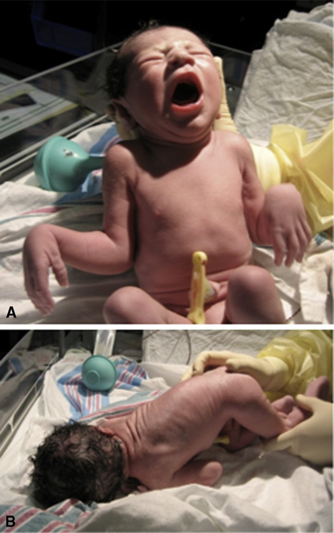
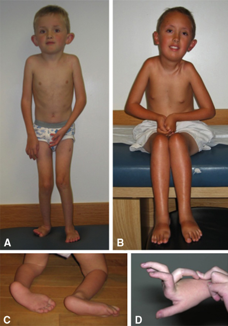
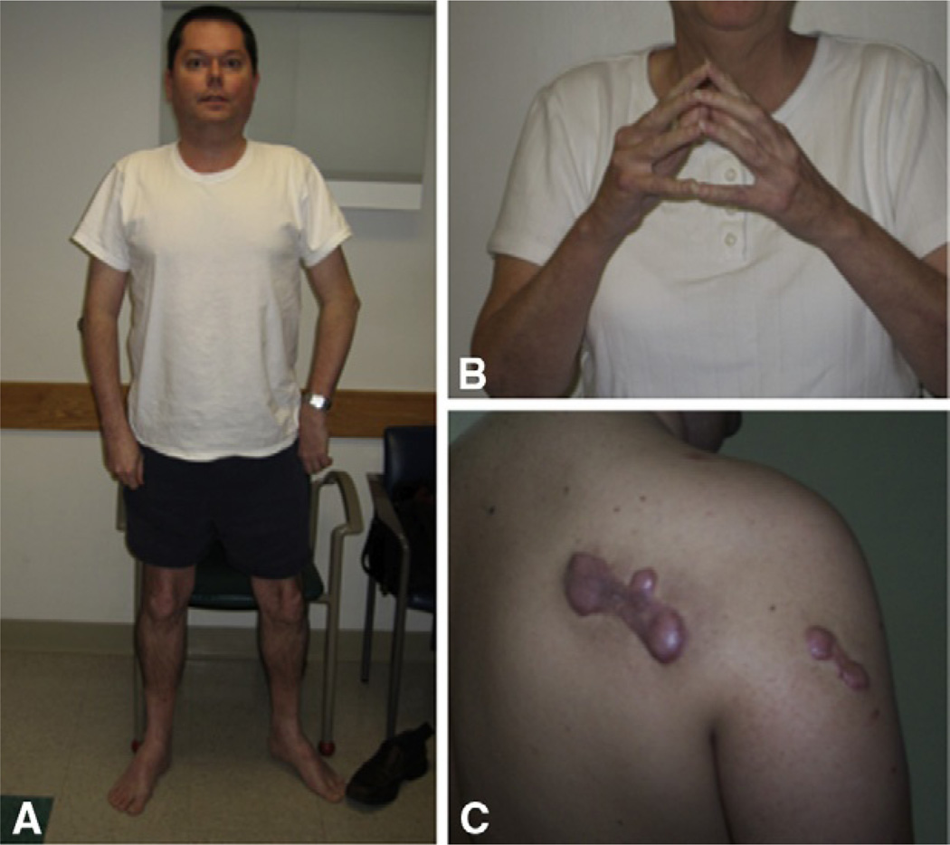
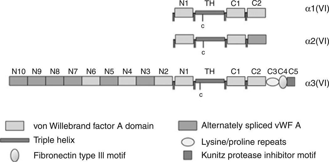
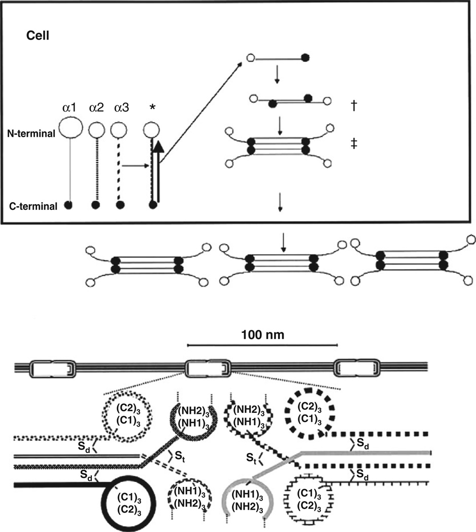
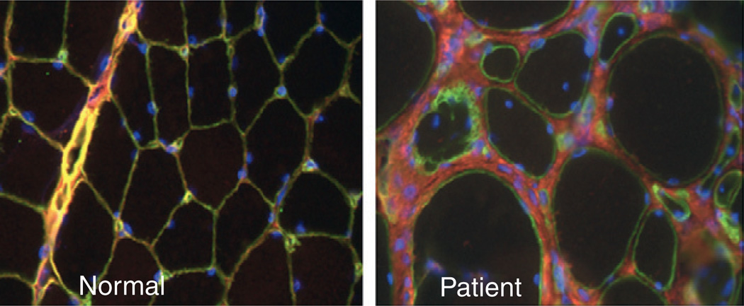
Mutations in the genes COL6A1, COL6A2, and COL6A3, coding for three α chains of collagen type VI, underlie a spectrum of myopathies, ranging from the severe congenital muscular dystrophy-type Ullrich (UCMD) to the milder Bethlem myopathy (BM), with disease manifestations of intermediate severity in between. UCMD is characterized by early-onset weakness, associated with pronounced distal joint hyperlaxity and the early onset or early progression of more proximal contractures. In the most severe cases ambulation is not achieved, or it may be achieved only for a limited period of time. BM may be of early or later onset, but is milder in its manifestations, typically allowing for ambulation well into adulthood, whereas typical joint contractures are frequently prominent. A genetic spectrum is emerging, with BM being caused mostly by dominantly acting mutations, although rarely recessive inheritance of BM is also possible, whereas both dominantly as well as recessively acting mutations underlie UCMD.
Copyright © 2011 Elsevier B.V. All rights reserved.
Figures
Similar articles
-
[Collagen VI-related muscle disorders].Brain Nerve. 2011 Nov;63(11):1169-78. Brain Nerve. 2011. PMID: 22068469 Review. Japanese.
-
Autosomal dominant Ullrich congenital muscular dystrophy due to a de novo mutation in COL6A3 gene. A case report.Acta Myol. 2022 Jun 30;41(2):95-98. doi: 10.36185/2532-1900-073. eCollection 2022 Jun. Acta Myol. 2022. PMID: 35832501 Free PMC article.
-
Exon skipping mutations in collagen VI are common and are predictive for severity and inheritance.Hum Mutat. 2008 Jun;29(6):809-22. doi: 10.1002/humu.20704. Hum Mutat. 2008. PMID: 18366090
-
Automated genomic sequence analysis of the three collagen VI genes: applications to Ullrich congenital muscular dystrophy and Bethlem myopathy.J Med Genet. 2005 Feb;42(2):108-20. doi: 10.1136/jmg.2004.023754. J Med Genet. 2005. PMID: 15689448 Free PMC article.
-
Collagen VI related muscle disorders.J Med Genet. 2005 Sep;42(9):673-85. doi: 10.1136/jmg.2002.002311. J Med Genet. 2005. PMID: 16141002 Free PMC article. Review.
Cited by
-
Examining the lineage autonomous role of β3-integrin in muscle regeneration.FASEB J. 2022 Jul;36(7):e22385. doi: 10.1096/fj.202200464. FASEB J. 2022. PMID: 35734962 Free PMC article.
-
The role of basement membranes in cardiac biology and disease.Biosci Rep. 2021 Aug 27;41(8):BSR20204185. doi: 10.1042/BSR20204185. Biosci Rep. 2021. PMID: 34382650 Free PMC article. Review.
-
Collagen VI-related myopathy with scoliosis alone: A case report and literature review.World J Clin Cases. 2021 Jul 6;9(19):5302-5312. doi: 10.12998/wjcc.v9.i19.5302. World J Clin Cases. 2021. PMID: 34307582 Free PMC article.
-
[Anesthesia for thoracic surgery in a female patient with Ullrich congenital muscular dystrophy].Anaesthesiologie. 2022 Oct;71(10):784-788. doi: 10.1007/s00101-022-01124-9. Epub 2022 May 30. Anaesthesiologie. 2022. PMID: 35925158 Free PMC article. German.
-
Non-invasive quantification of collagen turnover in renal transplant recipients.PLoS One. 2017 Apr 21;12(4):e0175898. doi: 10.1371/journal.pone.0175898. eCollection 2017. PLoS One. 2017. PMID: 28430784 Free PMC article.
References
-
- Atkinson JC, Ruhl M, Becker J, et al. Collagen VI regulates normal and transformed mesenchymal cell proliferation in vitro. Exp Cell Res. 1996;228:283–291. - PubMed
-
- Aumailley M, Specks U, Timpl R. Cell adhesion to type VI collagen. Biochem Soc Trans. 1991;19:843–847. - PubMed
-
- Baker NL, Morgelin M, Peat R, et al. Dominant collagen VI mutations are a common cause of Ullrich congenital muscular dystrophy. Hum Mol Genet. 2005;14:279–293. - PubMed
-
- Baker NL, Morgelin M, Pace RA, et al. Molecular consequences of dominant Bethlem myopathy collagen VI mutations. Ann Neurol. 2007;62:390–405. - PubMed
Publication types
MeSH terms
Substances
Supplementary concepts
Grants and funding
LinkOut - more resources
Full Text Sources
Miscellaneous
