. 2007 Jul 19;55(2):217-30.
doi: 10.1016/j.neuron.2007.06.029.

The transcription factor Yin Yang 1 is essential for oligodendrocyte progenitor differentiation

Affiliations

The transcription factor Yin Yang 1 is essential for oligodendrocyte progenitor differentiation

Ye He et al. Neuron. .

Abstract

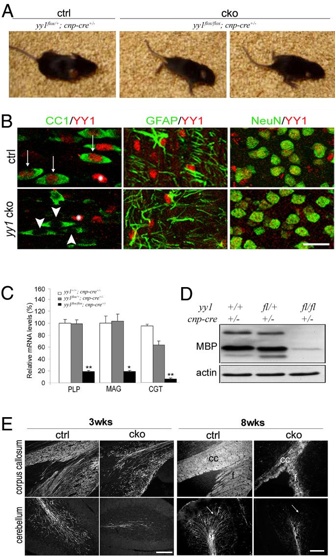
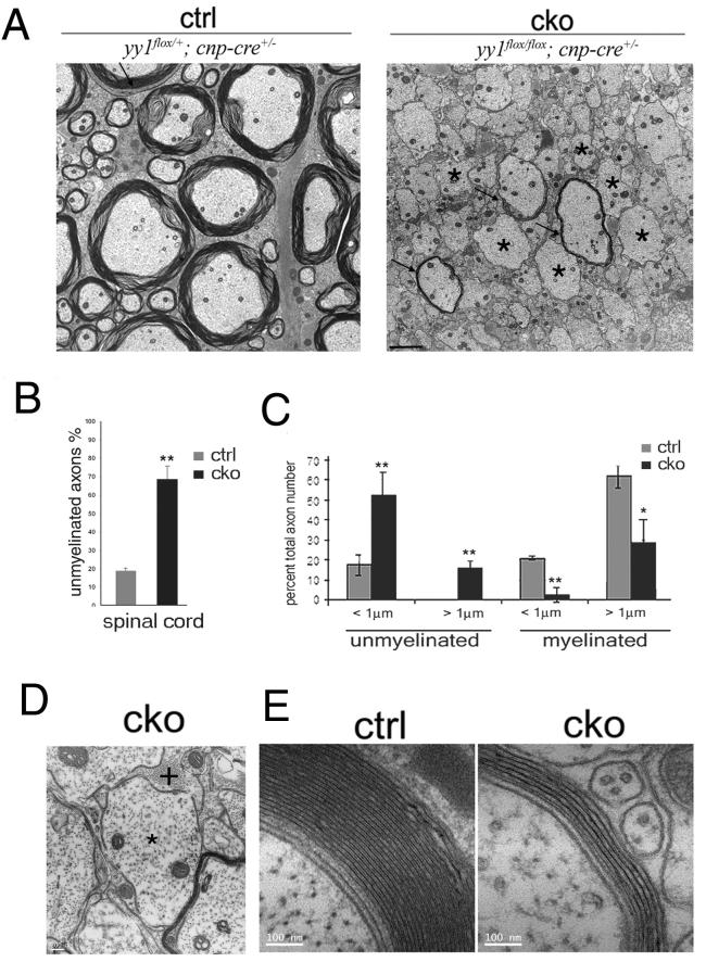
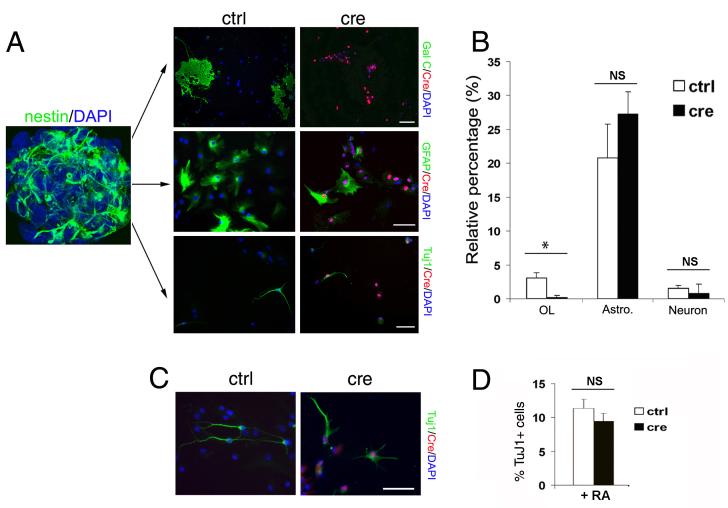
The progression of progenitors to oligodendrocytes requires proliferative arrest and the activation of a transcriptional program of differentiation. While regulation of cell cycle exit has been extensively characterized, the molecular mechanisms responsible for the initiation of differentiation remain ill-defined. Here, we identify the transcription factor Yin Yang 1 (YY1) as a critical regulator of oligodendrocyte progenitor differentiation. Conditional ablation of yy1 in the oligodendrocyte lineage in vivo induces a phenotype characterized by defective myelination, ataxia, and tremor. At the cellular level, lack of yy1 arrests differentiation of oligodendrocyte progenitors after they exit from the cell cycle. At the molecular level, YY1 acts as a lineage-specific repressor of transcriptional inhibitors of myelin gene expression (Tcf4 and Id4), by recruiting histone deacetylase-1 to their promoters during oligodendrocyte differentiation. Thus, we identify YY1 as an essential component of the transcriptional network regulating the transition of oligodendrocyte progenitors from cell cycle exit to differentiation.


Figures

Figure 1
Figure 1. Conditional ablation of YY1 in the oligodendrocyte lineage in mice results in a trembling phenotype
(A) Conditional yy1 knockout mice (cko), obtained by crossing yy1flox/flox with Cnp-cre mice displayed progressive tremor, ataxia and head wobbling during the second-third postnatal week. Examples of abnormal posturing and gait in the mutant mice (cko) at postnatal day 18 (p18) are shown in comparison with control siblings (ctrl). (B) Double immunostaining of cells in white matter tracts of p18 mouse sections stained for YY1 (red) and cell-specific markers (green): CC1 for oligodendrocytes, GFAP for astrocytes and NeuN for neurons. Note the specific deletion of YY1 in oligodendrocytes. Scale bar = 20 μm. (C) Quantitative real-time PCR of brain RNA from wild type (yy1+/+; cnp-cre+/-), heterozygotes (yy1flox/+; cnp-cre+/-) and conditional knockout (yy1flox/flox; cnpcre+/-) mice at p14. The levels of the indicated transcripts are normalized to GAPDH and the mRNA control levels are arbitrarily set as 100. The bar graph indicates decreased myelin gene transcripts in yy1 cko mice (black), but not in heterozygous siblings (gray) compared to controls (white) *p<0.05, **p<0.01. (D) Western blot analysis of protein lysates from the cortex of p14 mice revealed decreased myelin basic protein (MBP) expression in the mutants. Beta-actin serves as loading control. (E) Brain sagittal sections stained for PLP (3wks) and for MBP (8wks) reveal fewer myelinated fibers in the corpus callosum (cc), fornix (f) and cerebellum of yy1 cko mice compared to controls (ctrl). Scale bar = 100 μm.
Figure 2
Figure 2. Defective myelination in the spinal cord of yy1 conditional knockout mice
(A) Ultrastructural analysis of spinal cord sections shows lack of myelin (asterisks) and thinner myelin sheaths (arrows) in p18 yy1 cko mice compared to controls. Scale bar= 2 μm. (B) Bar graph shows increased % of unmyelinated axons relative to total number of axons in the spinal cord of yy1 cko mice (black) compared to controls (gray). (C) Bar graph shows the relative distribution of unmyelinated and myelinated axons in relation to axonal diameter in the spinal cord of yy1 cko mice (black) and controls (gray) at p18 *p<0.05; **p<0.01. (D) Representative EM picture of a large yy1 cko spinal cord axon (diameter >1.5μm) that lacks myelin (*) and is in contact with an astrocytic process (+). Scale bar = 200 nm. (E) Representative EM pictures of myelinated axons of equivalent diameter in control and yy1 cko siblings. Scale bar = 100nm.
Figure 3
Figure 3. Impaired oligodendrocyte progenitor differentiation in the spinal cord of yy1 conditional mutants
(A) Confocal image of lumbar spinal cord sections from controls (ctrl) and yy1 cko mice at postnatal day 2, 11 and 18, stained with antibodies specific for NG2 (green) to identify progenitors, CC1 (red) to identify oligodendrocytes and DAPI (blue) to counterstain nuclei. Optical sections (Z = 1.0 μm; X = 12 μm) of confocal epifluorescence images were sequentially acquired and LSM software was used to merge images. Examples of NG2+/DAPI+ and CC1+/DAPI+cells selected for counting are shown in the boxed areas (arrowheads) while their relative position is shown at low magnification (arrows). Scale bar=50μm, 10μm in inserts. (B) Quantification of the data shown in panel A. The values indicate mean ± SD of cell counts obtained in 3-4 mice of each genotype per time point. (C) Co-localization of progenitor markers PDGFRα (red) and NG2 (green) in the spinal cord of yy1 cko mice at p18. Scale bar=20μm
Figure 4
Figure 4. In vitro ablation of yy1 in oligodendrocyte progenitors prevents their differentiation into oligodendrocytes
(A) Primary oligodendrocyte progenitors isolated from the cortex of neonatal yy1flox/flox mice were transduced with adenoviral vectors expressing the recombinase Cre (CMV-Cre). Forty-eight hours later the cells were differentiated by mitogen withdrawal. Immunoreactivity for O4 (green) was assessed 3 days later, while immunoreactivity for MBP, GalC or PLP (green) was assessed 5 days after transduction. Note that the Cre+ yy1flox/flox cells (red) progressed to the O4+ stage, but were unable to differentiate into myelin-expressing cells. Scale bar = 50μm. (B) Quantification of three distinct experiments performed in duplicate. * p<0.05, ***p<0.001.
Figure 5
Figure 5. Lack of YY1 does not affect the ability of oligodendrocyte progenitors to exit from the cell cycle
(A) Confocal image of spinal cord sections of p2 yy1 cko and control siblings (ctrl) in vivo labeled with BrdU for 1 hour. Immunohistochemistry with YY1 (red), BrdU (green) and DAPI (blue) revealed a similar distribution BrdU+ cells in mice of the two genotypes. Scale bar = 20μm. (B) Quantification of BrdU+ cells in white matter tracts of the spinal cord at the indicated time points; *p<0.05. (C) Representative images of spinal cord sections of control and cko mice stained with antibodies against Ki-67 (green) to identify cells in any phase of the cell cycle except for G0 and BrdU (red) to identify cells in S phase at the time of labeling. (D) Cell cycle exit index was calculated by dividing the number of BrdU+/Ki-67 cells, by the total number of BrdU+ cells. Scale bar = 20μm.
Figure 6
Figure 6. YY1 is a repressor of oligodendrocyte differentiation inhibitors Id4 and Tcf4
(A) Validation of gene expression profiling studies in the cortex of yy1 cko mice and control littermates at p2, by semi-quantitative (A) and quantitative (B) RT-PCR. The RNA levels were normalized to the levels of GAPDH and the values in the control mice were arbitrarily set as 1. *p<0.05. (C) The expression level of Tcf4 and Id4 was down-regulated in oligodendrocyte progenitors transfected with pCX-yy1-EGFP (YY1) and not in cells transfected with pCX-EGFP vector as control. (D) Luciferase activity of immortalized oligodendrocyte progenitors (Oli-neu cells) co-transfected with Tcf4, and either myelin (MBP-luc, CGT-luc) or astrocytes-specific (GFAP-luc) promoters driving luciferase reporters. The luciferase activity in pCDNA3 transfected controls was arbitrarily set as 100; ** p<0.01.
Figure 7
Figure 7. In vitro ablation of YY1 in nestin+ precursor cells impairs oligodendrocyte differentiation in a cell type specific manner
(A) Subventricular zone (SVZ) cells were isolated from neonatal yy1flox/flox mice and cultured as nestin+ (green) neurospheres. Dissociated neurospheres without (ctrl) or with adenovirus-CMV-Cre transduction (cre) were allowed to differentiate into oligodendrocytes, astrocytes and neurons. Seven days later the cultures were stained for Cre (red) and for the lineage specific markers (green): Gal C (oligodendrocytes), GFAP (astrocytes) and Tuj1 (neurons). Scale bar=50μm. (B) Bar graphs indicate the percentage of immunoreactive cells relative to total cell number. * p<0.05. (C) The experiment was repeated in medium supplemented with retinoic acid (RA) to promote neuronal differentiation. (D) Bar graphs show that even in this culture condition no significant difference was detected between control and yy1- cells.
Figure 8
Figure 8. The YY1-dependent repression of Tcf4 and Id4 is lineage-specific and mediated by recruitment of HDAC1 to their promoters
(A) Quantitative RT-PCR of Tcf4, Id4 and REST in oligodendrocyte progenitors transfected with pCX-EGFP (vector) or pCX-yy1-EGFP (YY1) and induced to differentiate into astrocytes (astro) or oligodendrocytes (oligo). The transcript levels of each gene in vector transfected cells was arbitrarily set as 100; *p<0.05. (B) Co-immunoprecipitation of YY1 and HDAC-1 and -2. Western blot analysis of whole cell lysates (lysates) and YY1 immunoprecipitated protein extracts (IP:YY1) derived from undifferentiated progenitors (prog) and cells differentiated into oligodendrocytes (diff prog) or astrocytes (astro). (C) YY1 activity, measured by TransLucent vector reporter system. Note the increased activity of YY1 in cultures of progenitors differentiating into oligodendrocytes (diff. prog.) compared to undifferentiated cells (prog.) or cells differentiating into astrocytes (astro). **p<0.01. (D) Chromatin immunoprecipitation (ChIP) of samples isolated from progenitors (prog), differentiating oligodendrocytes (diff) or astrocytes (astro) and immunoprecipitated with antibodies against YY1 and HDAC1. The diagram shows the Tcf4 promoter with the relative position of the YY1 consensus sequences (black boxes) and the regions of DNA (roman numerals) amplified by specific primer pairs (arrows). Input DNA was used as positive control, while ChIP in the absence of antibodies or amplification of immunoprecipitated chromatin with primers for region IV were used as negative controls. (E) ChIP of samples isolated in the same conditions described above. The diagram shows the Id4 promoter, with the position of the YY1 consensus sequence (black box) and the regions of the promoter (roman numerals) amplified by specific primer pairs (arrows). YY1 was bound to the Id4 promoter in progenitors, but it recruited HDAC1 only when cells differentiated into oligodendrocytes. No binding was observed in progenitors differentiating into astrocytes. (F) Model of oligodendrocyte progenitor differentiation as two step event. First, proliferating progenitors exit from the cell cycle and remain in an undifferentiated state characterized by high levels of transcriptional inhibitors (Id4, Tcf4) and lack of myelin gene expression. As the progenitors begin differentiating, repressive complexes containing YY1 and HDAC1 are recruited to the promoter of these inhibitors. The decreased levels of these inhibitory molecules allow myelin gene expression to begin.

Similar articles

Cited by

References

    1. Affar el B, Gay F, Shi Y, Liu H, Huarte M, Wu S, Collins T, Li E, Shi Y. Essential dosage-dependent functions of the transcription factor yin yang 1 in late embryonic development and cell cycle progression. Mol Cell Biol. 2006;26:3565–3581. - PMC - PubMed
    1. Alvarez-Salas LM, Benitez-Hess ML, Dipaolo JA. YY-1 and c-Jun transcription factors participate in the repression of the human involucrin promoter. Int J Oncol. 2005;26:259–266. - PubMed
    1. Ballas N, Grunseich C, Lu DD, Speh JC, Mandel G. REST and its corepressors mediate plasticity of neuronal gene chromatin throughout neurogenesis. Cell. 2005;121:645–657. - PubMed
    1. Barres BA, Lazar MA, Raff MC. A novel role for thyroid hormone, glucocorticoids and retinoic acid in timing oligodendrocyte development. Development. 1994;120:1097–1108. - PubMed
    1. Belachew S, Chittajallu R, Aguirre AA, Yuan X, Kirby M, Anderson S, Gallo V. Postnatal NG2 proteoglycan-expressing progenitor cells are intrinsically multipotent and generate functional neurons. J Cell Biol. 2003;161:169–186. - PMC - PubMed

Publication types

Substances