. 2003 Oct;112(8):1255-63.
doi: 10.1172/JCI19159.

Autosomal dominant pseudohypoparathyroidism type Ib is associated with a heterozygous microdeletion that likely disrupts a putative imprinting control element of GNAS

Affiliations

Autosomal dominant pseudohypoparathyroidism type Ib is associated with a heterozygous microdeletion that likely disrupts a putative imprinting control element of GNAS

Murat Bastepe et al. J Clin Invest. 2003 Oct.

Abstract

Patients with pseudohypoparathyroidism type Ib (PHP-Ib) have hypocalcemia and hyperphosphatemia due to renal parathyroid hormone (PTH) resistance, but lack physical features of Albright hereditary osteodystrophy. PHP-Ib is thus distinct from PHP-Ia, which is caused by mutations in the GNAS exons encoding the G protein alpha subunit. However, an imprinted autosomal dominant form of PHP-Ib (AD-PHP-Ib) has been mapped to a region of chromosome 20q13.3 containing GNAS. Furthermore, loss of methylation at a differentially methylated region (DMR) of this locus, exon A/B, has been observed thus far in all investigated sporadic PHP-Ib cases and the affected members of multiple AD-PHP-Ib kindreds. We now report that affected members and obligate gene carriers of 12 unrelated AD-PHP-Ib kindreds and four apparently sporadic PHP-Ib patients, but not healthy controls, have a heterozygous approximately 3-kb microdeletion located approximately 220 kb centromeric of GNAS exon A/B. The deleted region, which is flanked by two direct repeats, includes three exons of STX16, the gene encoding syntaxin-16, for which no evidence of imprinting could be found. Affected individuals carrying the microdeletion show loss of exon A/B methylation but no epigenetic abnormalities at other GNAS DMRs. We therefore postulate that this microdeletion disrupts a putative cis-acting element required for methylation at exon A/B, and that this genetic defect underlies the renal PTH resistance in AD-PHP-Ib.


Figures

Figure 1
Figure 1
The GNAS locus and parent-of-origin–specific inheritance of hormone resistance. (a) GNAS gives rise, besides Gsα, to multiple other transcripts including NESP55, XLαs, the antisense (AS) and A/B (also referred to as 1A or 1′), all of which show differential methylation (asterisks) in their promoters and are expressed exclusively from the non-methylated paternal (p) or maternal (m) allele. Exons that lead to transcripts in the sense (right-pointing arrow) and antisense (left-pointing arrow) directions are depicted as black and gray boxes, respectively, and the splice patterns are indicated. Note that the promoter giving rise to the Gsα transcript, encoded by exons 1 through 13, does not show differential methylation. However, although expression is biallelic in most tissues, Gsα transcription from the paternal GNAS allele is proposed to be silenced in a limited number of cells (dotted arrow), including those in renal proximal tubules. (b) In kindreds with PHP-Ia and PPHP, as well as in those with AD-PHP-Ib, the genetic defect (black squares) leads to hormone resistance only if it is inherited from a female obligate carrier of the GNAS mutation. On the other hand, hormone resistance does not develop if the genetic defect is inherited from a male obligate carrier. Each black square indicates the mutation leading to PHP-Ia/PPHP or AD-PHP-Ib. Paternal and maternal alleles of the GNAS locus are depicted by white and gray rectangles, respectively. X’s indicate the silencing of paternal Gsα expression that occurs in a small number of cells, including those of the renal proximal tubules.
Figure 2
Figure 2
The AD-PHP-Ib critical interval. The AD-PHP-Ib genetic interval was determined by recombination events in kindreds W (centromeric [cen]) and F (telomeric [tel]). Boundary markers are underlined; microsatellites are in italics. While the distance between 907-Rep2 and 806M20-119516 is about 300 kb, the region between 806M20-98760 and 806M20-119516 has been previously excluded through direct sequence analysis (23). Thus, the critical interval comprises approximately 280 kb. Note that 261P9-CA1 is a dinucleotide repeat located within intron 6 of STX16. Known and predicted genes within the linked interval are depicted as filled or open boxes, respectively (note that individual exons are not shown). For GNAS, exons are shown with black (sense) and gray (antisense) boxes (N, exon NESP55; A, exons encoding an antisense transcript; X, exon XL; A/B, exon A/B; 1–13, exons encoding Gsα). MGC4294 (National Center for Biotechnology Information locus ID: 79160) and NPEPL1 (locus ID: 79716) are predicted genes. Note that the centromeric boundary of the AD-PHP-Ib locus was previously defined at D20S149 (22, 23). However, the daughter of W-II/9 in kindred W (23) was shown to have a loss of exon A/B methylation, making the recombination event in W-II/9 informative, which redefined the centromeric boundary at 907-Rep2.
Figure 3
Figure 3
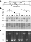
Identification of the 3-kb microdeletion in kindred F. (a) Genomic structure of the region surrounding the microdeletion. Filled boxes and connecting lines depict STX16 exons and introns, respectively. Horizontal bars represent the probes used in Southern blot analysis. Arrowheads indicate the location of the two 391-bp direct repeats (nucleotides 3,786–4,176 and nucleotides 6,764–7,154 of AL139349). Arrows indicate the approximate positions of the PCR primers used for amplification of the wild-type and mutant alleles. (b) Southern blot analysis of genomic DNA from selected members of kindred F (22). Affected individuals, filled symbols and bold identification numbers. Healthy individuals, open symbols and plain numbers. Unaffected obligate gene carrier, gray circles and bold italic numbers. Genomic DNA digested with either AvrII (top) or ClaI (bottom) was separated on 0.8% agarose and transferred onto nitrocellulose before hybridization with the radiolabeled probe. (c) PCR amplification of the wild-type (∼4.3 kb) and mutant (∼1.3 kb) alleles from genomic DNA of members of kindred F (22). Nucleotide sequence analysis of the 1.3-kb PCR product derived from the mutant allele revealed that 2,978 bp were deleted (based on AL139349 sequence), although this number varies due to polymorphisms including the dinucleotide repeat polymorphism 261P9-CA1 (see Supplemental Table 1; www.jci.org/cgi/content/full/112/8/1255/DC1).
Figure 4
Figure 4
Genomic structure of the human chromosomal region containing the AD-PHP-Ib microdeletion. The illustrated region, extending from nucleotides 3,600 to 7,600 of AL139349, contains STX16 exons 4–6 (black boxes), which are flanked by almost-identical 391-bp direct repeats (gray boxes); note that only two nucleotides differ between the two repeat sequences and that nucleotide 3,959 is a polymorphic C/T (see Supplemental Table 1; www.jci.org/cgi/content/full/112/8/1255/DC1). The approximate location of the dinucleotide repeat marker 261P9-CA1 is indicated. Horizontal bars represent uninterrupted sequence stretches showing more than 50% identity between human and mouse or human and rat. Note that the longest stretch of nucleotide sequence conservation, besides STX16 exons 4–6, is observed in a 98-bp segment located within intron 4 (nucleotides 5,460–5,557 of AL139349). Dot blots confirmed the location of this segment in intron 4 of mouse and rat Stx16 (not shown); sequences homologous to this region could not be found in GenBank. The 391-bp repeats are not present in the corresponding regions of mouse and rat genomes. No CpG islands are found in the deleted region. However, a 140-bp segment within exon 4 (white box; nucleotides 4,572–4,711 of AL139349) has an observed/expected (Obs/Exp) CpG > 0.6 and % (C + G) > 50; note that the corresponding regions in mouse and rat do not meet these criteria. Bisulfite PCR analysis indicated that all 15 CpG dinucleotides in this region were methylated (asterisks) on the maternal (m) and paternal (p) alleles, i.e., there was no evidence for differential, parent-specific methylation.
Figure 5
Figure 5
Possible molecular mechanism underlying the marked loss of Gsα expression in the renal proximal tubule in AD-PHP-Ib. Maternal inheritance of the microdeletion is associated with loss of methylation at exon A/B and no epigenetic abnormalities at the NESP55 and XL DMRs, while paternal inheritance of the same deletion is not associated with methylation defects. We thus postulate that the identified microdeletion, which includes STX16 exons 4–6, disrupts a putative cis-acting element regulating the exon A/B methylation imprint. It has been suggested that in the renal proximal tubules, a lack of exon A/B methylation and/or active transcription of A/B mRNA, both of which are normally seen on the paternal GNAS allele, mediate, in cis, the silencing of Gsα transcription (24). Therefore, the maternal loss of exon A/B methylation in AD-PHP-Ib is predicted to cause a marked reduction in Gsα expression levels in that tissue, thereby leading to resistance to PTH (and perhaps to other hormones), whose action is mediated by the PTH/PTHrP receptor, a predominantly Gsα-coupled receptor (43). Black and gray boxes depict exons of STX16 and GNAS in the sense or antisense direction, respectively. White boxes, NPEPL1; asterisks, CpG methylation; arrows, direction of active transcription.

Similar articles

Cited by

References

    1. Levine, M.A. 2002. Pseudohypoparathyroidism. In Principles of bone biology. J.P. Bilezikian, L.G. Raisz, and G.A. Rodan, editors. Academic Press. New York, New York, USA. 1137–1163.
    1. Weinstein LS, Yu S, Warner DR, Liu J. Endocrine manifestations of stimulatory G protein alpha-subunit mutations and the role of genomic imprinting. Endocr. Rev. 2001;22:675–705. - PubMed
    1. Kozasa T, Itoh H, Tsukamoto T, Kaziro Y. Isolation and characterization of the human Gsα gene. Proc. Natl. Acad. Sci. U. S. A. 1988;85:2081–2085. - PMC - PubMed
    1. Kehlenbach RH, Matthey J, Huttner WB. XLαs is a new type of G protein [erratum 1995, 375:253] Nature. 1994;372:804–809. - PubMed
    1. Pasolli H, Klemke M, Kehlenbach R, Wang Y, Huttner W. Characterization of the extra-large G protein alpha-subunit XLalphas. I. Tissue distribution and subcellular localization. J. Biol. Chem. 2000;275:33622–33632. - PubMed

Publication types

Substances