

Molecular and fluorescence in situ hybridization characterization of the breakpoints in 46 large supernumerary marker 15 chromosomes reveals an unexpected level of complexity
- PMID: 14560400
- PMCID: PMC1180486
- DOI: 10.1086/379155
Molecular and fluorescence in situ hybridization characterization of the breakpoints in 46 large supernumerary marker 15 chromosomes reveals an unexpected level of complexity
Abstract
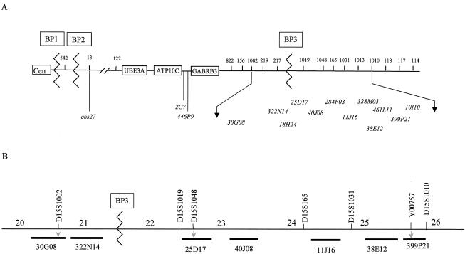
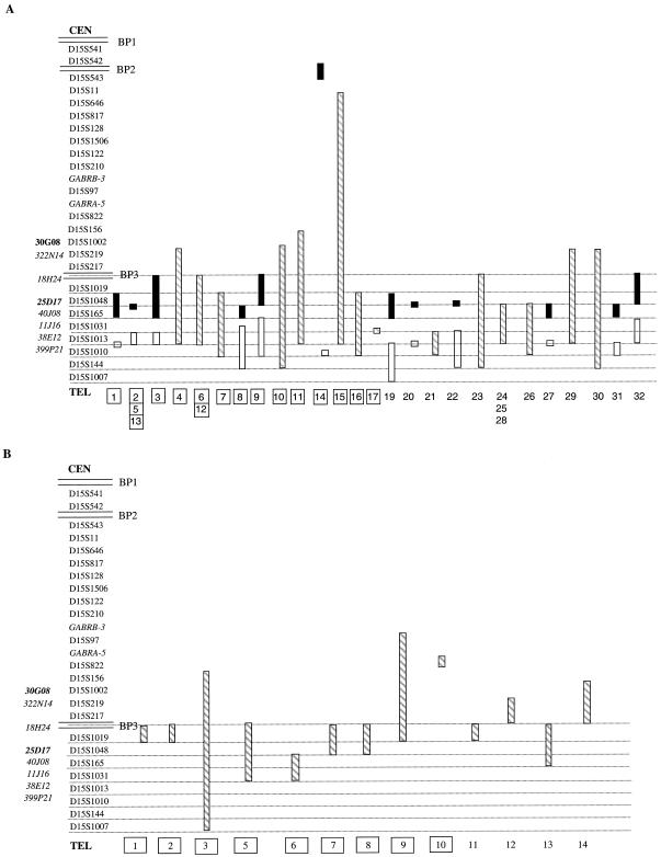
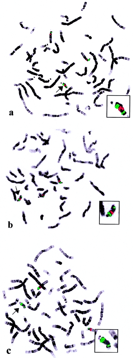
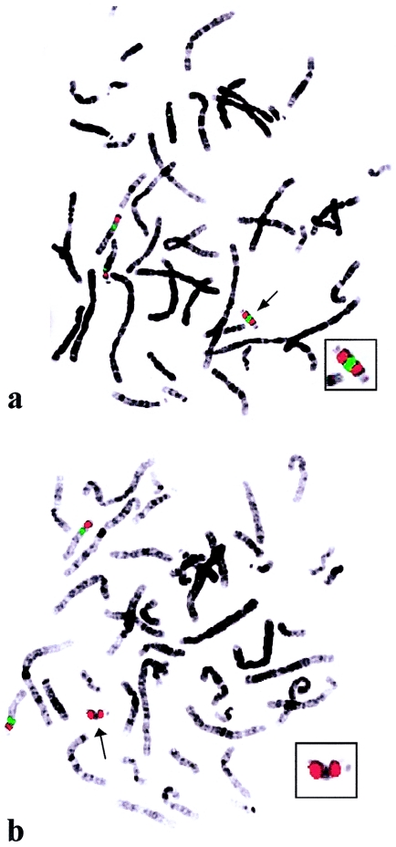
Supernumerary marker chromosomes (SMCs) of chromosome 15, designated "SMC(15)s," are the most common SMC in humans, accounting for as much as 60% of all those observed. We report the characterization of 46 large SMC(15)s, using both fluorescence in situ hybridization and polymerase chain reaction analysis within and distal to the Prader-Willi/Angelman syndrome critical region (PWACR). Our aim was to establish detailed information on origin, content, and breakpoints, to address the formation of SMC(15)s, and to facilitate genotype-phenotype correlations. For all patients in whom we were able to establish the parental origin, the SMC(15)s were maternally derived. Two patients were observed who had familial SMC(15)s, both inherited from the mother; however, in all remaining patients for whom parental samples were available, the SMC(15)s were shown to have arisen de novo. With one exception, all the SMC(15)s were shown to include the entire PWACR. Detailed investigations of the distal breakpoints categorized the SMC(15)s into two groups. Group A, representing approximately two-thirds of the SMC(15)s, had a breakpoint beyond the standard distal PWS/AS deletion breakpoint BP3, at a position close to the microsatellite marker D15S1010 and the bacterial artificial chromosome 10I10. The group B SMC(15)s were shorter, with more variable breakpoints located around BP3. The majority of the SMC(15)s were shown to have asymmetrical breakpoints, with the two inverted arms of the SMC being unequal in length. Our study revealed an unexpected level of complexity and heterogeneity among SMC(15)s that is not seen in other chromosome 15 rearrangements, such as deletions and duplications. This suggests that multiple mechanisms are involved in the formation of large SMC(15)s.
Figures
Similar articles
-
Chromosome breakage in the Prader-Willi and Angelman syndromes involves recombination between large, transcribed repeats at proximal and distal breakpoints.Am J Hum Genet. 1999 Aug;65(2):370-86. doi: 10.1086/302510. Am J Hum Genet. 1999. PMID: 10417280 Free PMC article.
-
Investigation of a cryptic interstitial duplication involving the Prader-Willi/Angelman syndrome critical region.Hum Genet. 1999 Nov;105(5):384-7. doi: 10.1007/s004390051120. Hum Genet. 1999. PMID: 10598802
-
Multiple forms of atypical rearrangements generating supernumerary derivative chromosome 15.BMC Genet. 2008 Jan 4;9:2. doi: 10.1186/1471-2156-9-2. BMC Genet. 2008. PMID: 18177502 Free PMC article.
-
Trisomy 17p10-p12 due to mosaic supernumerary marker chromosome: delineation of molecular breakpoints and clinical phenotype, and comparison to other proximal 17p segmental duplications.Am J Med Genet A. 2005 Oct 1;138A(2):175-80. doi: 10.1002/ajmg.a.30948. Am J Med Genet A. 2005. PMID: 16152635 Review.
-
Prenatal diagnosis of supernumerary marker 15 chromosomes and exclusion of uniparental disomy for chromosome 15.Prenat Diagn. 1999 Aug;19(8):721-6. Prenat Diagn. 1999. PMID: 10451515 Review.
Cited by
-
Microdeletion/duplication at 15q13.2q13.3 among individuals with features of autism and other neuropsychiatric disorders.J Med Genet. 2009 Apr;46(4):242-8. doi: 10.1136/jmg.2008.059907. Epub 2008 Sep 19. J Med Genet. 2009. PMID: 18805830 Free PMC article.
-
De novo unbalanced translocations in Prader-Willi and Angelman syndrome might be the reciprocal product of inv dup(15)s.PLoS One. 2012;7(6):e39180. doi: 10.1371/journal.pone.0039180. Epub 2012 Jun 14. PLoS One. 2012. PMID: 22720067 Free PMC article.
-
Supernumerary marker chromosome 15 in a male with azoospermia and open bite deformity.Asian J Androl. 2009 Sep;11(5):617-22. doi: 10.1038/aja.2009.37. Epub 2009 Aug 24. Asian J Androl. 2009. PMID: 19701220 Free PMC article.
-
Complex chromosomal rearrangement involving 15q11-q13 interstitial triplication and duplication: A new case report of dysmorphic and neuropsychiatric features.Clin Case Rep. 2022 May 15;10(5):e05835. doi: 10.1002/ccr3.5835. eCollection 2022 May. Clin Case Rep. 2022. PMID: 35600042 Free PMC article.
-
Possible Phenotypic Consequences of Structural Differences in Idic(15) in a Small Cohort of Patients.Int J Mol Sci. 2019 Oct 5;20(19):4935. doi: 10.3390/ijms20194935. Int J Mol Sci. 2019. PMID: 31590400 Free PMC article.
References
Electronic-Database Information
-
- Ensembl Genome Browser, http://www.ensembl.org (for BACs and microsatellite markers)
-
- Entrez, http://www.ncbi.nlm.nih.gov/entrez (for microsatellite markers)
-
- Genome Database, http://www.gdb.org (for microsatellite marker primer sequences)
-
- Online Mendelian Inheritance in Man (OMIM), http://www.ncbi.nlm.nih.gov/Omim/ (for AS, ATP10, HERC2, and PWS
-
- Unique—Rare Chromosome Disorder Support Group, http://www.rarechromo.org - PubMed
References
-
- Blennow E, Bui TH, Kristoffersson U, Vujic M, Anneren G, Holmberg E, Nordenskjold M (1994) Swedish survey on extra structurally abnormal chromosomes in 39,105 consecutive prenatal diagnoses: prevalence and characterisation by fluorescence in situ hybridisation. Prenat Diagn 14:1019–1028 - PubMed
-
- Buckton KE, Spowart G, Newton MS, Evans HJ (1985) Forty-four probands with an additional “marker” chromosome. Hum Genet 69:353–370 - PubMed
-
- Cassidy S, Conroy J, Becker L, Schwartz S (1996) Paternal triplication of 15q11-q13 in a hypotonic, developmentally delayed child without Prader-Willi or Angelman syndrome. Am J Med Genet 62:205–212
-
- Choo KH, Earle E, Vissel B, Filby RG (1990) Identification of two distinct subfamilies of α satellite DNA that are highly specific for human chromosome 15. Genomics 7:143–151. - PubMed
Publication types
MeSH terms
LinkOut - more resources
Full Text Sources
Research Materials
