. 2001 Nov;29(3):321-5.
doi: 10.1038/ng753.

A 1.5 million-base pair inversion polymorphism in families with Williams-Beuren syndrome

Affiliations

A 1.5 million-base pair inversion polymorphism in families with Williams-Beuren syndrome

L R Osborne et al. Nat Genet. 2001 Nov.

Abstract

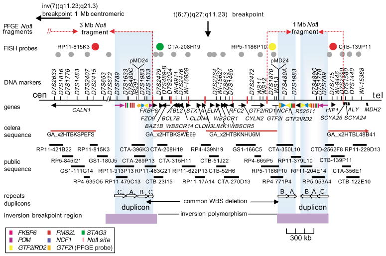
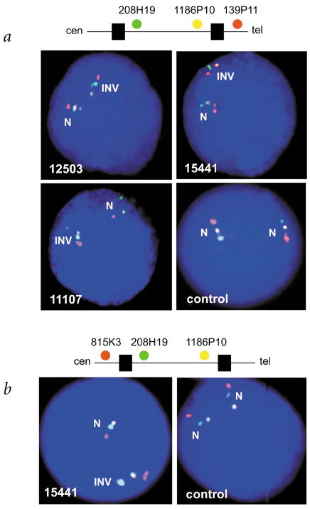
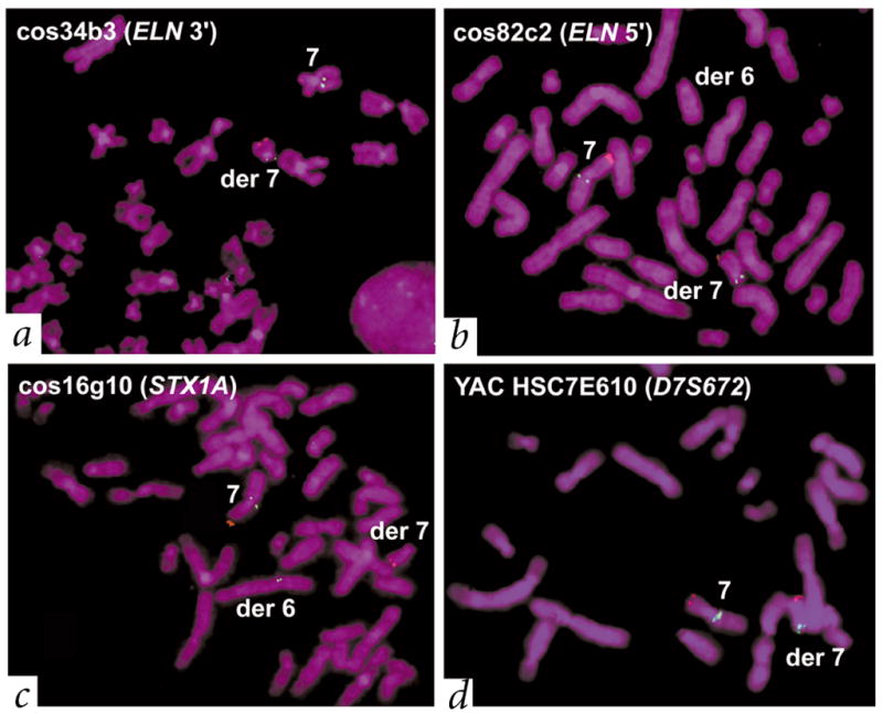
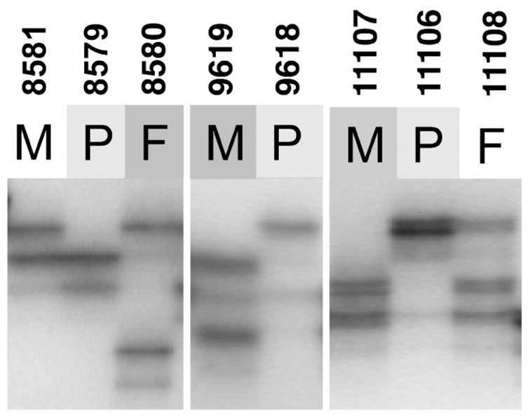
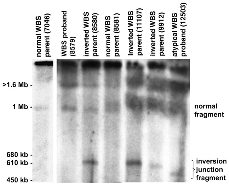
Williams-Beuren syndrome (WBS) is most often caused by hemizygous deletion of a 1.5-Mb interval encompassing at least 17 genes at 7q11.23 (refs. 1,2). As with many other haploinsufficiency diseases, the mechanism underlying the WBS deletion is thought to be unequal meiotic recombination, probably mediated by the highly homologous DNA that flanks the commonly deleted region. Here, we report the use of interphase fluorescence in situ hybridization (FISH) and pulsed-field gel electrophoresis (PFGE) to identify a genomic polymorphism in families with WBS, consisting of an inversion of the WBS region. We have observed that the inversion is hemizygous in 3 of 11 (27%) atypical affected individuals who show a subset of the WBS phenotypic spectrum but do not carry the typical WBS microdeletion. Two of these individuals also have a parent who carries the inversion. In addition, in 4 of 12 (33%) families with a proband carrying the WBS deletion, we observed the inversion exclusively in the parent transmitting the disease-related chromosome. These results suggest the presence of a newly identified genomic variant within the population that may be associated with the disease. It may result in predisposition to primarily WBS-causing microdeletions, but may also cause translocations and inversions.


Figures

Fig. 1
Fig. 1
The WBS region at 7q11.23. The rearrangement breakpoints in translocation patient 11719 and inversion patient 15441, as determined by FISH, are shown (top). We determined the locations of NotI sites for PFGE on the basis of both DNA sequence analysis and published work. The four probes used for interphase FISH are represented in color as they appear in Fig. 2. The 18 probes used to fine-map the inversion breakpoints and to test for subtle chromosome rearrangements are indicated by gray circles. (left to right: RP11-421B22, RP5-845I21, RP4-635O5, HSC7E610, CTB-23I15, CTA-208H19, CTA-315H11, cos16g10, cos82c2, cos34b3, RP11-122H9, cos209c11, RP11-267N24, RP11-54H15, CTB-139P11, CTA-356E1, CTB-122E10, HSC7E139). Genes are depicted as arrows where the transcriptional orientation (5′ to 3′) is known, and as blocks when it is not known. An additional seven genes mapping between WBSCR14 and ELN were recently reported at the 2001 International Congress of Human Genetics (L.F. Magano et al.). DNA sequence scaffolds from Celera (component 3 assembly) and the public genome project are shown. Repetitive gene sequences within the duplicons are color-coded as in the legend; the duplicons themselves are presented as large vertical boxes shaded blue. The duplicons consist of actively transcribed genes (FKBP6, GTF2I, GTF2IRD2 and NCF1), highly conserved pseudogenes with near-identical genomic structure (GTF2IP1, GTF2IP2, NCF1P1, NCF1P2, GTF2IRD2P1, FKBP6P1, FKBP6P2) and pseudogenes corresponding to ancestral progenitors found at other sites on chromosome 7 (PMS2-like genes, three STAG3 pseudogenes and POM pseudogenes)–,. Blocks of direct and inverted repeats exist between the duplicons. These are represented as A, B and C according to established nomenclature. They include directly repeated blocks of DNA sequences greater than 65 kb in length with 98% identity within clones CTA-269P13 and CTA-350L10, and larger, inverted blocks spanning more than 120 kb, also with 98% identity within clones RP11-313P13 and RP5-953A4. We also identified a 1-kb sequence with 85% similarity to the pMD24 telomere-associated sequence in each WBS duplicon (in the same orientation as GTF2I/GTF2IP1). All probes are available upon request; additional information can be found at http://www.genet.sickkids.on.ca/chromosome7/. The minimal regions to which the inversion breakpoints could be localized, based on our analysis, are depicted by horizontal boxes at the bottom of the figure.
Fig. 2
Fig. 2
Detection and characterization of the 1.5-Mb inversion in families with WBS by three-color interphase FISH. The inversion polymorphism is seen on one chromosome 7 in individuals with atypical WBS (12503, 15441) and from a parent transmitting WBS (11107), but not in a control individual. We used two different clone sets for FISH, both with two probes from within the common WBS-deletion interval, but with the third either telomeric (a) or centromeric (b) to the region. The order of probes along a normal chromosome 7 are shown above each figure (the black boxes along the line represent duplicons). a, The probes, from centromere to telomere, are CTA-208H19 (green), RP5-1186P10 (yellow) and CTB-139P11 (red; see Fig. 1). On the normal chromosome (N), the signals appear in the expected order. On the inverted chromosome (INV), the green signal appears between the red and yellow, indicating that an inversion of the region has occurred. b, The probes are, from centromere to telomere, RP11-815K3 (red), CTA-208H19 (green) and RP5-1186P10 (yellow). On the inverted chromosome (INV), the yellow signal appears between the red and green, indicating an inversion of the region. From our combined data, which include the FISH shown here, and by using additional probes, we show that the inversion breakpoints reside within the duplicon region (Fig. 1).
Fig. 3
Fig. 3
Individual with atypical WBS (11719) with a t(6;7)(q27;q11.23) translocation also carried the WBS inversion. The father (11976) of this affected individual also carries the inversion but not the translocation (Table 1). We used a control probe on chromosome 7p22 (RP11-13N3 containing LFNG, red) to identify the derivative chromosome 7. We used multiple test probes (green) to determine the site of the translocation and the extent of the inversion in patient 11719. a, b, We mapped the translocation breakpoint to the immediate 5′ end of ELN, on the basis of either the presence or absence of signals on the derivative chromosomes (see Fig. 1 for location; note that no deletions in the region were detectable). Characterization of the WBS inversion. c, d, We observed that probes cos34b3 and cos82c2 had the same pattern of hybridization to the translocation chromosomes as HSC7E610 (hybridizing to the derivative chromosome 7), whereas cos16g10 hybridized to the derivative chromosome 6. This suggests a probe order of HSC7E610 (D7S672)–cos34b3 (3′ ELN)–cos82c2 (5′ ELN)–cos16g10 (STX1A). On a normal chromosome, however, the known order of probes is 7cen–HSC7E610 (D7S672)–duplicon–cos16g10 (STX1A)–cos82c2 (5′ ELN)–cos34b3 (3′ ELN)–duplicon–7qter (see Fig. 1). Thus, patient 11719 carried the WBS inversion. The combined results of testing 20 probes using the same strategy indicate that the inversion breakpoints in this patient occurred within the duplicons.
Fig. 4
Fig. 4
Polymorphic DNA marker analysis in families with WBS. Analysis of the WBS-deletion region at 7q11.23 identifies the microdeletion-containing chromosome in WBS probands to be inherited from the parent carrying the inversion. Representative results are shown for three families with WS13 (D7S3197), a polymorphic (TAGA)n repeat marker that resides within the unique 5′ end of GTF2I and, therefore, within the WBS microdeletion (Fig. 1). Proband 8579 shows loss of the paternal allele (8580), whereas probands 9618 and 11106 show loss of the maternal allele (9619 and 11107). In each case, these are the parents that carry the inversion chromosome (see Table 1). P, proband; F, father; M, mother.
Fig. 5
Fig. 5
A new NotI–PFGE restriction fragment in individuals carrying the WBS inversion. NotI-digested genomic DNA from families with WBS was fractionated by PFGE and examined by blot-hybridization analysis using a GTF2I-specific probe (corresponding to the 3′ UTR of GTF2I; nt 2134–2638 of GenBank NM_032999). A representative result with resolution of fragments in the 450 kb–1.6 Mb range is shown. We observed a new NotI junction fragment only in those individuals (8580, 11107, 9912, 12503) shown by FISH to carry the WBS inversion. In normal individuals, the GTF2I probe should detect NotI fragments 3 Mb and 1 Mb in size on the centromeric and telomeric side of the WBS region, respectively. Note that GTF2I is present at each end of the WBS region and therefore hybridizes to two NotI fragments (see Fig. 1). Our results (lane 2, WBS proband 8579) and those previously published show that the 1.5-Mb microdeletion observed in individuals with WBS leads to the formation of a 4-Mb junction fragment, in addition to the 3-Mb and 1-Mb NotI fragments present on nondeleted (normal) chromosomes. The 3-Mb and 4-Mb NotI fragments remain in the compression zone on this gel; the 1-Mb band is visible (lane 2). In carriers of the WBS inversion, the NotI junction fragment is in the 500–600 kb range. This size is consistent with what would be predicted if the inversion breakpoints occurred within the duplicons, as was known to be the case based on our FISH results (see Fig. 1). Such an event would lead to a reduction in size of the 1-Mb NotI-fragment on the rearranged chromosome, as we observed when probing with GTF2I (the identity of the new 500–600-kb fragment was also confirmed by hybridization with an HIP1-gene probe). The extent in reduction of size of the 1-Mb NotI fragment would depend on the site of the inversion breakpoint(s) within the duplicon. In parent 8581 with WBS (lane 4), who does not carry the inversion, we observed a 1.1-Mb NotI fragment in addition to the normal 1-Mb fragment. This may be due to size polymorphism within the WBS region occurring on one chromosome.

Similar articles

Cited by

References

    1. Ewart AK, et al. Hemizygosity at the elastin locus in a developmental disorder, Williams syndrome. Nature Genet. 1993;5:11–16. - PubMed
    1. Osborne LR. Williams-Beuren syndrome—unraveling the mysteries of a microdeletion disorder. Mol Genet Metab. 1999;67:1–10. - PubMed
    1. Baumer A, et al. High level of unequal meiotic crossovers at the origin of the 22q11. 2 and 7q11.23 deletions. Hum Mol Genet. 1998;7:887–894. - PubMed
    1. Greenberg F. Williams syndrome professional symposium. Am J Med Genet Suppl. 1990;6:85–88. - PubMed
    1. Pober BR, Dykens EM. Williams syndrome: an overview of medical, cognitive, and behavioural features. Child Adoles Psychiatr Clin N Am. 1996;5:929–943.

Publication types

Substances