

Multiple leiomyomas of the esophagus, lung, and uterus in multiple endocrine neoplasia type 1
- PMID: 11549605
- PMCID: PMC1850469
- DOI: 10.1016/S0002-9440(10)61788-9
Multiple leiomyomas of the esophagus, lung, and uterus in multiple endocrine neoplasia type 1
Abstract
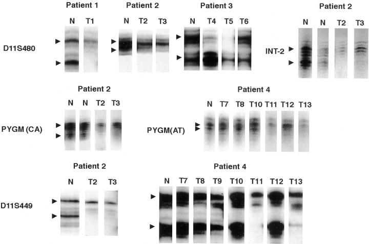
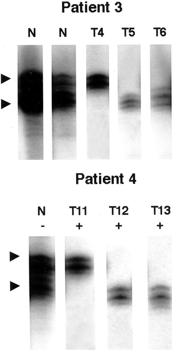
Multiple endocrine neoplasia type 1 (MEN1) is an autosomal dominant hereditary disorder characterized by multiple parathyroid, pancreatic, duodenal, and pituitary neuroendocrine tumors. Nonendocrine mesenchymal tumors, such as lipomas, collagenomas, and angiofibromas have also been reported. MEN1-associated neuroendocrine and some mesenchymal tumors have documented MEN1 gene alterations on chromosome 11q13. To test whether the MEN1 gene is involved in the pathogenesis of multiple smooth muscle tumors, we examined the 11q13 loss of heterozygosity (LOH) and clonality patterns in 15 leiomyomata of the esophagus, lung, and uterus from five patients with MEN1. Forty sporadic uterine leiomyomata were also studied for 11q13 LOH. LOH analysis was performed using four polymorphic DNA markers at the MEN1 gene locus; D11S480, PYGM, D11S449, and INT-2. 11q13 LOH was detected in 10 of 12 (83%) MEN1-associated esophageal and uterine smooth muscle tumors. In contrast, LOH at the MEN1 gene locus was demonstrated only in 2 of 40 (5%) sporadic uterine tumors. LOH at 11q13 was not documented in three lung smooth muscle tumors from a single patient with MEN1. Ten tumors from two female patients were additionally assessed for clonality by X-chromosome inactivation analysis. The results demonstrated different clonality patterns in multiple tumors in the same organ in each individual patient. The data indicate that leiomyomata of the esophagus and uterus in MEN1 patients arise as independent clones, develop through MEN1 gene alterations, and are an integral part of MEN1. However, the MEN1 gene is not a significant contributor to the tumorigenesis of sporadic uterine leiomyomata.
Figures
Similar articles
-
Allelic deletions on chromosome 11q13 in multiple tumors from individual MEN1 patients.Cancer Res. 1996 Nov 15;56(22):5272-8. Cancer Res. 1996. PMID: 8912868
-
Loss of heterozygosity in 11q13-14 regions in gastric neuroendocrine tumors not associated with multiple endocrine neoplasia type 1 syndrome.Lab Invest. 1999 Jun;79(6):671-7. Lab Invest. 1999. PMID: 10378509
-
Eighteen new polymorphic markers in the multiple endocrine neoplasia type 1 (MEN1) region.Hum Genet. 1997 Nov;101(1):102-8. doi: 10.1007/s004390050595. Hum Genet. 1997. PMID: 9385379
-
Multiple endocrine neoplasia type 1: clinical and genetic topics.Ann Intern Med. 1998 Sep 15;129(6):484-94. doi: 10.7326/0003-4819-129-6-199809150-00011. Ann Intern Med. 1998. PMID: 9735087 Review.
-
Thoracic and duodenopancreatic neuroendocrine tumors in multiple endocrine neoplasia type 1: natural history and function of menin in tumorigenesis.Endocr Relat Cancer. 2014 May 6;21(3):R121-42. doi: 10.1530/ERC-13-0482. Print 2014 Jun. Endocr Relat Cancer. 2014. PMID: 24389729 Review.
Cited by
-
Clinical aspects of multiple endocrine neoplasia type 1.Nat Rev Endocrinol. 2021 Apr;17(4):207-224. doi: 10.1038/s41574-021-00468-3. Epub 2021 Feb 9. Nat Rev Endocrinol. 2021. PMID: 33564173 Review.
-
Causes of death and prognostic factors in multiple endocrine neoplasia type 1: a prospective study: comparison of 106 MEN1/Zollinger-Ellison syndrome patients with 1613 literature MEN1 patients with or without pancreatic endocrine tumors.Medicine (Baltimore). 2013 May;92(3):135-181. doi: 10.1097/MD.0b013e3182954af1. Medicine (Baltimore). 2013. PMID: 23645327 Free PMC article.
-
Multiple Uterine Leiomyomas in Multiple Endocrine Neoplasia Type 1 with a Novel MEN1 Gene Mutation.J Hum Reprod Sci. 2020 Jan-Mar;13(1):75-77. doi: 10.4103/jhrs.JHRS_42_19. Epub 2020 Apr 7. J Hum Reprod Sci. 2020. PMID: 32577074 Free PMC article.
-
Mutations in the fumarate hydratase gene cause hereditary leiomyomatosis and renal cell cancer in families in North America.Am J Hum Genet. 2003 Jul;73(1):95-106. doi: 10.1086/376435. Epub 2003 May 22. Am J Hum Genet. 2003. PMID: 12772087 Free PMC article.
-
Multiple endocrine neoplasia type 1 involving both the liver and lung: a case report.World J Surg Oncol. 2022 May 10;20(1):151. doi: 10.1186/s12957-022-02622-1. World J Surg Oncol. 2022. PMID: 35538538 Free PMC article.
References
-
- Larsson C, Skogseid B, Öberg K, Nakamura Y, Nordenskjöld M: Multiple endocrine neoplasia type 1 gene maps to chromosome 11 and is lost in insulinoma. Nature 1988, 332:85-87 - PubMed
-
- Chandrasekharappa SC, Guru SC, Manickam P, Olufemi SE, Collins FS, Emmert-Buck MR, Debelenko LV, Zhuang Z, Lubensky IA, Liotta LA, Crabtree JS, Wang Y, Roe BA, Weisemann J, Boguski MS, Agarwal SK, Kester MB, Kim YS, Heppner C, Dong Q, Speigel AM, Burns AL, Marx SJ: Positional cloning of the gene for multiple endocrine neoplasia-type 1. Science 1997, 276:404-407 - PubMed
-
- Lubensky IA, Debelenko LV, Zhuang Z, Emmert-Buck MR, Dong Q, Chandrasekharappa SC, Guru SC, Manickam P, Olufemi S-E, Marx SJ, Spiegel AM, Collins FS, Liotta LA: Allelic deletions on chromosome 11q13 in multiple tumors from individual MEN1 patients. Cancer Res 1996, 56:5272-5278 - PubMed
-
- Knudson AG, Jr: Hereditary cancer, oncogenes, and antioncogenes. Cancer Res 1985, 45:1437-1443 - PubMed
-
- Komminoth P: Review: multiple endocrine neoplasia type 1, sporadic neuroendocrine tumors, and MENIN. Diagn Mol Pathol 1999, 8:107-112 - PubMed
MeSH terms
LinkOut - more resources
Full Text Sources
Medical
