

Cerebral autosomal dominant arteriopathy with subcortical infarcts and leukoencephalopathy: decrease in regional cerebral blood volume in hyperintense subcortical lesions inversely correlates with disability and cognitive performance
- PMID: 11498413
- PMCID: PMC7975190
Cerebral autosomal dominant arteriopathy with subcortical infarcts and leukoencephalopathy: decrease in regional cerebral blood volume in hyperintense subcortical lesions inversely correlates with disability and cognitive performance
Abstract
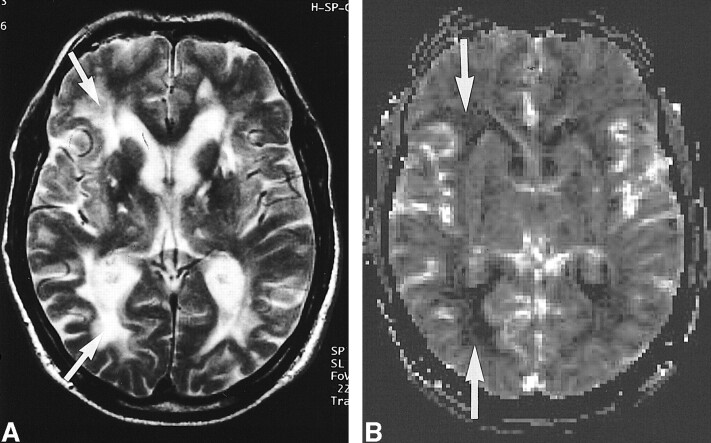
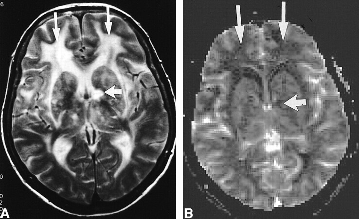
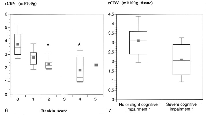
Background and purpose: Cerebral autosomal dominant arteriopathy with subcortical infarcts and leukoencephalopathy (CADASIL) is an arteriopathic syndrome related to a genetic defect on chromosome 19. Characteristic changes in CADASIL can be observed onT2-weighted MR images in the subcortical white matter. The purpose of this study was to measure changes of regional cerebral blood volume (rCBV) with dynamic contrast-enhanced MR imaging and to correlate the changes to disability and cognitive performance.
Methods: We obtained rCBV measurements of 24 individuals with proven CADASIL on a 1.5-T MR imaging unit. A susceptibility-weighted MR imaging sequence was used for bolus tracking. Principles of the indicator dilution theory were applied to estimate values of absolute rCBV (mL/100 g). Disability was determined by using the Rankin scale, and overall cognitive performance was assessed by using the Mini-Mental State Examination.
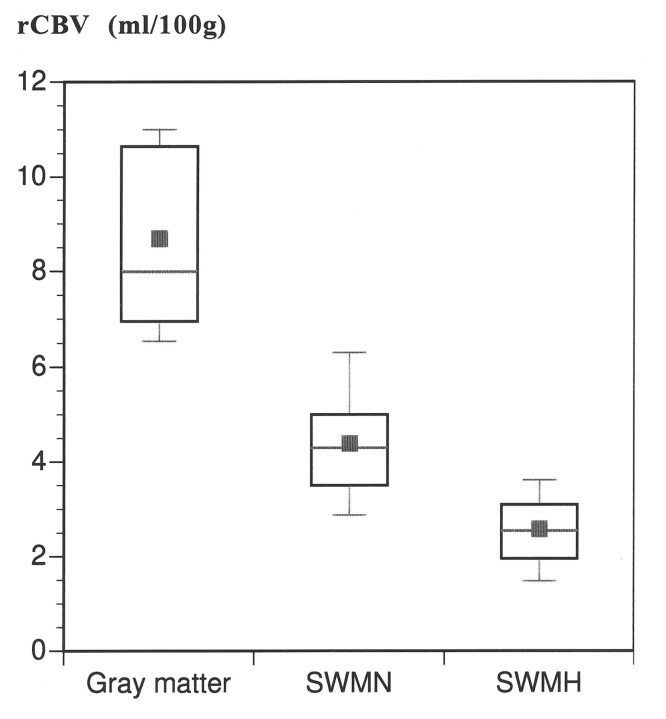
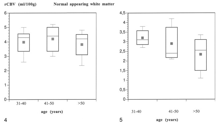
Results: The mean rCBV in the subcortical white matter that was hyperintense on the T2-weighted images (2.7 +/- 0.8 mL/100 g) was significantly lower than the rCBV in the white matter that appeared normal on the T2-weighted images (4.4 +/- 1.3 mL/100 g) (P <.05). The mean rCBV in the gray matter was within the normal range (8.3 +/- 1.7 mL/100 g). Both cognitive impairment and disability negatively correlated with rCBV in the subcortical white matter that was hyperintense (P <.05) but not with rCBV in the normal appearing white matter. rCBV did not correlate with age.
Conclusion: rCBV measured in the hyperintense subcortical white matter in individuals with CADASIL was decreased and inversely correlated with disability and cognitive impairment.
Figures
Similar articles
-
Role of subvoxel free fluid on diffusion parameters in brain tissue with cerebral autosomal dominant arteriopathy with subcortical infarcts and leukoencephalopathy and its correlation with physical disability: histogram analysis of standard and fluid-attenuated MR diffusion.AJNR Am J Neuroradiol. 2003 Jun-Jul;24(6):1083-9. AJNR Am J Neuroradiol. 2003. PMID: 12812930 Free PMC article.
-
Lacunar lesions are independently associated with disability and cognitive impairment in CADASIL.Neurology. 2007 Jul 10;69(2):172-9. doi: 10.1212/01.wnl.0000265221.05610.70. Neurology. 2007. PMID: 17620550
-
Correlations between clinical findings and magnetization transfer imaging metrics of tissue damage in individuals with cerebral autosomal dominant arteriopathy with subcortical infarcts and leukoencephalopathy.Stroke. 2001 Mar;32(3):643-8. doi: 10.1161/01.str.32.3.643. Stroke. 2001. PMID: 11239180 Clinical Trial.
-
Notch3 mutations in cerebral autosomal dominant arteriopathy with subcortical infarcts and leukoencephalopathy (CADASIL), a mendelian condition causing stroke and vascular dementia.Ann N Y Acad Sci. 1997 Sep 26;826:213-7. doi: 10.1111/j.1749-6632.1997.tb48472.x. Ann N Y Acad Sci. 1997. PMID: 9329692 Review.
-
CADASIL: hereditary disease of arteries causing brain infarcts and dementia.Neuropathol Appl Neurobiol. 1999 Aug;25(4):257-65. doi: 10.1046/j.1365-2990.1999.00198.x. Neuropathol Appl Neurobiol. 1999. PMID: 10476042 Review.
Cited by
-
Regional cerebral blood flow and blood volume in patients with subcortical arteriosclerotic encephalopathy (SAE).Eur Radiol. 2007 Oct;17(10):2483-90. doi: 10.1007/s00330-007-0617-y. Epub 2007 Mar 6. Eur Radiol. 2007. PMID: 17340101
-
Genetic animal models of cerebral vasculopathies.Prog Mol Biol Transl Sci. 2012;105:25-55. doi: 10.1016/B978-0-12-394596-9.00002-0. Prog Mol Biol Transl Sci. 2012. PMID: 22137428 Free PMC article. Review.
-
Role of neuroimaging in our understanding of the pathogenesis of primary headaches.Curr Pain Headache Rep. 2004 Oct;8(5):404-9. doi: 10.1007/s11916-996-0015-1. Curr Pain Headache Rep. 2004. PMID: 15361326 Review.
-
CADASIL and CARASIL.Brain Pathol. 2014 Sep;24(5):525-44. doi: 10.1111/bpa.12181. Brain Pathol. 2014. PMID: 25323668 Free PMC article. Review.
-
CADASIL: a common form of hereditary arteriopathy causing brain infarcts and dementia.Brain Pathol. 2002 Jul;12(3):371-84. doi: 10.1111/j.1750-3639.2002.tb00451.x. Brain Pathol. 2002. PMID: 12146805 Free PMC article. Review.
References
-
- Chabriat H, Vahedi K, Iba-Zizen MT, et al. Clinical spectrum of CADASIL: a study of 7 families: cerebral autosomal dominant arteriopathy with subcortical infarcts and leukoencephalopathy. Lancet 1995;346:934-939 - PubMed
-
- Tournier-Lasserve E, Iba-Zizen MT, Romero N, Bousser MG. Autosomal dominant syndrome with strokelike episodes and leukoencephalopathy. Stroke 1991;22:1297-1302 - PubMed
-
- Tournier-Lasserve E, Joutel A, Melki J, et al. Cerebral autosomal dominant arteriopathy with subcortical infarcts and leukoencephalopathy maps to chromosome 19ql2. Nat Genet 1993;3:256-259 - PubMed
-
- Joutel A, Corpechot C, Ducros A, et al. Notch3 mutations in CADASIL, a hereditary adult-onset condition causing stroke and dementia. Nature 1996;383:707-710 - PubMed
-
- Joutel A, Vahedi K, Corpechot C, et al. Strong clustering and stereotyped nature of Notch3 mutations in CADASIL patients. Lancet 1997;350:1511-1515 - PubMed
Publication types
MeSH terms
LinkOut - more resources
Full Text Sources
Medical