In the visual system, thousands of retinal ganglion cells (RGCs) relay signals to precise locations in the brain via delicate projections called axons. As the visual system develops, migrating axons diverge in an area of the brain called the optic chasm. Some axons cross over to the other side of the brain as they make those deeper connections. Others don't.
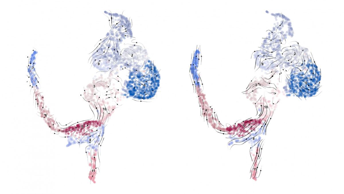
These images depict cell clusters at the edges of mouse retinas, each color representing distinct stages of maturity for different cells as determined by variations in their genetic activity. The arrows indicate predicted developmental courses of certain retinal cells in normal (left) and albino (right) mice, which exhibit poor depth perception. Credit: Mason Lab/Zuckerman Institute.
Carol Mason and colleagues at Columbia University found that cell birth date influences the trajectory of retinal ganglion cell (RGC) axons as they form connections with the brain during development. Using an albino mouse model, they found that when timing is off-kilter, axons migrate to the wrong brain region, impairing depth perception. Treating young mouse pups, however, redirected axons, resulting in improved depth perception.