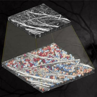
The graphic shows an image generated by AO-OCT (top), and the result of WeakGCSeg algorithms to identify and trace the shapes of the ganglion cells in the eye (bottom). Image credit: Sina Farsiu, Duke University
A new combination of optical coherence tomography (OCT), adaptive optics and deep neural networks should enable better diagnosis and monitoring for neuron-damaging eye and brain diseases like glaucoma.
Biomedical engineers at Duke University led a multi-institution consortium to develop the process, which easily and precisely tracks changes in the number and shape of retinal ganglion cells in the eye.
In their new paper, Sina Farsiu and Somayyeh Soltanian-Zadeh, a postdoctoral researcher in Farsiu’s lab, develop a highly adaptive and easy-to-train deep learning-based algorithm that is the first to identify and trace the shapes of ganglion cells from adaptive optics OCT scans.