Vision loss from diseases like glaucoma is typically irreversible, due to the death of neurons in the light-sensing retina. But not so in certain species of salamanders and fish, which can regrow their retina and regain vision. The National Eye Institute (NEI) Audacious Goals Initiative (AGI) is exploring the possibility that the natural world holds the keys to restorative therapies that might unlock regenerative powers in humans.
In 2016, the NEI funded 6 research projects as part of the AGI Discovery Factors Consortium. Their collective goal was to search for genes and other biological factors that might activate regenerative potential in cells of the retina. Each set out to explore various facets of the regenerative process, exploring light-sensing photoreceptors; retinal ganglion cells (RGCs), which carry visual signals through the optic nerve to the brain; and various interneurons between the photoreceptors and the RGCs.

Mike Dyer and Paul Sieving, former NEI director who launched the AGI.
“These projects were super high-risk,” said Michael Dyer, a researcher at St. Jude Children's Research Hospital, who is a member of the AGI discovery consortium’s external scientific oversight committee and is coordinating construction of a database to house consortium data. “The investigators asked bold questions, knowing answers would not come easily. I credit NEI with structuring the consortium in a way that encouraged collaboration. The various teams met regularly to share their successes and challenges. Most projects changed course mid-stream, but in the end the consortium generated an incredible amount of data which we are in the process of making publicly available.”
Three years in, the projects are making progress and reporting findings.
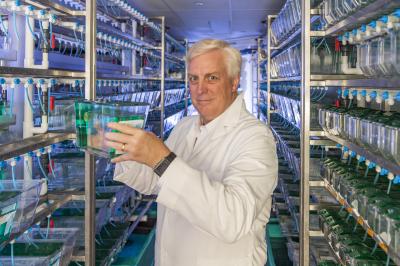
David Hyde, University of Notre Dame.
David Hyde, University of Notre Dame, with collaborators at the University of Florida, Ohio State, and Johns Hopkins University, compared gene expression in mice, zebrafish, and chicks. They compared gene activity (transcriptomics) and modifications to genomic DNA (epigenomics) during development and in response to retinal damage to determine if regeneration mimicked retinal development. Hyde’s team homed in on non-neuronal retinal support cells called Müller glia, which in zebrafish and chicks respond to retinal damage by reverting to an immature progenitor state and then developing into replacement neurons. Hyde found that gene expression of reprogrammed Müller glia is nearly identical to immature retinal neurons. Additionally, they identified both evolutionarily conserved and species-specific transcriptomic and epigenomic events that occur in Müller glia following retinal injury. Their comparison studies also revealed that a critical point in regeneration is gliosis—a response by glial cells to injury. Hyde and colleagues demonstrated that zebrafish and chick Müller glia progress through gliosis to enter neurogenesis, while mammalian Müller glia either lack factors that push toward regeneration or possess repressors that prevent neurodegeneration. Further, they demonstrated that expression of specific genes in the damaged mouse retina could drive some of the Müller glia to regenerate retinal neurons.
Edward Levine and collaborators at Vanderbilt University are also searching for factors that reprogram Müller glia into neuronal precursor cells. The team screened extracellular vesicles—tiny secreted blebs found in blood and other bodily fluids—and turned up a previously uncharacterized epidermal growth factor that stimulates Müller glia proliferation. The team is now determining the role of this protein during induction of regeneration.
Three projects searched for factors that control regeneration of the nerve fiber bundle known as the optic nerve. Each fiber, otherwise known as an axon, extends from one RGC in the retina and exits the eye as part of the optic nerve. Like a string between two cans, each axon extends from one RGC to a specific target in the brain. Optic neuropathies, including glaucoma, sever axons, cutting off communication and in turn causing the death of corresponding RGCs.
Jeffrey Goldberg, Stanford University, and collaborators at Harvard University and Scripps Research Institute screened for factors that influence RGC axon regeneration. Their team developed a method to measure RGC gene expression during the days following optic nerve damage as well as proteins that are transported into the optic nerve before and after damage. Starting with 450 candidate genes, Stephen Strittmatter and his team at Yale University identified 20 genes in mice whose role in RGC axon regeneration was previously unrecognized. And working with cultured mouse RGCs, Kevin Park and his team at the University of Miami isolated 20 genes that affect axonal regeneration after screening more than 400 candidates.
Donald Zack, Johns Hopkins University, and David Gamm, University of Wisconsin
Coaxing regenerated photoreceptors to hook up with other retinal neurons is the focus of a project led by Don Zack, Johns Hopkins University, and David Gamm, University of Wisconsin. Gamm and others have generated photoreceptors from induced pluripotent stem cells; however, to enable vision, photoreceptors must sprout root-like structures called neurites. Zack’s team screened more than 5,000 compounds for their ability to stimulate neurite outgrowth. The team has also developed a technique to trace connections between stem cell-derived photoreceptors and other retinal cell types to study how retinal cells communicate.
In addition to the Discovery Factors Consortium, the NEI funds a functional imaging consortium, which is developing tools to see the eye and optic nerve in unprecedented detail, and a translational-enabling models consortium, which funds several projects that are developing research models that mimic important aspects of vision and blinding diseases in order to test new therapies.
Launched in 2013, the AGI is an effort to push the boundaries of vision science and restore vision through regeneration of the retina. By facilitating cross-disciplinary research, NEI is tackling the most devastating and difficult-to-treat eye diseases. Learn more about the AGI at www.nei.nih.gov/AGI.
###
This press release describes a basic research finding. Basic research increases our understanding of human behavior and biology, which is foundational to advancing new and better ways to prevent, diagnose, and treat disease. Science is an unpredictable and incremental process— each research advance builds on past discoveries, often in unexpected ways. Most clinical advances would not be possible without the knowledge of fundamental basic research.
NEI leads the federal government’s research on the visual system and eye diseases. NEI supports basic and clinical science programs to develop sight-saving treatments and address special needs of people with vision loss. For more information, visit https://www.nei.nih.gov.
About the National Institutes of Health (NIH): NIH, the nation’s medical research agency, includes 27 Institutes and Centers and is a component of the U.S. Department of Health and Human Services. NIH is the primary federal agency conducting and supporting basic, clinical, and translational medical research, and is investigating the causes, treatments, and cures for both common and rare diseases. For more information about NIH and its programs, visit https://www.nih.gov/.
NIH…Turning Discovery Into Health®