. 2024 Nov 8;386(6722):667-672.
doi: 10.1126/science.adp7201. Epub 2024 Nov 7.

Regulated N-glycosylation controls chaperone function and receptor trafficking

Affiliations

Regulated N-glycosylation controls chaperone function and receptor trafficking

Mengxiao Ma et al. Science. .

Abstract

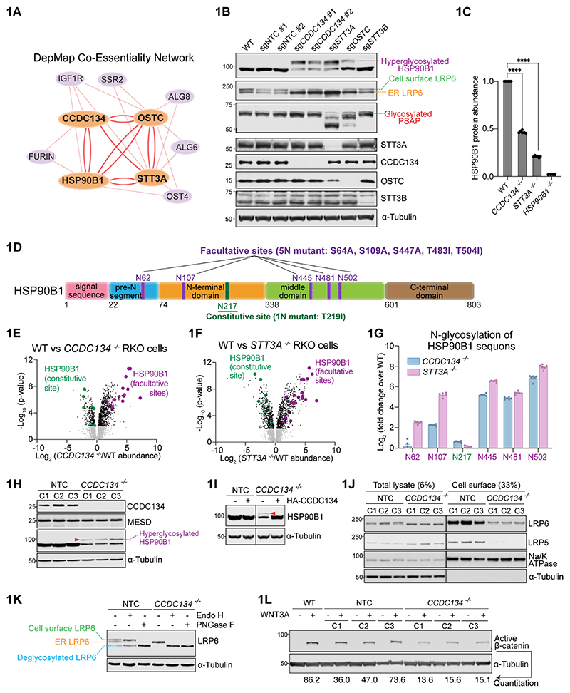
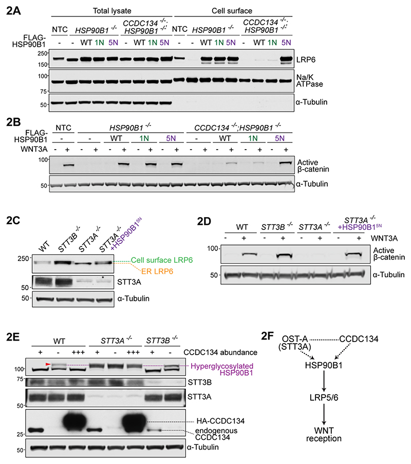
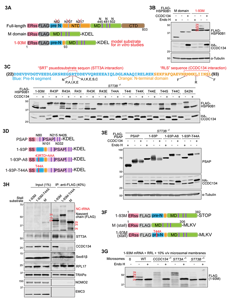
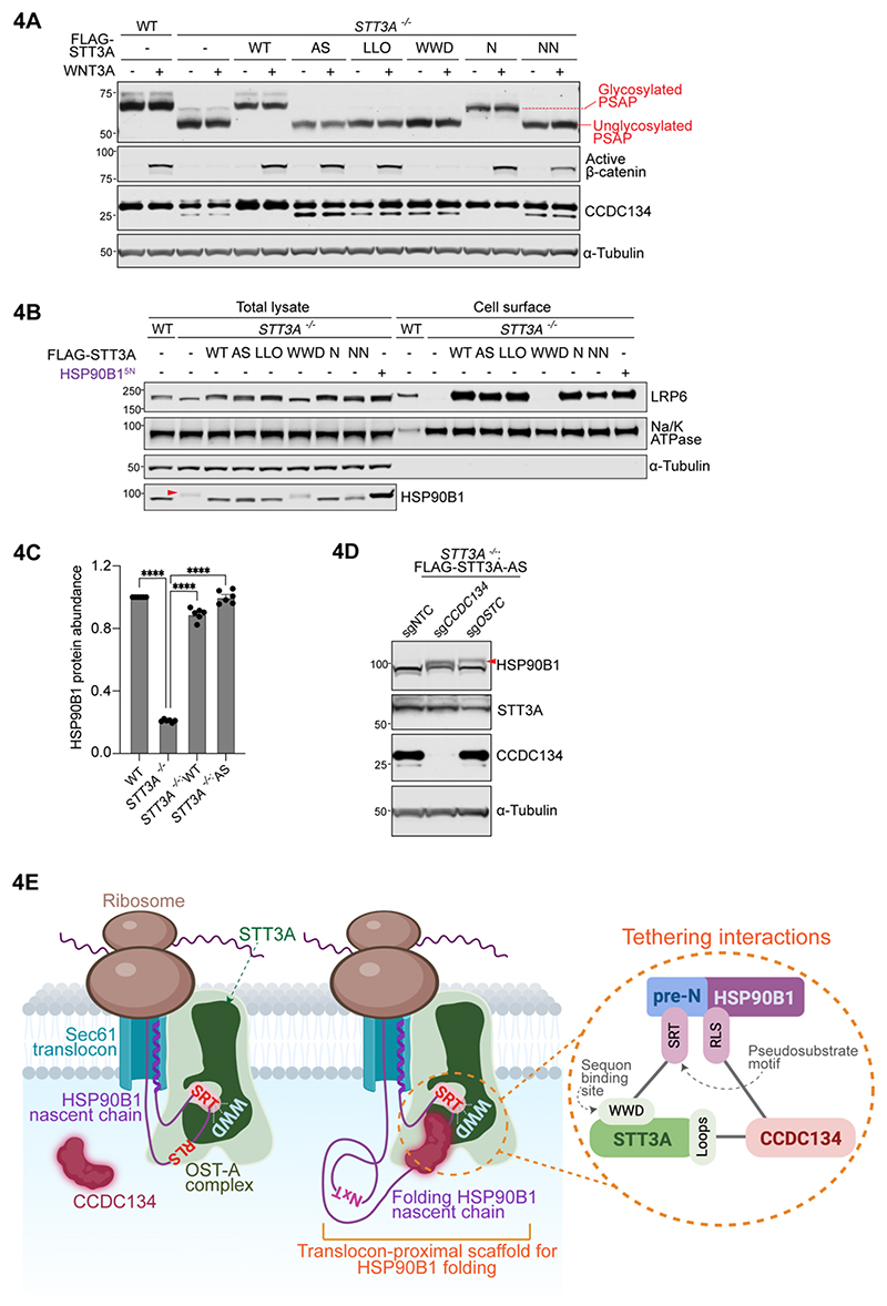
One-fifth of human proteins are N-glycosylated in the endoplasmic reticulum (ER) by two oligosaccharyltransferases, OST-A and OST-B. Contrary to the prevailing view of N-glycosylation as a housekeeping function, we identified an ER pathway that modulates the activity of OST-A. Genetic analyses linked OST-A to HSP90B1, an ER chaperone for membrane receptors, and CCDC134, an ER luminal protein. During its translocation into the ER, an N-terminal peptide in HSP90B1 templates the assembly of a translocon complex containing CCDC134 and OST-A that protects HSP90B1 during folding, preventing its hyperglycosylation and degradation. Disruption of this pathway impairs WNT and IGF1R signaling and causes the bone developmental disorder osteogenesis imperfecta. Thus, N-glycosylation can be regulated by specificity factors in the ER to control cell surface receptor signaling and tissue development.


Conflict of interest statement

Competing Interests

The authors declare no competing interests.

Figures

Figure 1
Figure 1. Regulation of HSP90B1 N-glycosylation and WNT signaling by an ER protein network.
(A) DepMap co-essentiality relationships between CCDC134, STT3A, HSP90B1, and OSTC visualized using Fireworks (16). The bi-directional edges between these four genes (bold orange lines) indicate concordant effects on cell growth across >1000 cell lines. (B) Abundances and glycosylation status of HSP90B1, its client LRP6, and PSAP (an exclusive OST-A substrate) in lysates from cells expressing a control (NTC) sgRNA or sgRNA’s against the indicated genes. (C) HSP90B1 protein abundance in wild-type (WT), CCDC134-/-, STT3A-/-, and HSP90B1-/- RKO cell lines measured by mass spectrometry, normalized to WT cells. Bars represent the mean abundance +/- SEM from six independent mass spectrometry runs, represented as individual data points. Statistical significance was determined by one-way ANOVA with Dunnett’s multiple comparisons test; **** p<0.0001. (D) Domain architecture of HSP90B1. The constitutive and facultative N-glycosylation sites are labeled in green and purple, respectively. The point mutations used to disrupt the one constitutive sequon (1N mutant) and the five facultative sequons (5N mutant) are listed. fig.S2A shows these features on the HSP90B1 three dimensional structure using the same coloring scheme. (E, F) Abundances of glycosylated peptides in CCDC134-/- and STT3A-/- cells measured using global, unbiased N-glycoprotemics. Each data point represents the fold change in abundance of a distinct glycopeptide (defined by sequence and glycan structure) in mutant compared to wild-type cells. Glycopeptides that include the constitutive and facultative sequons in HSP90B1 are colored green and purple, respectively. Full dataset in Data S2. (G) Enrichment of glycopeptides (normalized to total HSP90B1 protein abundance) that include each of the facultative and constitutive sequons of HSP90B1 in CCDC134-/- or STT3A-/- cells compared to WT cells. Bars show the mean +/- SEM from six independent mass spectrometry runs, represented as individual data points. The abundances of all peptides that include each HSP90B1 sequon were integrated using individual peptide data summarized in 1E and 1F and provided in Data S2. (H, I) Abundance and glycosylation status of HSP90B1 in lysates from three (C1-C3) independent control (NTC) and CCDC134-/- clonal cell lines (H), or after the stable expression of HA-tagged CCDC134 in CCDC134-/- cells (I). The hyperglycosylated form of HSP90B1 is denoted by a red arrowhead in this and all subsequent panels. (J) LRP6 and LRP5 abundances in total lysate or at the plasma membrane from three independently derived (C1-C3) control (NTC) or CCDC134-/- clonal cell lines. The cell surface protein Na/K ATPase and cytoplasmic protein α-tubulin serve as controls, both for loading and for the specificity of cell surface biotinylation. (K) Glycosidase sensitivity in conjunction with mobility on SDS-PAGE gels was used to measure the ER or cell-surface pools of LRP6 in control or CCDC134-/- cells. Endoglycosidase H (Endo H) can remove glycans added in the ER but not the complex glycan modifications added in the Golgi; Peptide-N-Glycosidase F (PNGase F) can remove glycans on both ER and cell-surface proteins. (L) Active (non-phosphorylated) β-catenin abundance (a metric of WNT signaling strength) was measured (+/- WNT3A) using immunoblots in wild-type cells or clonal cell lines expressing a control (NTC) sgRNA or an sgRNA targeting CCDC134.
Figure 2
Figure 2. Hyperglycosylation of HSP90B1 regulates WNT signaling strength.
(A-D) Abundances of cell-surface LRP6 (A,C) or active β-catenin abundance (B,D) in clonally derived cell lines of the indicated genotypes stably expressing wild-type (WT) FLAG-HSP90B1 or variants carrying mutations in the one constitutive (1N) or all five facultative (5N) sites (see Fig.1D). HSP90B1 variants were expressed at comparable levels (fig.S5A). (E) N-glycosylation status of HSP90B1 in STT3B-/- and STT3A-/- cells expressing different levels of CCDC134: No CCDC134 (-), endogenous CCDC134(+), stably overexpressed 3xHA-CCDC134 (+++) on top of endogenous CCDC134. (F) A provisional pathway diagram constructed based on genetic interactions (dotted lines) uncovered in our work and physical interactions (solid lines) described in the literature.
Figure 3
Figure 3. The pre-N segment of HSP90B1 inhibits its own N-glycosylation by recruiting CCDC134 to an OST-A containing secretory translocon.
(A) Variants of HSP90B1 used for cell-based and in vitro assays. Key features include the ERss, ER signal sequence; FLAG, 3xFLAG tag; pre-N, unstructured segment; NTD, N-terminal domain; MD, middle domain; CTD, C-terminal domain. N-glycosylation sites in pre-N and NTD were eliminated to allow easy assessment of the glycan modification of the three sequons in the M domain by gel shifts (see 3B and text). (B) Glycosylation status of HSP90B1 variants shown in 3A was assessed using gel shifts and Endo H sensitivity after transient co-expression in HEK293T cells with WT CCDC134 (+) or a non-functional variant (-) lacking its ER signal sequence (see Fig.S3J). The four predicted N-glycoforms (carrying 0, 1, 2 or 3 glycans) are labeled 0N-3N in red lettering. (C) Glycosylation status of the 1-93M variant (see 3A) of HSP90B1 carrying the indicated mutations in the “SRT” pseudosubstrate motif found in the pre-N segment. Each construct was co-expressed with functional (+) or non-functional (-) CCDC134. See fig.S6C and S6D for deletion analysis and alanine scanning mutagenesis of the pre-N segment. (D,E) Glycosylation status of chimeric proteins (3D) constructed by fusing variants of the 1-93 segment of HSP90B1 to the obligate OST-A substrate PSAP, which contains five N-glycosylation sites shown in 3D. T44A changes “SRT” to “SRA” and A8 changes “RTD” to “AAA” (see 3C and fig.S6D). All chimeras carry the ERss of HSP90B1. (F) Constructs used for in vitro translation experiments. To stably stall translation, the STOP codon was removed and a MLKV peptide sequence appended at the C-terminus. (G) Glycosylation status of the 1-93M variant of HSP90B1 (see 3A) translated in rabbit reticulocyte lysate (RRL) in the presence of rough microsomal membranes generated from wild-type or clonally-derived CCDC134-/-, STT3A-/-, or STT3B-/- HEK293T cells and concentrated by immunoprecipitation on anti-FLAG beads. The four N-glycoforms of 1-93M are labeled (compare to 3B) and show sensitivity to Endo H treatment. (H) Association of endogenous CCDC134 with stalled 1-93M, M domain alone or a 1-93M variant carrying a T44A mutation in the “SRT” pseudosubstrate site (see 3C). The stalled nascent chain, immunoprecipitated (IP) using anti-FLAG beads, associates with the ribosome (RPL17) and known components of the secretory translocon (STT3A, the SEC61 channel, and the TRAP complex), but not with components of the multi-pass translocon (NOMO2) or the ER-Membrane Protein Complex (EMC3) (34, 41). NC-tRNA: nascent chain-tRNA conjugates.
Figure 4
Figure 4. The oligosaccharyltransferase activity of OST-A is not required to promote HSP90B1 stability and WNT signaling.
(A,B) Glycosylation status and abundances of PSAP (A), active β-catenin (A), CCDC134 (A), HSP90B1 (B) and cell-surface LRP6 (B) in wild-type (WT) cells or STT3A-/- cells stably expressing FLAG-STT3A variants carrying mutations in various sites involved in catalytic transfer of the glycan from the lipid-linked oligosaccharide to the asparagine in sequons. Variants (shown on a structure in fig.S9A) carry mutations in residues involved in active site chemistry (AS), lipid-linked oligosaccharide binding (LLO), sequon binding (WWD) or N-glycosylation of STT3A itself (N and NN). Glycosylation of PSAP (A) was used to assess OST-A activity in cells. See fig.S9B. (C) HSP90B1 protein abundance in wild-type (WT) cells, STT3A-/- cells, and STT3A-/- cells stably expressing wild-type or catalytically inactive (AS) STT3A. The STT3A-AS variant carries mutations in four residues involved in the chemical step of glycan transfer (fig.S9A). Statistical significance was determined by one-way ANOVA with Dunnett’s multiple comparisons test; **** p<0.0001. (D) Abundance and glycosylation status (red arrowhead) of endogenous HSP90B1 in STT3A-/- cells stably expressing (1) catalytically inactive FLAG-STT3A-AS carrying mutations in active site residues (fig.S9A) and (2) sgRNAs targeting CCDC134 or OSTC. sgNTC=non-targeting control sgRNA. (E) During translation, HSP90B1 is tethered to a specialized CCDC134-containing translocon that forms a specialized microenvironment for its folding. Tethering interactions are shown in the circular inset: CCDC134 interacts both with STT3A and HSP90B1, while the pre-N domain of HSP90B1 itself binds to the sequon binding site of STT3A. This translocon-proximal scaffold prevents STT3A from recognizing sequons in HSP90B1 and also sterically prevents access of these sequons to OST-B during folding. See fig.S10.

Similar articles

References

    1. Khoury GA, Baliban RC, Floudas CA. Proteome-wide post-translational modification statistics: frequency analysis and curation of the swiss-prot database. Sci Rep. 2011;1 doi: 10.1038/srep00090. - DOI - PMC - PubMed
    1. Zielinska DF, Gnad F, Wiśniewski JR, Mann M. Precision mapping of an in vivo N-glycoproteome reveals rigid topological and sequence constraints. Cell. 2010;141:897–907. - PubMed
    1. Reily C, Stewart TJ, Renfrow MB, Novak J. Glycosylation in health and disease. Nat Rev Nephrol. 2019;15:346–366. doi: 10.1038/s41581-019-0129-4. - DOI - PMC - PubMed
    1. Gagneux P, Hennet T, Varki A. In: Essentials of Glycobiology. Varki A, Cummings RD, Esko JD, Stanley P, Hart GW, Aebi M, Mohnen D, Kinoshita T, Packer NH, Prestegard JH, Schnaar RL, Seeberger PH, editors. Cold Spring Harbor Laboratory Press; Cold Spring Harbor (NY): 2022. Biological Functions of Glycans. - PubMed
    1. Ruiz-Canada C, Kelleher DJ, Gilmore R. Cotranslational and posttranslational N-glycosylation of polypeptides by distinct mammalian OST isoforms. Cell. 2009;136:272–283. doi: 10.1016/j.cell.2008.11.047. - DOI - PMC - PubMed

LinkOut - more resources