Review
. 2023 Jun 22:22:520-555.
doi: 10.17179/excli2023-6221. eCollection 2023.

The bridge between cell survival and cell death: reactive oxygen species-mediated cellular stress

Affiliations
Review

The bridge between cell survival and cell death: reactive oxygen species-mediated cellular stress

Nese Vardar Acar et al. EXCLI J. .

Abstract

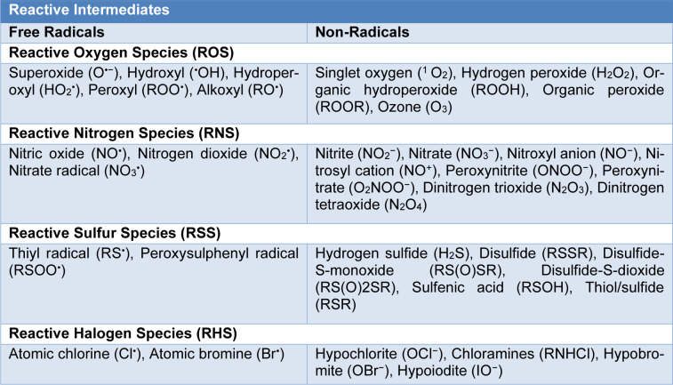
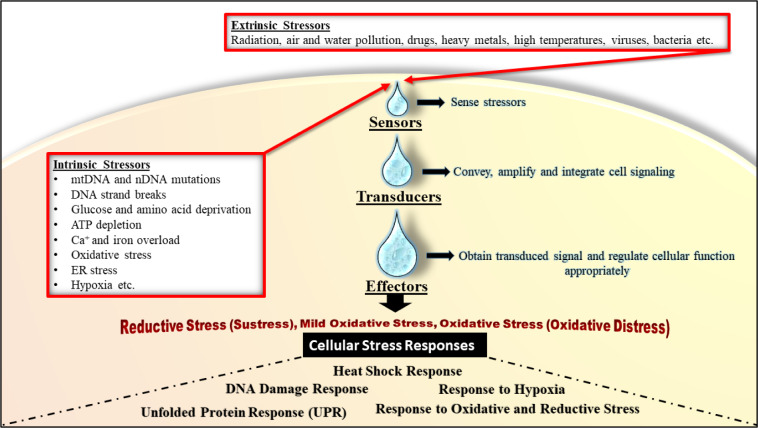
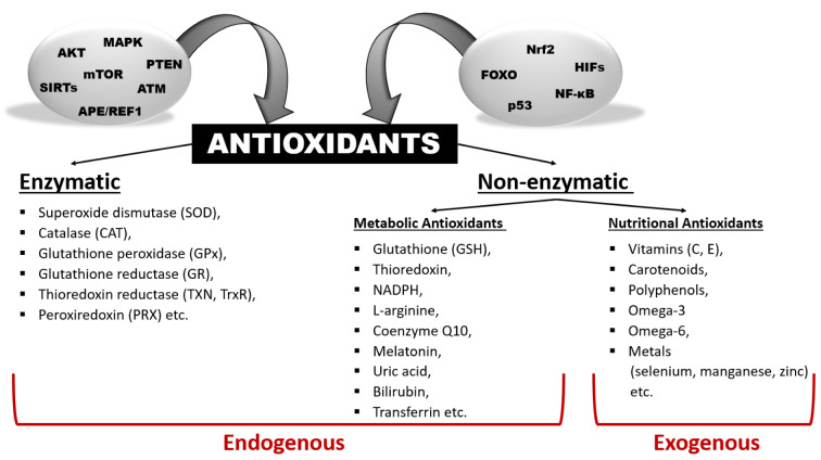
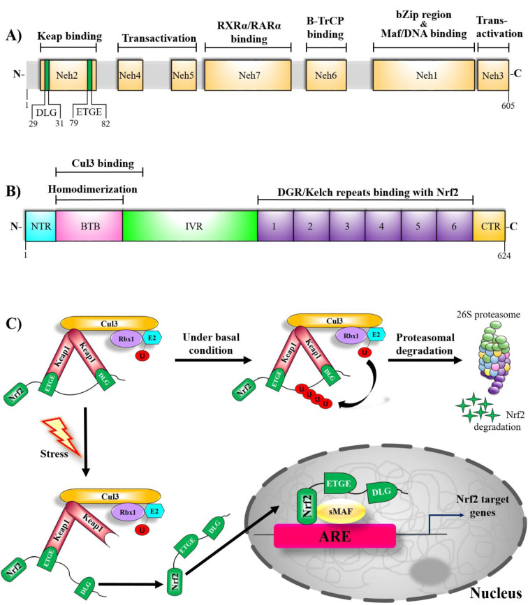
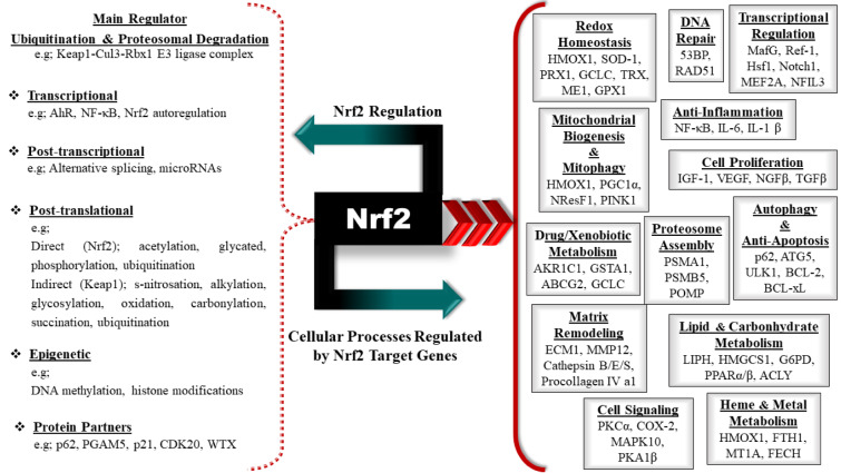
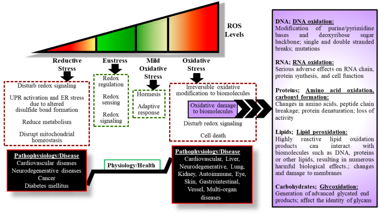
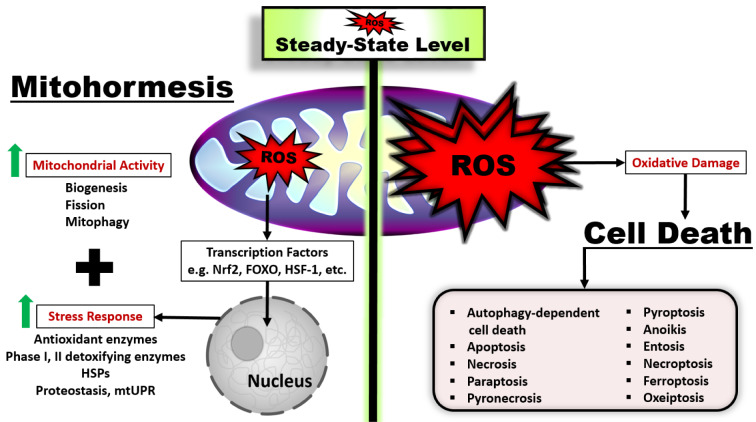
As a requirement of aerobic metabolism, regulation of redox homeostasis is indispensable for the continuity of living homeostasis and life. Since the stability of the redox state is necessary for the maintenance of the biological functions of the cells, the balance between the pro-oxidants, especially ROS and the antioxidant capacity is kept in balance in the cells through antioxidant defense systems. The pleiotropic transcription factor, Nrf2, is the master regulator of the antioxidant defense system. Disruption of redox homeostasis leads to oxidative and reductive stress, bringing about multiple pathophysiological conditions. Oxidative stress characterized by high ROS levels causes oxidative damage to biomolecules and cell death, while reductive stress characterized by low ROS levels disrupt physiological cell functions. The fact that ROS, which were initially attributed as harmful products of aerobic metabolism, at the same time function as signal molecules at non-toxic levels and play a role in the adaptive response called mithormesis points out that ROS have a dose-dependent effect on cell fate determination. See also Figure 1(Fig. 1).

Keywords: Nrf2; antioxidant defense systems; cell death pathways; mitohormesis; oxidative and reductive stress; redox homeostasis.


Figures

Table 1
Table 1. Instance of free radical and non-radical species [Phaniendra et al., 2015; Martemucci et al., 2022; Tanaka and Vécsei, 2020]
Figure 1
Figure 1. Graphical abstract
Figure 2
Figure 2. Extrinsic and intrinsic stressors, signal transduction and cellular stress response. The cells are exposed to numerous stress factors, both extrinsic and intrinsic. Stress factors are sensed by the cells, transported, replicated and integrated into the cells. The maintenance of cellular homeostasis is ensured by the regulation of cellular functions through the most appropriate cellular stress response to the transformed stress signal within the cells.
Figure 3
Figure 3. Antioxidant defense systems. Redox balance is tightly controlled through many enzymes and transcription factors, which are directly or indirectly mediate redox homeostasis, and enzymatic and non-enzymatic antioxidants. (Abbreviations: MAPK, mitogen-activating protein kinase; AKT, protein kinase B; APE/REF1, apurinic/apyrimidinic endonuclease 1/redox factor 1; ATM, ataxia-telangiectasia mutated kinase; mTOR, mammalian target of rapamycin; PTEN, phosphate and tensin homologue; SIRTs, sirtuins; FOXO, forkhead box O; NF-κB, nuclear factor-kappa B; p53, tumor suppressor p53; HIFs, hypoxia inducible factors; Nrf2, nuclear factor erythroid 2-related factor 2).
Figure 4
Figure 4. Domain structure of human Nrf2 and Keap1 and schematic representation of the Nrf2/Keap1 signaling mechanism. A) Nrf2, a 605 amino acid protein, comprises seven functional domains called Neh1-Neh7. The N-terminal domain Neh2 contains two motifs, DLG and ETGE, which are responsible for binding KEAP1 homodimer for performing ubiquitin-dependent proteasomal degradation of Nrf2. The Neh4 and Neh5 domains recruit transcriptional co-activators, CREB-binding protein (CBP), and/or repressor-associated coactivator (RAC) for the transactivation activity of NRF2. The Neh7 domain binds retinoid X (RXR) and retinoic acid (RAR) receptors that mediates repression of Nrf2. The Neh6 domain contains two motifs (DSGIS and DSAPGS) interacting with β-transducin repeat-containing protein (β-TrCP) for the β-TrCP-mediated proteasomal degradation. The Neh1, containing serine-rich domain, is responsible for dimerization with small musculoaponeurotic fibrosarcoma (Maf), which is the heterodimeric partner for Nrf2 to recognize the ARE sequence in target gene promoters. The C-terminal domain, Neh3, is a transcriptional co-activator that recruits chromodomain helicase DNA-binding domain protein 6 (CHD6). B) A 624-amino acid Keap1, the repressor of Nrf2, contains three functional domains in addition to the N-terminal and C-terminal domains. The broad complex/tram track/bric-a-brac (BTB) domain regulates Keap1 homodimerization and interaction with the Cul3-based ubiquitin E3 ligase complex for NRF2 ubiquitination. Intervening region (IVR) domain acts as a sensor for NRF2 inducers through highly-reactive cysteine residues. the double-glycine repeats (DGR)/Kelch domain is important for binding with the Neh2 domain of NRF2. C) Under basal conditions, Nrf2 binds to Keap1 via ETGE and DLG motifs in the cytosol and activates Cul3-mediated ubiquitination through interacting with the Cul3-RBX1 E3 ubiquitin ligase. Thus, ubiquitinated Nrf2 is degraded by the 26S proteasome. Under stressful conditions, Nrf2 dissociates from Keap1 and accumulates due to conformational changes in Keap1 as a result of modifications in cysteine residues of Keap1.Then, Nrf2 translocates into the nucleus, forms a heterodimer with sMaf proteins and binds to ARE to initiate the transcription of Nrf2 target genes. (Abbreviations: Nrf2, nuclear factor erythroid 2-related factor 2; Keap1, Kelch-like ECH-associated protein 1; Cul3, Cullin 3; Rbx1, Ring box 1; E2, ubiquitin-conjugating enzyme; ARE, antioxidant response elemen; U, Ubiquitin)
Figure 5
Figure 5. Regulation of Nrf2 and Nrf2 target genes. The target genes and cellular processes regulated by Nrf2 are illustrated with instances. Also, the regulation of this complex and multifunctional transcription factor is also explained with examples. (Abbreviations: Keap1-Cul3-Rbx1, Kelch-like ECH-associated protein 1-Cullin 3-Ring box 1; AhR, aryl hydrocarbon receptor; NF-κB, nuclear factor-kappa B; PGAM5, PGAM family member 5, mitochondrial serine/threonine protein phosphatase; CDK20, cyclin dependent kinase 20; WTX, Wilms tumor gene on the X chromosome; HMOX1, heme oxygenase 1; SOD-1, superoxide dismutase-1; PRX1, peroxiredoxin 1; GCLC, glutamate-cysteine ligase catalytic subunit; TRX, thioredoxin; ME1, malic enzyme 1; GPX1, glutathione peroxidase 1; 53BP, p53-binding protein 1; RAD51, DNA repair protein RAD51 homolog 1; MafG, V-Maf avian musculoaponeurotic fibrosarcoma oncogene homolog G; Ref-1, redox effector factor-1; Hsf1, heat shock transcription factor 1; Notch1, neurogenic locus notch homolog protein 1; MEF2A, myocyte enhancer factor 2; NFIL3, nuclear factor interleukin (IL)-3 regulated; PGC1α, peroxisome proliferator-activated receptor gamma activator 1 alpha; NResF1, nuclear respiratory factor 1; PINK1, PTEN-induced kinase 1; IL-6, Interleukin 6; IL-1 β, Interleukin-1 beta; IGF-1, growth factors [insulin like growth factor-1; VEGFα, vascular endothelial growth factor alpha; NGFβ, nerve growth factor beta; TGFβ, transforming growth factor beta; AKR1C1, aldo-keto reductase family 1 member C1; GSTA1, glutathione S-transferase alpha 1; ABCG2, ATP binding cassette subfamily G member 2 (Junior blood group); GCLC, glutamate-cysteine ligase catalytic subunit; PSMA1, proteasome subunit alpha 1; PSMB5, proteasome subunit beta 5; POMP, proteasome maturation protein; ATG5, autophagy-related 5; ULK1, Unc-51-like kinase 1; BCL-2, B-cell lymphoma 2; Bcl-xL, B-cell lymphoma-extra large; ECM1, extracellular matrix protein 1; MMP12, matrix metallopeptidase 12; LIPH, lipase member H; HMGCS1, 3-hydroxy-3-methylglutaryl-CoA synthase 1; G6PD, glucose-6-phosphate dehydrogenase; PPARα/β, peroxisome proliferator-activated receptor alpha/beta; ACLY, ATP citrate lyase; PKCα, protein kinase C, alpha; COX-2, cytochrome c oxidase subunit II; MAPK10, mitogen-activated protein kinase 10; PKA1β, protein kinase a1 beta; FTH1, ferritin heavy chain 1; MT1A, metallothionein 1A; FECH, ferrochelatase.
Figure 6
Figure 6. ROS levels are the bridge between cell survival and death response, resulting in physiology/ health and pathology/diseases.
Figure 7
Figure 7. The levels of ROS determine the decision of the cells between survival and death. Mitohormesis is a pro-survival adaptive response that results in increased health and vitality in a cell, tissue, or organism through mild ROS levels released by the induction of reduced amounts of mitochondrial stress. To maintain cell survival, the mild levels of ROS induce the activation of a retrograde mitochondrial-nucleus signaling mechanism, leading to a stress response in the cell and causing an increase in mitochondrial activity. High levels of ROS act as second messengers that cause different cell death mechanisms to be engaged in the cells.

Similar articles

Cited by

References

    1. Agledal L, Niere M, Ziegler M. The phosphate makes a difference: cellular functions of NADP. Redox Rep. 2010;15(1):2–10. - PMC - PubMed
    1. Al-Sawaf O, Clarner T, Fragoulis A, Kan YW, Pufe T, Streetz K, et al. Nrf2 in health and disease: current and future clinical implications. Clin Sci. 2015;129:989–999. - PubMed
    1. Amici DR, Ansel DJ, Metz KA, Smith RS, Phoumyvong CM, Gayatri S, et al. C16orf72/ HAPSTR1 is a molecular rheostat in an integrated network of stress response pathways. Proc Natl Acad Sci U S A. 2022;119(27):e2111262119. - PMC - PubMed
    1. Ashrafian H, Czibik G, Bellahcene M, Aksentijević D, Smith AC, Mitchell SJ, et al. Fumarate is cardioprotective via activation of the Nrf2 antioxidant pathway. Cell Metab. 2012;15:361–371. - PMC - PubMed
    1. Audousset C, McGovern T, Martin JG. Role of Nrf2 in disease: novel molecular mechanisms and therapeutic approaches–pulmonary disease/asthma. Front Physiol. 2021;12:727806. - PMC - PubMed

LinkOut - more resources