. 2023 May 6;24(9):8379.
doi: 10.3390/ijms24098379.

Vitamin C Regulates the Profibrotic Activity of Fibroblasts in In Vitro Replica Settings of Myocardial Infarction

Affiliations

Vitamin C Regulates the Profibrotic Activity of Fibroblasts in In Vitro Replica Settings of Myocardial Infarction

Yichen Xu et al. Int J Mol Sci. .

Abstract

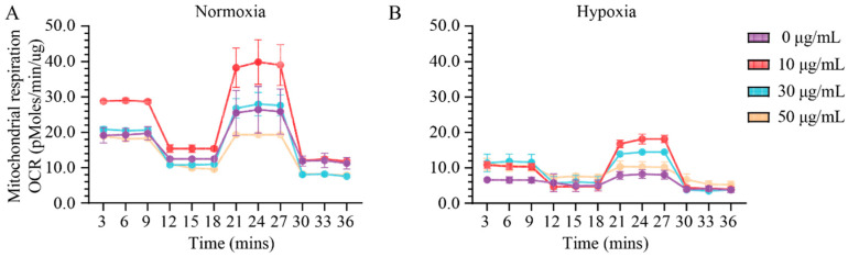
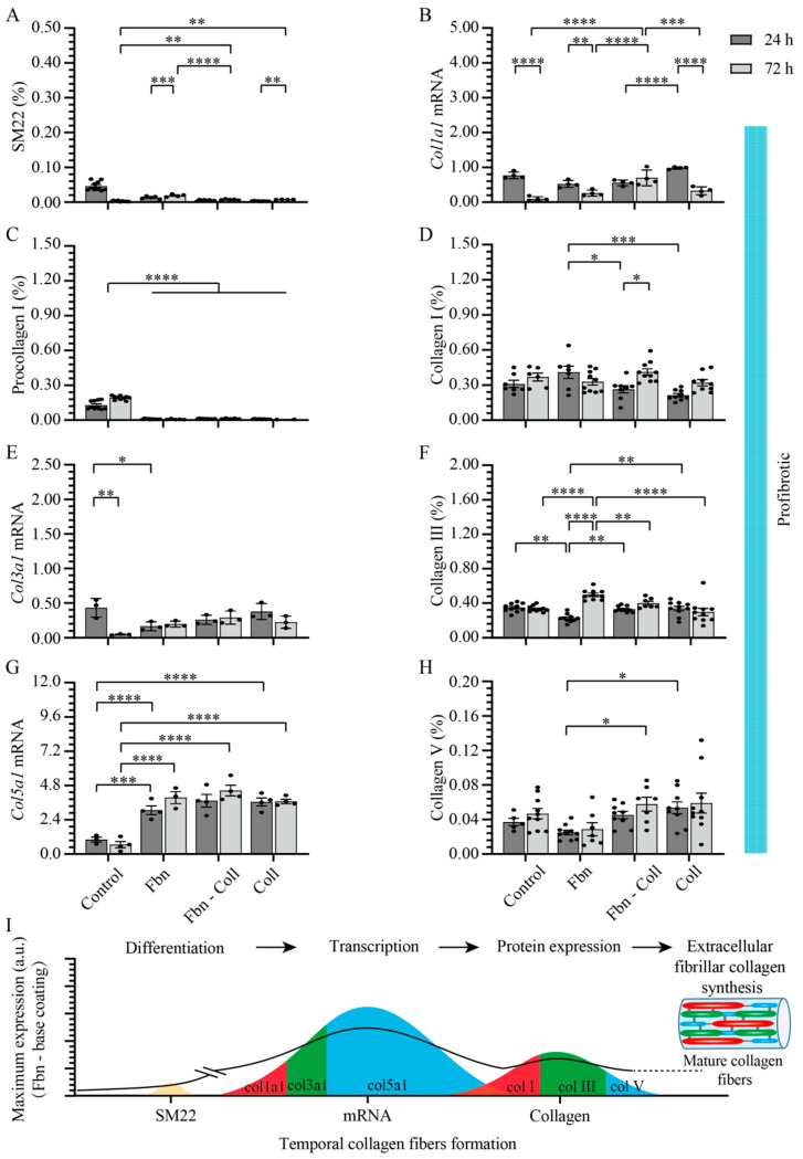
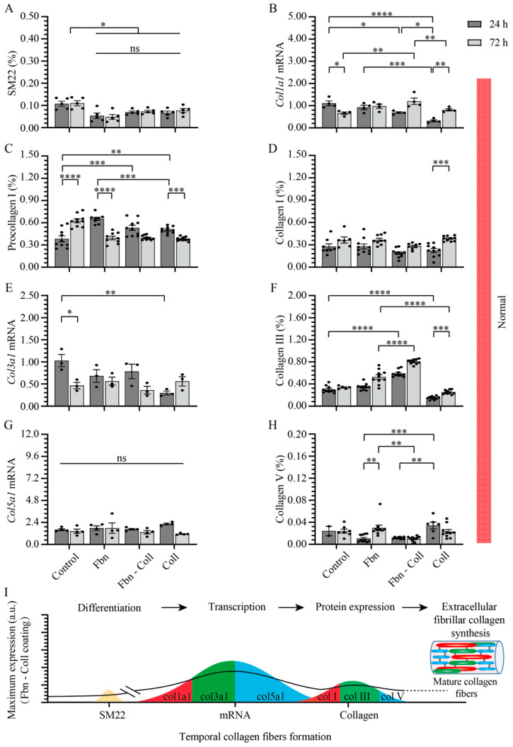
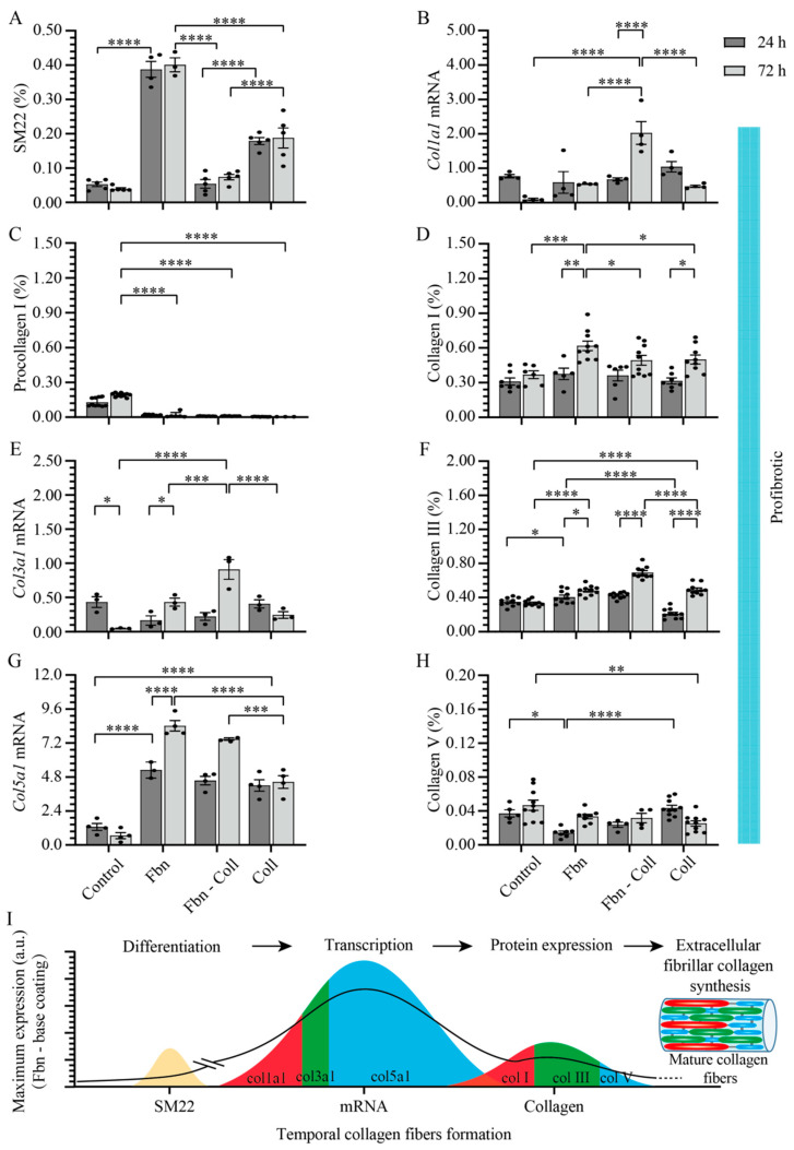
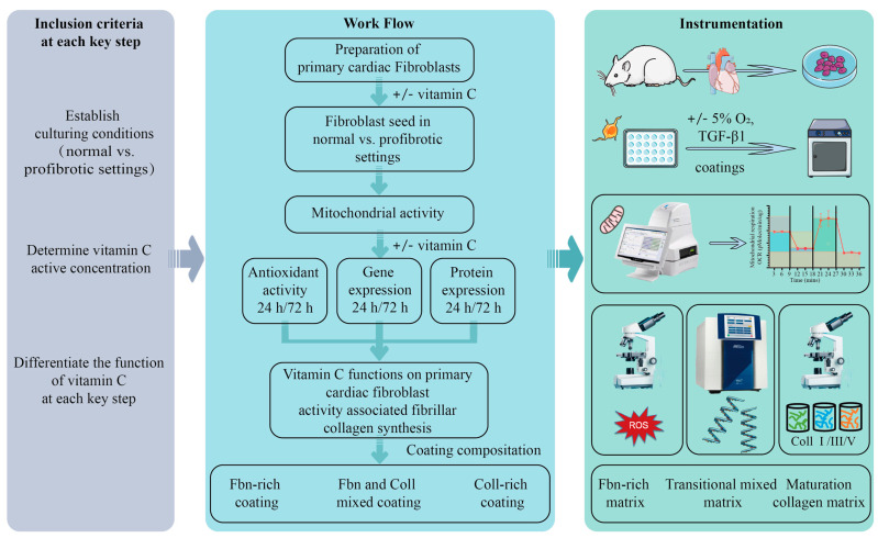
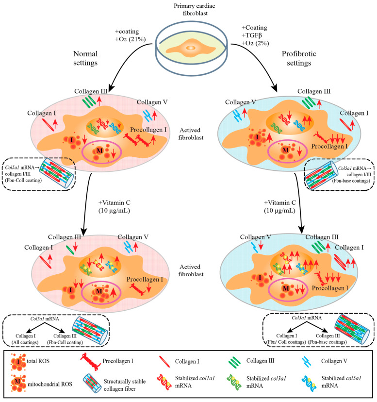
Extracellular collagen remodeling is one of the central mechanisms responsible for the structural and compositional coherence of myocardium in patients undergoing myocardial infarction (MI). Activated primary cardiac fibroblasts following myocardial infarction are extensively investigated to establish anti-fibrotic therapies to improve left ventricular remodeling. To systematically assess vitamin C functions as a potential modulator involved in collagen fibrillogenesis in an in vitro model mimicking heart tissue healing after MI. Mouse primary cardiac fibroblasts were isolated from wild-type C57BL/6 mice and cultured under normal and profibrotic (hypoxic + transforming growth factor beta 1) conditions on freshly prepared coatings mimicking extracellular matrix (ECM) remodeling during healing after an MI. At 10 μg/mL, vitamin C reprogramed the respiratory mitochondrial metabolism, which is effectively associated with a more increased accumulation of intracellular reactive oxygen species (iROS) than the number of those generated by mitochondrial reactive oxygen species (mROS). The mRNA/protein expression of subtypes I, III collagen, and fibroblasts differentiations markers were upregulated over time, particularly in the presence of vitamin C. The collagen substrate potentiated the modulator role of vitamin C in reinforcing the structure of types I and III collagen synthesis by reducing collagen V expression in a timely manner, which is important in the initiation of fibrillogenesis. Altogether, our study evidenced the synergistic function of vitamin C at an optimum dose on maintaining the equilibrium functionality of radical scavenger and gene transcription, which are important in the initial phases after healing after an MI, while modulating the synthesis of de novo collagen fibrils, which is important in the final stage of tissue healing.

Keywords: antioxidant capacity; collagen modulator; fibroblast; gene transcription regulator; vitamin C.


Conflict of interest statement

The authors declare that the manuscript was prepared in the absence of any conflict of interest.

Figures

Figure 1
Figure 1
Odd and even effects of low and high vitamin C concentrations stimulate or disrupt the bioenergetic status in primary cardiac fibroblasts. A concentration of 10 μg/mL of vitamin C selectively increases the respiration profile of mitochondria, whereas at higher concentrations, the nonmitochondrial respiration is significantly reduced in normoxia (A) and in hypoxic conditions (B). Data are normalized to total protein. Error bars indicate standard error of the mean; n = 3 independent experiments per condition.
Figure 2
Figure 2
Fibronectin potentiates the radical scavenging role of vitamin C under normal settings. iROS formation is shortly upregulated on fibronectin-containing coatings (A). Vitamin C significantly potentiates the formation of iROS at the late incubation time, independent of coating composition (B). Similarly, mROS formation is shortly upregulated, independent of coating composition (C). Vitamin C scavenges timely mROS only on fibronectin coating, but not in primary cardiac fibroblasts cultured on collagen-containing coatings (D). Both iROS and mROS were detected via DHE–mitoSOX–DAPI co-staining of primary cardiac fibroblasts (10,000 cells/well). Error bars indicate standard error of the mean; * p ≤ 0.05; ** p ≤ 0.01; *** p ≤ 0.001; **** p ≤ 0.0001. n = 3 experiments per condition. Fbn (Fibronectin); Coll (Collagen); mROS (mitochondrial reactive oxygen species); iROS (intracellular reactive oxygen species).
Figure 3
Figure 3
Profibrotic conditions potentiate the radical scavenging functionality of vitamin C. iROS formation is upregulated in a timely manner (A). Vitamin C downregulates iROS formation at the late incubation time (B). Similarly, mROS follows a decay trend in time (C), and it vanishes even more in the presence of vitamin C (D). Likewise, iROS and mROS were detected via DHE–mitoSOX–DAPI co-staining of primary cardiac fibroblasts (10,000 cells/well). Error bars indicate standard error of the mean; * p ≤ 0.05; ** p ≤ 0.01; *** p ≤ 0.001; **** p ≤ 0.0001. n = 3 experiments per condition. Fbn (Fibronectin); Coll (Collagen); mROS (mitochondrial reactive oxygen species); iROS (intracellular reactive oxygen species).
Figure 4
Figure 4
Synthesis of fibrillar collagen depends moderately on substrate composition and is not associated with primary cardiac fibroblasts’ activity. In normal settings, SM22 expression levels are significantly upregulated in control group and decrease systematically with the incubation time on different substrates (A). Col1a1 mRNA expression was upregulated at the early stage of incubation and decreased later only on collagen-based coatings (B). Type I procollagen expression remained constant on fibronectin coatings (C), whereas at the protein level, independent of coatings’ composition, type I collagen expression increases at 72 h (D). Fibronectin-based coatings significantly potentiate both col3a1 mRNA (E) and type III collagen expressions (F). Col5a1 mRNA expression is upregulated at 24 h on fibronectin–collagen mix (G) whereas collagen coatings promote the upregulation of type V collagen at the later incubation stage (H). Schematic overview of results over time. The solid line towards mature collagen fibers formation serves as a guide to the reader (I). Error bars indicate standard error of the mean; ns stands for not significant p-value, * p ≤ 0.05; ** p ≤ 0.01; *** p ≤ 0.001; **** p ≤ 0.0001. n = 3 experiments per condition. Fbn (Fibronectin); Coll (Collagen).
Figure 5
Figure 5
Profibrotic settings stimulate col5a1 collagen gene transcription. Profibrotic settings stimulated SM22 expression over time mostly on fibronectin coatings (A). The mix of fibronectin–collagen coatings barely influenced the expression of col1a1 mRNA in time, but it decreased on fibronectin and collagen coatings in a timely manner (B). The expression level of type I procollagen barely varied over time, but it decreased more then that of the control group (C). Type I collagen increased in a timely manner only on collagen-containing coatings (D). Col3a1 mRNA expression was suppressed in activated primary cardiac fibroblasts on each substrate (E), whereas fibronectin and non-collagen-based coatings potentiated the expression type III collagen at the later time point (F). Strikingly, col5a1 mRNA expression increased about 4-fold more than that of control group on each coating (G), whereas on collagen-based coatings, type V collagen protein expression stayed elevated (H). Schematic overview of results over time. The solid line towards mature collagen fibers formation serves as a guide to the reader (I). Error bars indicate standard error of the mean; * p ≤ 0.05; ** p ≤ 0.01; *** p ≤ 0.001; **** p ≤ 0.0001. n = 3 experiments per condition. Fbn (Fibronectin); Coll (Collagen).
Figure 6
Figure 6
Vitamin C potentiates type III collagen turnover at the later time point under normal settings. SM22 expression levels is significantly upregulated in control group and decreases after incubation on different substrates (A). Col1a1 mRNA expression is enhanced, mostly on fibronectin–collagen mix and collagen coatings (B). Type I procollagen expression downregulates at the later time point, independent of coating composition (C). Type I collagen expression increases over time, peaking on fibronectin–collagen mix (D). The expression levels of col3a1 mRNA are constant over time, independent of coating composition (E), whereas the amount of type III collagen significantly increases over time (F). Col5a1 mRNA expression displays constant levels over time, independent of coating composition (G). Protein conversion levels of type V collagen reaches maximum values at the later time point on collagen coatings (H). Schematic overview of results, in time. The solid line towards mature collagen fibers formation serves as a guide to the reader (I). Error bars indicate standard error of the mean; ns stands for not significant p-value, * p ≤ 0.05; ** p ≤ 0.01; *** p ≤ 0.001; **** p ≤ 0.0001. n = 3 experiments per condition. Fbn (Fibronectin); Coll (Collagen).
Figure 7
Figure 7
Collagen synthesis is modulated in a timely manner by vitamin C. Under profibrotic settings, vitamin C downregulates SM22 expression in primary cardiac fibroblasts cultured on collagen-based coatings (A). Col1a1 mRNA expression decreases in time on collagen coatings, but is significantly upregulated at the later time point on the fibronectin–collagen mix coating (B). Type I procollagen expression remains elevated in time on fibronectin coatings and exhibits the lowest expression value on collagen coatings (C), which is associated with the increased levels of type I collagen, mostly on fibronectin coatings (D). Vitamin C significantly increases the expression levels of col3a1 mRNA over time (E), which is associated with the elevated expression of type III collagen on fibronectin–collagen mix (F). Vitamin C significantly increases the col5a1 mRNA expression over time, exhibiting a maximum value on fibronectin coatings (G). This finding is associated with the increased levels of type V collagen expression on fibronectin coatings, but not on collagen substrates (H). Schematic overview of results over time. The solid line towards mature collagen fibers formation serves as a guide to the reader (I). Error bars indicate standard error of the mean; * p ≤ 0.05; ** p ≤ 0.01; *** p ≤ 0.001; **** p ≤ 0.0001. n = 3 experiments per condition. Fbn (Fibronectin); Coll (Collagen).
Figure 8
Figure 8
Overview of the workflow, including the inclusion criteria and methodologies.
Figure 9
Figure 9
The synergistic and complementary functionalities of vitamin C in in vitro MI replica on fibrillar collagen formation becomes evident at the later incubation time point. In normal conditions, primary cardiac fibroblasts regulate naturally the internal ROS formation to low levels, which are relevant for optimal functionality of cells. The radical scavenger role of vitamin C is highly potentiated over time at the level of the oxidative protection of primary cardiac fibroblasts activation under normal conditions. The synergistic activity of vitamin C and coating composition is clearly visible through the massive upregulation of col5a1 mRNA and downregulation of ROS under profibrotic vs. normal settings. The increase expression of col5a1 mRNA translates into structurally stable type I and III collagen fibers, which is a process that is mainly potentiated by (i) fibronectin–collagen mixture coating in normal settings and (ii) fibronectin and collagen coatings, respectively, in profibrotic conditions. Altogether, optimum supplementation doses of cell media with vitamin C are required to prove its radical scavenging activity and gene and protein stabilizer/modulator role for the synthesis of structurally and compositionally stable collagen fibrils.

Similar articles

Cited by

References

    1. Virani S.S., Alonso A., Benjamin E.J., Bittencourt M.S., Callaway C.W., Carson A.P., Chamberlain A.M., Chang A.R., Cheng S., Delling F.N., et al. Heart Disease and Stroke Statistics-2020 Update: A Report From the American Heart Association. Circulation. 2020;141:e139–e596. doi: 10.1161/CIR.0000000000000757. - DOI - PubMed
    1. Rusu M., Hilse K., Schuh A., Martin L., Slabu I., Stoppe C., Liehn E.A. Biomechanical assessment of remote and postinfarction scar remodeling following myocardial infarction. Sci. Rep. 2019;9:16744. doi: 10.1038/s41598-019-53351-7. - DOI - PMC - PubMed
    1. Zhang W., Liang J., Han P. Cardiac cell type-specific responses to injury and contributions to heart regeneration. Cell Regen. 2021;10:4. doi: 10.1186/s13619-020-00065-1. - DOI - PMC - PubMed
    1. Liu Z., Liu L., Cheng J., Zhang H. Risk Prediction Model Based on Biomarkers of Remodeling in Patients with Acute Anterior ST-Segment Elevation Myocardial Infarction. Med. Sci. Monit. Int. Med. J. Exp. Clin. Res. 2021;27:e927404. doi: 10.12659/MSM.927404. - DOI - PMC - PubMed
    1. Silva A.C., Pereira C., Fonseca A., Pinto-do-Ó P., Nascimento D.S. Bearing My Heart: The Role of Extracellular Matrix on Cardiac Development, Homeostasis, and Injury Response. Front. Cell Dev. Biol. 2020;8:621644. doi: 10.3389/fcell.2020.621644. - DOI - PMC - PubMed