Review
. 2020 Nov 24;44(6):701-724.
doi: 10.1093/femsre/fuaa019.

A decade of research on the second messenger c-di-AMP

Affiliations
Review

A decade of research on the second messenger c-di-AMP

Wen Yin et al. FEMS Microbiol Rev. .

Abstract

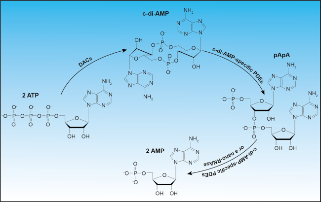
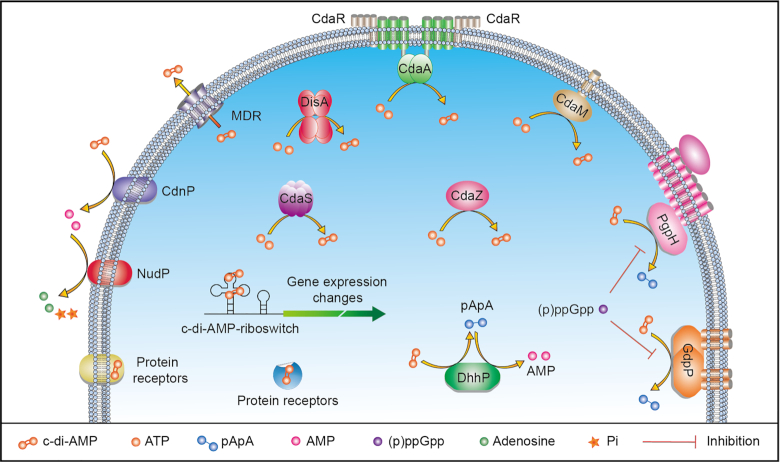
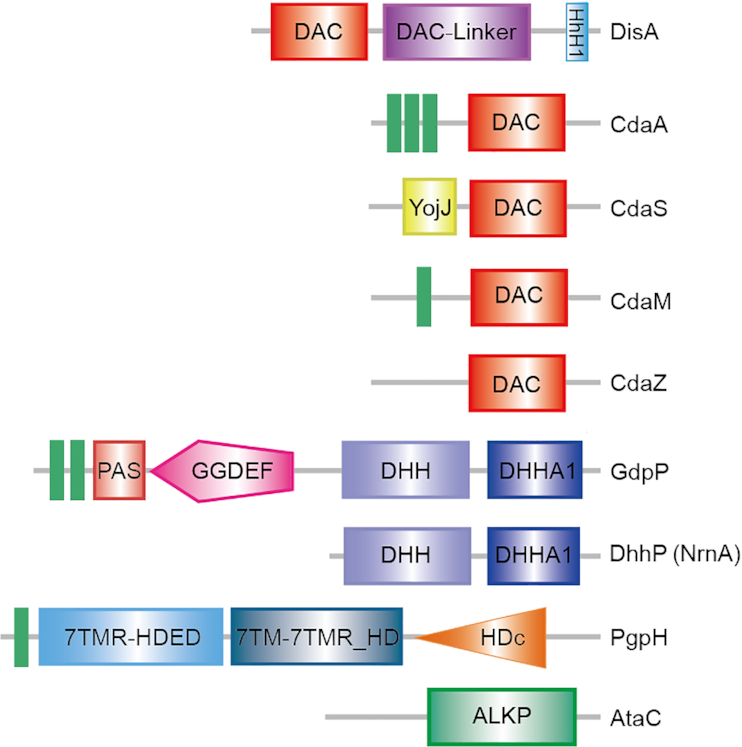
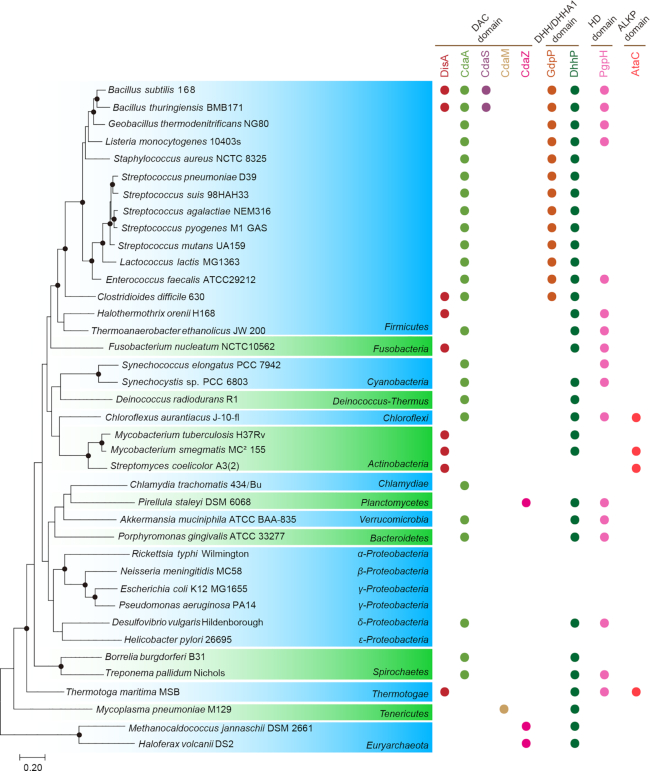
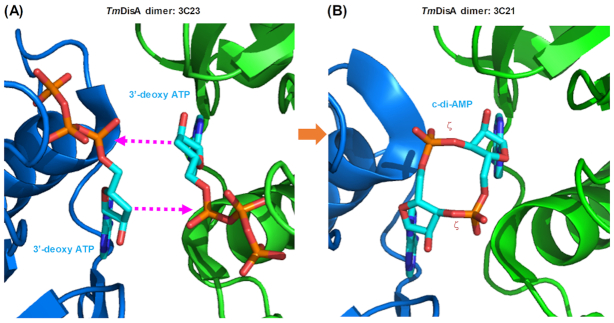
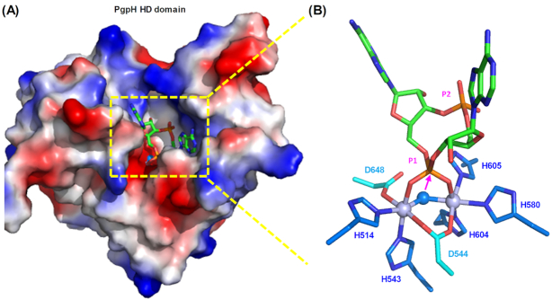
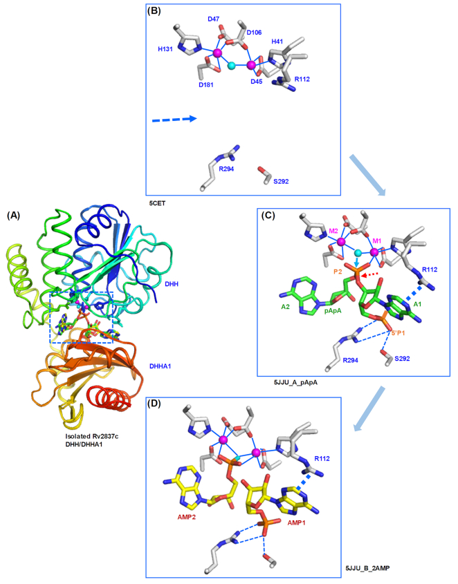
Cyclic dimeric adenosine 3',5'-monophosphate (c-di-AMP) is an emerging second messenger in bacteria and archaea that is synthesized from two molecules of ATP by diadenylate cyclases and degraded to pApA or two AMP molecules by c-di-AMP-specific phosphodiesterases. Through binding to specific protein- and riboswitch-type receptors, c-di-AMP regulates a wide variety of prokaryotic physiological functions, including maintaining the osmotic pressure, balancing central metabolism, monitoring DNA damage and controlling biofilm formation and sporulation. It mediates bacterial adaptation to a variety of environmental parameters and can also induce an immune response in host animal cells. In this review, we discuss the phylogenetic distribution of c-di-AMP-related enzymes and receptors and provide some insights into the various aspects of c-di-AMP signaling pathways based on more than a decade of research. We emphasize the key role of c-di-AMP in maintaining bacterial osmotic balance, especially in Gram-positive bacteria. In addition, we discuss the future direction and trends of c-di-AMP regulatory network, such as the likely existence of potential c-di-AMP transporter(s), the possibility of crosstalk between c-di-AMP signaling with other regulatory systems, and the effects of c-di-AMP compartmentalization. This review aims to cover the broad spectrum of research on the regulatory functions of c-di-AMP and c-di-AMP signaling pathways.

Keywords: c-di-AMP; metabolic enzyme; osmotic balance; physiological functions; protein receptor; riboswitch.


Figures

Figure 1.
Figure 1.
Synthesis and degradation of c-di-AMP. c-di-AMP is synthesized from two molecules of ATP through the action of DACs and is degraded by c-di-AMP-specific phosphodiesterases (PDEs) into a linear molecule of pApA or further to two molecules of AMP.
Figure 2.
Figure 2.
Conformation of cAMP (A, B) compared to the E-type (extended) (C, D) and U-type (E, F) conformations of c-di-AMP. (A) cAMP is synthesized by adenylyl cyclase via a cyclization reaction starting from an intramolecular attack of the ribose 3′-OH of ATP on its own α-phosphoryl group (indicated by a magenta arrow) to form a ζ bond with the release of a pyrophosphate moiety. The six torsional angles, α, β, γ, δ, ε and ζ, as well as the C3′ and C4′ atoms of cAMP, are marked. The oxygen atoms are shown in red, nitrogen atoms in blue, phosphorus atoms in orange and carbon atoms in green. (B) cAMP shown in a space-filling model and colored as in (A). Due to the ring rigidity, cAMP only exhibits two main conformations, syn- and anti-, that differ in the position of the adenine base with respect to the glycosidic bond (Tutar 2008). (C) c-di-AMP is synthesized by DAC via attacks of both ribose 3′-OHs (indicated by two magenta arrows) on the α-phosphoryl groups of two ATP molecules, and thus contains two adenine bases with a large cyclic phosphate lactone ring of twelve atoms, comprising two sets of torsional angles (with one set marked). Since these bonds are more flexible and freer to adopt various torsional angles, c-di-AMP can form many different conformations, ranging from the highly extended conformation (C, D) to the highly stacked conformation (E, F). Other conformations have also been observed (He et al. 2020). The conformational flexibility of c-di-AMP allows it to interact with a wide variety of receptors.
Figure 3.
Figure 3.
The signaling network mediated by c-di-AMP in bacteria. C-di-AMP is synthesized from two molecules of ATP by cytosolic DACs DisA, CdaS, CdaZ, membrane-bound CdaM and CdaA. Among them, CdaA activity can be inhibited by interacting with the regulatory protein CdaR. On the other hand, c-di-AMP can be hydrolyzed by membrane-bound phosphodiesterases PgpH and GdpP to pApA, or by cytosolic DhhP to pApA, which is further degraded to AMP by the same or paralogous DhhP (NrnA). PgpH and GdpP activities can be inhibited by the stringent response alarmone (p)ppGpp. C-di-AMP regulates various bacterial physiological functions by binding to either a riboswitch receptor or transmembrane or cytoplasmic protein receptors. It can be potentially secreted out of the bacterial cell and released into the host cell by multidrug efflux pumps. Extracellular c-di-AMP can also be hydrolyzed to AMP by CdnP, followed by degradation into adenosine and phosphate by NudP.
Figure 4.
Figure 4.
Domain architecture of DACs and c-di-AMP-specific PDEs. There are five types of DACs (DisA, CdaA, CdaS, CdaM and CdaZ) and four classes of PDEs (GdpP, DhhP, PgpH and AtaC). All five synthetases contain the enzymatic DAC domain, as well as other unique domains, according to the Pfam database (El-Gebali et al. 2019). Besides the DAC domain, DisA contains the DNA-binding HhH1 domain and a DisA-linker domain that connects the DAC and HhH1 domains; CdaA contains three transmembrane (TM) segments; CdaS includes a YojJ domain; CdaM contains a single TM segment, while CdaZ possess an N-terminal α/β domain (not shown). Among the four c-di-AMP-degrading enzymes, GdpP consists of a 2TM transmembrane domain, a PAS domain, a degenerate GGDEF domain and a combined C-terminal DHH-DHHA1 domain. DhhP contains only a combined DHH-DHHA1 domain. The nano-RNAse NrnA has the same domain architecture as DhhP. PgpH belongs to the 7TMR-HD family and consists of a signal peptide, followed by a 7TM-HD extracellular (7TM-HDED) domain, a transmembrane domain with 7 TM segments (7TM-7TMR-HD), and the phosphodiesterase HD-type catalytic domain (HDc). The recently described AtaC-type c-di-AMP PDE consists of a single alkaline phosphatase (ALKP) domain (Latoscha et al. 2020). See the Pfam database (El-Gebali et al. 2019) entry DisA_N (http://pfam.xfam.org/family/PF02457#tabview=tab1) for additional domain architectures that involve the DAC domain.
Figure 5.
Figure 5.
Distribution of the predicted DACs and c-di-AMP-specific PDEs in the bacterial strains studied so far. The maximum likelihood phylogenetic tree of sequenced bacterial genomes was constructed by MEGA6.0 software (Tamura et al. 2013) from a concatenated alignment of 12 ribosomal proteins, L1-L6, L9-L10 and S2-S5, as described previously (Yutin and Galperin 2013). The nodes with bootstrap support over 50% are marked with black dots. The presence of enzymes with specific DAC and PDE domain architectures was assigned according to the Pfam and COG databases and/or identified through BLAST searches. Most strains encoded only a single DAC and two or three types of c-di-AMP-specific PDEs. The PDEs that comprise only the DHH/DHHA1 domain pair are usually referred to as either DhhP or NrnA. This figure specifically excludes the single-stranded-DNA-specific exonuclease RecJ (COG0608), which combines the DHH/DHHA1 domain pair with a C-terminal RecJ_OB (PF17768) domain and is encoded by nearly all bacteria and archaea. See Table S1 in He et al. (2020) for phylogenetic profiles of c-di-AMP-related enzymes in many other species.
Figure 6.
Figure 6.
The likely mechanism of c-di-AMP synthesis by DisA. (A) The active site structure of the DisA/3′-deoxy ATP complex (PDB: 3C23), with the two DisA monomers from T. maritima (Witte et al. 2008) shown in ribbon form and colored in blue and green, respectively. The 3′-deoxy ATP is shown in stick representation with its carbon atoms colored in cyan. (B) The active site structure of the DisA/c-di-AMP complex (PDB: 3C21). The two newly formed ζ torsional angles are labeled.
Figure 7.
Figure 7.
The likely mechanism of c-di-AMP hydrolysis by the PgpH HD domain. (A) Electrostatic surface model of the monomeric PgpH HD domain from the bacterial pathogen L. monocytogenes in complex with c-di-AMP (PDB: 4S1B; Huynh et al. 2015). The c-di-AMP is accommodated in a deep binding site (boxed in a yellow rectangle and expanded in (B) with the two adenine bases pointing outward in a ‘V-type’ conformation (He et al. 2020) and one water molecule (colored in light blue in (B)) deeply buried in the cavity in panel (A).
Figure 8.
Figure 8.
The mechanism of c-di-AMP hydrolysis by the DHH/DHHA1 domains (type I) of the Rv2837c protein from M. tuberculosis, an example of lock-and-key interaction between two domains. The structures of DHH/DHHA1 domains of Rv2837c with metal ions only (PDB: 5CET), with an extra pApA molecule bound (PDB: 5JJU_A) and with two extra AMP molecules bound (PDB: 5JJU_B) could be superimposed very well with little conformational change (with r.m.s.d. of only 0.15–0.17 Å) and are colored in rainbow. There are two proteins in the unit cell of isolated Rv2837c DHH/DHHA1 protein, with protein A containing a pApA dinucleotide and protein B containing two AMP molecules (PDB: 5JJU, He et al. 2016). See text for details of parts (AD).
Figure 9.
Figure 9.
The likely mechanism of c-di-AMP hydrolysis by the GdpP DHH/DHHA1 domains (type II) in dynamic fluctuation mode. (A) The superimposed complex structures of DHH/DHHA1 domains with two metal ions (Mn2+) only (PDB: 5XSI), with an extra c-di-AMP bound (PDB: 5XSN), or with extra pApA bound (PDB: 5XSP) (Wang et al. 2018), are colored in magenta, green and blue, respectively. The metal ions in the DHH domain are in a blue box, while the c-di-AMP and pApA ligands in the DHHA1 domain are in a red box. These regions are further enlarged in (B), (C), and (D), respectively.
Figure 10.
Figure 10.
Distribution of the c-di-AMP riboswitch within bacterial phyla. The distribution profile of c-di-AMP riboswitches was collected and assembled from the Rfam database (Kalvari et al. 2018). The ordinate shows the total number of predicted c-di-AMP riboswitches found in each phylum.
Figure 11.
Figure 11.
Functional annotation of the genes located downstream of the c-di-AMP riboswitches. Genes associated with the c-di-AMP riboswitches are grouped into COG functional categories (Galperin et al. 2015) as noted on the right.
Figure 12.
Figure 12.
Cyclic di-AMP receptors involved in osmotic pressure regulation. (A) S. aureus c-di-AMP protein receptor CpaA is a transporter of K+ ion or Na+ ion, and c-di-AMP binding with its C-terminal RCK_C domain affects its ion transport properties. (B) L. lactis c-di-AMP receptors KupA and KupB belong to a class of K+ ion transporters, and their transport activities are inhibited upon c-di-AMP binding. (C) S. aureus c-di-AMP receptor KtrA is a cytoplasmic gating component of the Ktr-type K+ ion transport system, and c-di-AMP binding to its C-terminal RCK_C domain affects the K+ ion transport by the KtrAB system. (D) B. subtilis c-di-AMP receptor KtrC is a cytoplasmic gating component of the Ktr-type K+ ion transport system, and c-di-AMP binding to its C-terminal domain RCK_C domain affects the K+ ion transport by the KtrCD system. (E) S. pneumoniae c-di-AMP receptor CabP can bind c-di-AMP to influence the formation of the CabP-SPD_0076 complex, which in turn alters the transport of K+ ion. (F) S. aureus c-di-AMP receptor KdpD can bind c-di-AMP with its universal stress protein (USP)-like domain to change the phosphorylation status of KdpD and downstream response regulator KdpE, which controls the expression of the KdpFABC K+ ion transport system. (G) S. aureus c-di-AMP receptor OpuCA forms a complex with OpuCBD, which is involved in the transport of the osmolytes carnitine and glycine betaine. (H) Group B Streptococcus c-di-AMP-binding receptor BusR is a transcriptional factor that regulates the expression of the busAB genes encoding a transporter of betaine. (I) C-di-AMP riboswitch can bind c-di-AMP to affect the transcription or translation of downstream ion transport-related genes, including ktr, trk, kup, kimA, kdpFABC and some unidentified gene(s). See text for references and further details.
Figure 13.
Figure 13.
Sensing of c-di-AMP in the mammalian innate immune system. Five c-di-AMP sensors in mammalian host cells have been discovered to date: NLRP3, DDX41, STING, ERAdP and RECON. NLRP3 promotes interleukin-1β (IL-1β) production in response to c-di-NMP binding, while DDX41 binds c-di-NMP to promote the formation of a stable DDX41–STING complex, thereby activating the immune response. After binding c-di-AMP, c-di-GMP, or 2′,3′-cGAMP that is synthesized from ATP and GTP by the host cyclic GMP-AMP synthetase (cGAS) under foreign dsDNA segments activation, STING activates two downstream kinases TBK1 and IKK, thereby stimulating phosphorylation of a) IRF3 (interferon regulatory factor 3) to promote the production of Type I interferons (IFN-β), and b) of the inhibitor IκB to release NF-κB, leading ultimately to the production of pro-inflammatory cytokines. ERAdP activates downstream kinase TAK1 in response to c-di-AMP binding, which causes dissociation of NF-κB to promote the production of pro-inflammatory cytokines. In contrast, RECON inhibits NF-κB activity, but c-di-AMP binding attenuates this activity to indirectly activate NF-κB, also leading to the promotion of pro-inflammatory cytokine production.

Similar articles

Cited by

References

    1. Abdul-Sater AA, Tattoli I, Jin L et al. Cyclic-di-GMP and cyclic-di-AMP activate the NLRP3 inflammasome. EMBO Rep. 2013;14:900–6. - PMC - PubMed
    1. Andrade WA, Firon A, Schmidt T et al. Group B Streptococcus degrades cyclic-di-AMP to modulate STING-dependent type I interferon production. Cell Host Microbe. 2016;20:49–59. - PMC - PubMed
    1. Bai Y, Yang J, Eisele LE et al. Two DHH subfamily 1 proteins in Streptococcus pneumoniae possess cyclic di-AMP phosphodiesterase activity and affect bacterial growth and virulence. J Bacteriol. 2013;195:5123–32. - PMC - PubMed
    1. Bai Y, Yang J, Zarrella TM et al. Cyclic di-AMP impairs potassium uptake mediated by a cyclic di-AMP binding protein in Streptococcus pneumoniae. J Bacteriol. 2014;196:614–23. - PMC - PubMed
    1. Barrick JE, Corbino KA, Winkler WC et al. New RNA motifs suggest an expanded scope for riboswitches in bacterial genetic control. Proc Natl Acad Sci USA. 2004;101:6421–6. - PMC - PubMed

Publication types

MeSH terms

Substances