

Craniofacial features of cleidocranial dysplasia
- PMID: 30895069
- PMCID: PMC6395362
- DOI: 10.1016/j.jds.2017.07.002
Craniofacial features of cleidocranial dysplasia
Abstract
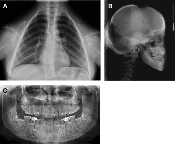
Cleidocranial dysplasia (CCD) is an autosomal-dominant malformation syndrome affecting bones and teeth. The most common skeletal and dental abnormalities in affected individuals are hypoplastic/aplastic clavicles, open fontanelles, short stature, retention of primary teeth, delayed eruption of permanent teeth, supernumerary teeth, and multiple impacted teeth. Treatment of CCD requires a multidisciplinary approach that may include dental corrections, orthognathic surgery and cranioplasty along with management of any complications of CCD. Early diagnosis of this condition enables application of the treatment strategy that provides the best quality of life to such patients. Notably, Runx2 gene mutations have been identified in CCD patients. Therefore, further elucidation of the molecular mechanism of supernumerary teeth formation related to Runx2 mutations may improve understanding of dental development in CCD. The insights into CCD pathogenesis may assist in the development of new treatments for CCD.
Keywords: Runx2; cleidocranial dysplasia; mutation; supernumerary teeth.
Figures
Similar articles
-
Genetic Pattern, Orthodontic and Surgical Management of Multiple Supplementary Impacted Teeth in a Rare, Cleidocranial Dysplasia Patient: A Case Report.Medicina (Kaunas). 2021 Dec 10;57(12):1350. doi: 10.3390/medicina57121350. Medicina (Kaunas). 2021. PMID: 34946295 Free PMC article.
-
Cleidocranial dysplasia: A report of two cases with brief review.Intractable Rare Dis Res. 2016 May;5(2):117-20. doi: 10.5582/irdr.2016.01022. Intractable Rare Dis Res. 2016. PMID: 27195196 Free PMC article.
-
Diversity of supernumerary tooth formation in siblings with cleidocranial dysplasia having identical mutation in RUNX2 : possible involvement of non-genetic or epigenetic regulation.Orthod Craniofac Res. 2007 Nov;10(4):222-5. doi: 10.1111/j.1601-6343.2007.00404.x. Orthod Craniofac Res. 2007. PMID: 17973689
-
Cleidocranial dysplasia: etiology and stomatognathic and craniofacial abnormalities.Minerva Stomatol. 2010 Mar;59(3):117-27. Minerva Stomatol. 2010. PMID: 20357738 Review. English, Italian.
-
Tooth formation and eruption - lessons learnt from cleidocranial dysplasia.Eur J Oral Sci. 2018 Oct;126 Suppl 1:72-80. doi: 10.1111/eos.12418. Eur J Oral Sci. 2018. PMID: 30178560 Review.
Cited by
-
Clinical and radiological findings in a severe case of cleidocranial dysplasia.BMJ Case Rep. 2018 Nov 12;2018:bcr2018226671. doi: 10.1136/bcr-2018-226671. BMJ Case Rep. 2018. PMID: 30420564 Free PMC article.
-
Genetic Pattern, Orthodontic and Surgical Management of Multiple Supplementary Impacted Teeth in a Rare, Cleidocranial Dysplasia Patient: A Case Report.Medicina (Kaunas). 2021 Dec 10;57(12):1350. doi: 10.3390/medicina57121350. Medicina (Kaunas). 2021. PMID: 34946295 Free PMC article.
-
Imaging studies used as aid in the diagnosis of cleidocranial dysplasia. A review.Rev Cient Odontol (Lima). 2021 Jun 21;9(2):e063. doi: 10.21142/2523-2754-0902-2021-063. eCollection 2021 Apr-Jun. Rev Cient Odontol (Lima). 2021. PMID: 38465273 Free PMC article. Review.
-
A novel perspective of calvarial development: the cranial morphogenesis and differentiation regulated by dura mater.Front Cell Dev Biol. 2024 Jun 24;12:1420891. doi: 10.3389/fcell.2024.1420891. eCollection 2024. Front Cell Dev Biol. 2024. PMID: 38979034 Free PMC article. Review.
-
Cranial Base Synchondrosis: Chondrocytes at the Hub.Int J Mol Sci. 2022 Jul 15;23(14):7817. doi: 10.3390/ijms23147817. Int J Mol Sci. 2022. PMID: 35887171 Free PMC article. Review.
References
-
- McNamara C.M., O'Riordan B.C., Blake M., Sandy J.R. Cleidocranial dysplasia: radiological appearances on dental panoramic radiography. Dentomaxillofac Radiol. 1999;28:89–97. - PubMed
-
- Martin S. Sur un déplacement natural de la clavicule. J Med Chir Pharmacol. 1765;23:456–460.
-
- Scheuthauer G. Kombination rudimentärer Schüsselbeine mit Anomalien des, Schädels biem erwachsenen Menschen. Allg Wien Med Ztg. 1871;16:293–295.
-
- Marie P., Sainton P. Sur la dysostose cléido-cranienne héréditaire. Rev Neurol. 1898;6:835–838. - PubMed
-
- McKusick V.A., Scott C.I. A nomenclature for constitutional disorders of bone. J Bone Jt Surg Am. 1971;53A:978–986. - PubMed
Publication types
LinkOut - more resources
Full Text Sources
