. 2018 Dec 20;201(2):e00473-18.
doi: 10.1128/JB.00473-18. Print 2019 Jan 15.

Regulation of Biofilm Aging and Dispersal in Bacillus subtilis by the Alternative Sigma Factor SigB

Affiliations

Regulation of Biofilm Aging and Dispersal in Bacillus subtilis by the Alternative Sigma Factor SigB

M Bartolini et al. J Bacteriol. .

Abstract

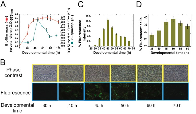
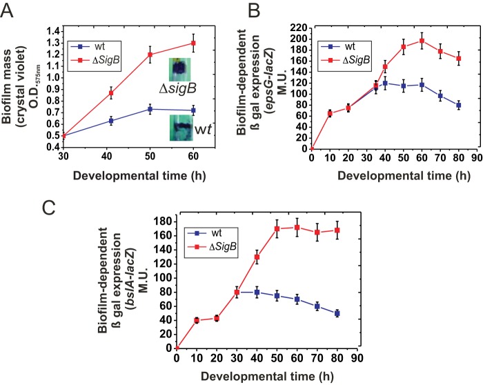
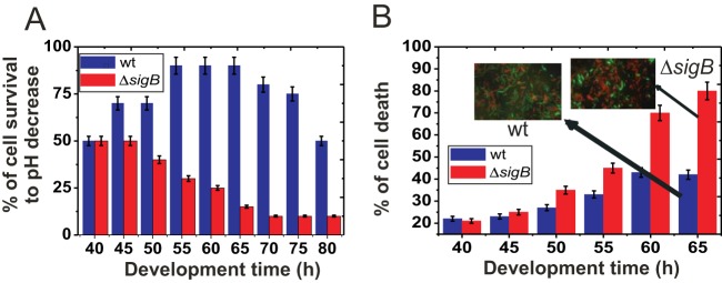
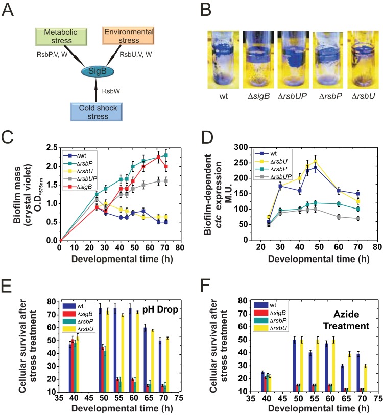
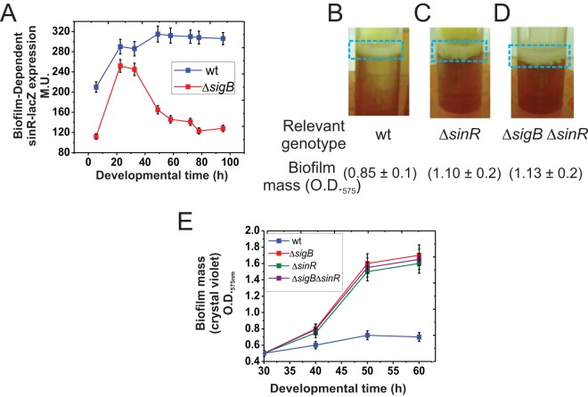
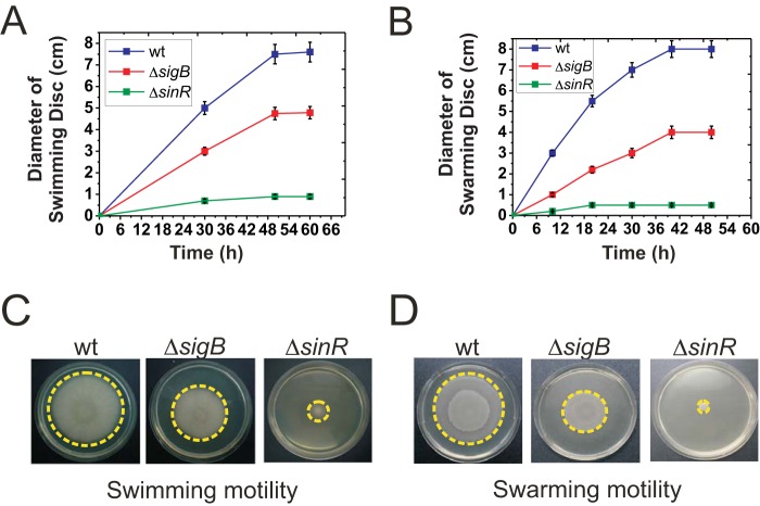
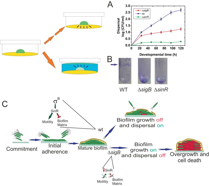
Bacterial biofilms are important in natural settings, biotechnology, and medicine. However, regulation of biofilm development and its persistence in different niches is complex and only partially understood. One key step during the biofilm life cycle is dispersal, when motile cells abandon the mature biofilm to spread out and colonize new niches. Here, we show that in the model bacterium Bacillus subtilis the general stress transcription factor SigB is essential for halting detrimental overgrowth of mature biofilm and for triggering dispersal when nutrients become limited. Specifically, SigB-deficient biofilms were larger than wild-type biofilms but exhibited accelerated cell death, significantly greater sensitivity to different stresses, and reduced dispersal. Interestingly, the signal detected by SigB to limit biofilm growth was transduced through the RsbP-dependent metabolic arm of the SigB regulatory cascade, which in turn positively controlled expression of SinR, the master regulator of biofilm formation and cell motility. This novel SigB-SinR regulatory circuit might be important in controlling the fitness of biofilms (either beneficial or harmful) in diverse environments.IMPORTANCE Biofilms are crucial for bacterial survival, adaptation, and dissemination in natural, industrial, and medical systems. Sessile cells embedded in the self-produced extracellular matrix of the biofilm benefit from a division of labor and are protected from environmental insults. However, as the biofilm ages, cells become stressed because of overcrowding, starvation, and accumulation of waste products. How does the sessile biofilm community sense and respond to stressful conditions? Here, we show that in Bacillus subtilis, the transcription factors SigB and SinR control whether cells remain in or leave a biofilm when metabolic conditions become unfavorable. This novel SigB-SinR regulatory circuit might be important for controlling the fitness of biofilms (either beneficial or harmful) in diverse environments.

Keywords: Bacillus subtilis; biofilm aging; biofilm dispersal; sigma B; stress activation.


Figures

FIG 1
FIG 1
SigB expression during the biofilm cell cycle. (A) Kinetics of biofilm formation and expression of the SigB-dependent ctc gene in biofilm-supporting LBY static culture. O.D.575nm, optical density at 575 nm; β-Gal, β-galactosidase. (B) Representative photographs from inner biofilm regions of GFP-expressing isogenic JH642 cells (strain MR655) at the indicated development times. Cells from exterior regions of the biofilm did not exhibit significant SigB-directed fluorescence (data not shown). Representative results from three independent experiments performed in parallel are shown. (C) Relative fluorescence intensity, indicated as a percentage of maximal (100%) SigB-directed gfp expression by the JH642 isogenic strain MR655 (amyE::PsigB-gfp::cat). Maximal fluorescence (100%) was considered to occur at 45 h of biofilm incubation. All of the other relative fluorescence levels, at the different times, were calculated as a percentage of the maximum fluorescence observed at 45 h of biofilm development. (D) Percentage of cells taken from inner regions of the biofilm that express GFP (MR655 strain). Each data point shown in panels A, C, and D is the mean ± SEM from a representative experiment performed in triplicate.
FIG 2
FIG 2
SigB regulates biofilm growth. (A) Rate of biofilm (floating pellicle) formation by wild-type (wt) JH642 and isogenic ΔsigB cells grown in static LBY broth. Inset photographs are representative of typical biofilms developed for 50 h and stained with crystal violet. (B) β-Galactosidase activity of the biofilm-reporter fusion epsG-lacZ::amyE in wild-type and isogenic ΔsigB cells grown in static LBY broth. A typical output of three independent experiments performed in parallel is shown. (C) β-Galactosidase activity of the biofilm-reporter fusion bslA-lacZ::amyE in wild-type and isogenic ΔsigB cells grown in static LBY broth. Each data point is the mean ± SEM. A typical output of three independent experiments performed in parallel is shown.
FIG 3
FIG 3
SigB regulates biofilm fitness. (A) Cell survival in wild-type JH642 and ΔsigB (strain MR644) biofilms after a drastic decrease in pH. At the indicated developmental times, wild-type and ΔsigB biofilms were exposed to a pH of 3.0 for 1 h before viable cells were counted, as indicated in Materials and Methods. A typical output of three independent experiments performed in parallel is shown. (B) Cell survival in wild-type JH642 and ΔsigB (strain MR644) biofilms of different ages were measured as the percentage of the total number of fluorescent cells observed in 10 different fields after staining with the LIVE/DEAD BacLight kit (green and red cells are alive and dead cells, respectively). Inset photographs are representative of several pictures taken at the indicated time. Each data point is the mean ± SEM from a representative experiment performed in triplicate.
FIG 4
FIG 4
SigB input during the biofilm cell cycle. (A) Cartoon summarizing the three possible routes of SigB activation (see the text for details). (B) Fifty-hour-old biofilms, as observed after crystal violet staining, of wild-type JH642 and isogenic mutants with altered routes of SigB activation: strain MR644 (ΔsigB), strain RG5572 (ΔrsbU), strain RG5573 (ΔrsbP), and strain RG5574 (ΔrsbUP) (Table 1). Representative pictures of five independent experiments are shown. (C) Rate of biofilm (pellicle) formation by the wild type and different isogenic mutants with altered SigB activity shown in panel B. A typical output of three independent experiments performed in parallel is shown. (D) β-Galactosidase activity of the SigB-dependent reporter fusion ctc-lacZ::amyE in wild-type and isogenic mutants with altered SigB activity grown in static LBY broth. (E and F) Cell survival in biofilms of wild-type JH642 and isogenic mutants with altered SigB activity after a drastic decrease in pH (E) or treatment with azide (F). The percentage of cellular survival is referenced to the number of CFU of biofilms of the same age that had not been exposed to stress. For panels B to F, typical output ± SEM from five independent experiments is shown.
FIG 5
FIG 5
SigB regulates sinR expression. (A) β-Galactosidase activity from the sinR-lacZ::amyE reporter fusion in wild-type and isogenic ΔsigB cells grown in static LBY. Typical output from one of four independent experiments is shown. (B to D) SinR and SigB do not generate additive effects on biofilm growth. Fifty-hour-old representative biofilms developed by standing cultures of the wild type (strain JH642) and isogenic mutants with altered SinR (ΔsinR, strain RG432) and/or SigB (ΔsigB ΔsinR, strain RG5578) activity. Biofilm mass was measured using the crystal violet technique, as indicated. Similar results were observed in several independent experiments. (E) Rate of biofilm formation by the wild type and different isogenic mutants with altered SigB, SinR, and SigB-SinR activities. Each data point in panels A and E is an average ± SEM from five independent representative experiments.
FIG 6
FIG 6
SigB regulates flagellum-dependent motilities in B. subtilis. (A) Swimming proficiencies of wild-type (strain JH642) and isogenic mutants with altered SigB or SinR activity (strains MR644 and RG432, respectively). (B) Swarming proficiencies of the wild type (strain NCIB3610) and isogenic mutants with altered SigB or SinR activity (strains RG5568 and RG4576, respectively). For the swarming experiments, the NCIB3610 strain was utilized instead of the JH642 strain, because the latter strain does not swarm. (C and D) Representative pictures from several independent experiments of top-viewed swimming (C) or swarming (D) cells after 48 h of incubation are shown. Each data point in panels A and B is the mean ± SEM from a representative experiment performed in triplicate.
FIG 7
FIG 7
SigB regulates biofilm dispersal. (A and B) Quantification of cell dispersal from wild-type and ΔsigB biofilms. (A) Colonies of JH642 and isogenic ΔsigB and ΔsinR derivatives (strains MR644 and RG432, respectively) developed on 2-cm-diameter wells poured with LBY medium (yellow in the cartoon) prepared with 0.8% agar and incubated for 30 h at 37°C, as indicated in Materials and Methods. At the indicated times, the agar (yellow in the cartoon) surrounding each solid biofilm (green in the cartoon) was carefully removed, and trapped cells were eluted and washed before appropriate serial dilutions were plated on LB agar plates as described in Materials and Methods. After 24 h of incubation at 37°C, the number of CFU ml−1 was calculated and plotted. The means ± SEM from 10 independent experiments are shown. (B) Colonies of wild-type (JH642), ΔsigB (MR644), and ΔsinR (RG432) strains were developed for 30 h at 37°C on 0.2 ml of LBY medium solidified with 2% agar at the bottom of a glass tube. The colonies were then carefully covered with 2.5 ml of LBY broth (blue in the cartoon) and incubated for another 15 h at 25°C to visualize the cellular ring (new green pellicle biofilm in the cartoon) produced at the liquid-air interface by the dispersed cells (see Materials and Methods for details). Ring cells adhered to the well walls were visualized by crystal violet staining (arrow). In the case of ΔsigB and ΔsinR biofilms, the staining at the bottom of the tubes represents the dispersal-defective SigB and SinR cells adhered to the glass bottom. A typical result from 10 independent experiments is shown. (C) Proposed model for the role of SigB during the biofilm life cycle and its regulation on sinR (see the text for details).

Similar articles

Cited by

References

    1. Hall-Stoodley L, Costerton JW, Stoodley P. 2004. Bacterial biofilms: from the natural environment to infectious diseases. Nat Rev Microbiol 2:95–108. doi:10.1038/nrmicro821. - DOI - PubMed
    1. Flemming HC, Wingender J, Szewzyk U, Steinberg P, Rice SA, Kjelleberg S. 2016. Biofilms: an emergent form of bacterial life. Nat Rev Microbiol 14:563–575. doi:10.1038/nrmicro.2016.94. - DOI - PubMed
    1. Flemming HC, Wingender J. 2010. The biofilm matrix. Nat Rev Microbiol 8:623–633. doi:10.1038/nrmicro2415. - DOI - PubMed
    1. Abee T, Kovács AT, Kuipers OP, van der Veen S. 2011. Biofilm formation and dispersal in Gram-positive bacteria. Curr Opin Biotechnol 22:172–179. doi:10.1016/j.copbio.2010.10.016. - DOI - PubMed
    1. McDougald D, Rice SA, Barraud N, Steinberg P, Kjelleberg S. 2011. Should we stay or should we go: mechanisms and ecological consequences for biofilm dispersal. Nat Rev Microbiol 10:39–50. doi:10.1038/nrmicro2695. - DOI - PubMed

Publication types

MeSH terms

LinkOut - more resources