. 2018 Jul 12;23(7):1700.
doi: 10.3390/molecules23071700.

Understanding the Role of Anti-PEG Antibodies in the Complement Activation by Doxil in Vitro

Affiliations

Understanding the Role of Anti-PEG Antibodies in the Complement Activation by Doxil in Vitro

Barry W Neun et al. Molecules. .

Abstract

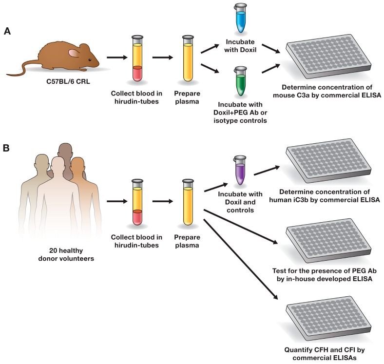
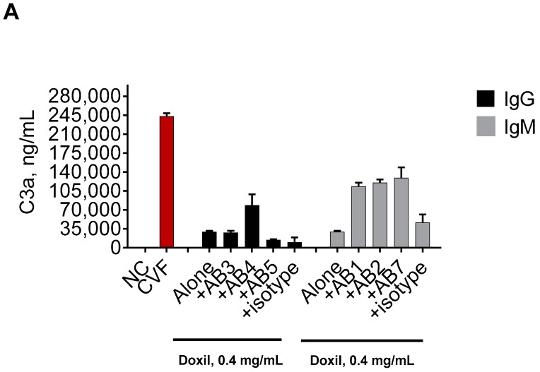
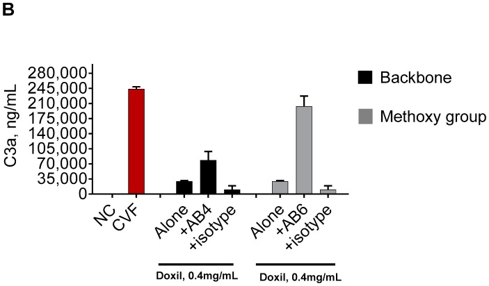
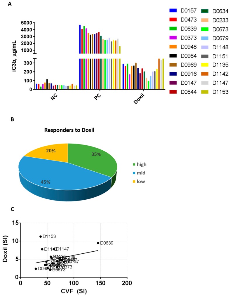
Infusion reactions (IRs) are common immune-mediated side effects in patients treated with a variety of drug products, including, but not limited to, nanotechnology formulations. The mechanism of IRs is not fully understood. One of the best studied mechanisms of IRs to nanomedicines is the complement activation. However, it is largely unknown why some patients develop reactions to nanomedicines while others do not, and why some nanoparticles are more reactogenic than others. One of the theories is that the pre-existing anti-polyethylene glycol (PEG) antibodies initiate the complement activation and IRs in patients. In this study, we investigated this hypothesis in the case of PEGylated liposomal doxorubicin (Doxil), which, when used in a clinical setting, is known to induce IRs; referred to as complement activation-related pseudoallergy (CARPA) in sensitive individuals. We conducted the study in vitro using plasma derived from C57BL/6 mice and twenty human donor volunteers. We used mouse plasma to test a library of well-characterized mouse monoclonal antibodies with different specificity and affinity to PEG as it relates to the complement activation by Doxil. We determined the levels of pre-existing polyclonal antibodies that bind to PEG, methoxy-PEG, and PEGylated liposomes in human plasma, and we also assessed complement activation by Doxil and concentrations of complement inhibitory factors H and I in these human plasma specimens. The affinity, specificity, and other characteristics of the human polyclonal antibodies are not known at this time. Our data demonstrate that under in vitro conditions, some anti-PEG antibodies contribute to the complement activation by Doxil. Such contribution, however, needs to be considered in the context of other factors, including, but not limited to, antibody class, type, clonality, epitope specificity, affinity, and titer. In addition, our data contribute to the knowledge base used to understand and improve nanomedicine safety.

Keywords: CARPA; anti-PEG antibody; complement activation; hypersensitivity; immunotoxicity; infusion reaction; liposomes; nanoparticles.


Conflict of interest statement

The authors declare no conflict of interest.

Figures

Figure 1
Figure 1
Study design. (A) The role of well-characterized mouse antibody in the complement activation by Doxil in mouse plasma; (B) detecting and understanding the role of pre-existing anti-PEG antibodies in the complement activation by Doxil in human plasma. Ab—antibody; ELISA—enzyme-linked immunosorbent assay; CFI—complement factor I; CFH—complement factor H; PEG—polyethylene glycol; CRL—Charles River Laboratories.
Figure 2
Figure 2
Influence of the anti-PEG antibodies on the complement activation by Doxil in murine plasma. Mouse plasma anticoagulated with hirudin was spiked with either Doxil or Doxil mixed with anti-PEG antibodies. The concentration of Doxil was 0.4 mg/mL of doxorubicin and was constant between all samples. The concentration of all antibodies was 10 μg/mL. Complement activation was assessed by ELISA measuring levels of the murine C3a component of the complement. (A) Role of antibody class. Doxil mixed with a PEG-specific IgM (AB1, AB2, or AB7), regardless of the antibodies’ affinity, stimulated a higher level of complement activation than Doxil alone. Doxil combined with one PEG-specific IgG (AB4) stimulated a higher level of the complement activation than Doxil alone. Doxil mixed with two other PEG-specific IgG (AB3 or AB5) induced complement activation comparable to that induced by Doxil alone; (B) Role of epitope specificity. Doxil incubated with IgG reactive to the PEG backbone or IgG reactive to the PEG terminal methoxy group induced stronger complement activation than Doxil alone. However, in the presence of IgG specific to the methoxy group, the complement activation by Doxil was stronger than that in the presence of PEG backbone-reactive IgG. Mouse IgG and mouse IgM, as specified in the materials and methods, were used as isotype controls and did not show a physiologically significant (i.e., two-fold or higher) influence on the complement activation by Doxil. Each bar shows mean and standard deviation (n = 3). All antibodies were in storage buffer containing 50% glycerol. When tested alone, 50% glycerol storage buffer did not result in C3a levels above those in the negative control (data not shown). DXR—doxorubicin; AB—antibody; numbers following “AB” refer to the antibodies described in Table 1; NC—negative control (phosphate-buffered saline—PBS); CVF—cobra venom factor used as the assay positive control.
Figure 2
Figure 2
Influence of the anti-PEG antibodies on the complement activation by Doxil in murine plasma. Mouse plasma anticoagulated with hirudin was spiked with either Doxil or Doxil mixed with anti-PEG antibodies. The concentration of Doxil was 0.4 mg/mL of doxorubicin and was constant between all samples. The concentration of all antibodies was 10 μg/mL. Complement activation was assessed by ELISA measuring levels of the murine C3a component of the complement. (A) Role of antibody class. Doxil mixed with a PEG-specific IgM (AB1, AB2, or AB7), regardless of the antibodies’ affinity, stimulated a higher level of complement activation than Doxil alone. Doxil combined with one PEG-specific IgG (AB4) stimulated a higher level of the complement activation than Doxil alone. Doxil mixed with two other PEG-specific IgG (AB3 or AB5) induced complement activation comparable to that induced by Doxil alone; (B) Role of epitope specificity. Doxil incubated with IgG reactive to the PEG backbone or IgG reactive to the PEG terminal methoxy group induced stronger complement activation than Doxil alone. However, in the presence of IgG specific to the methoxy group, the complement activation by Doxil was stronger than that in the presence of PEG backbone-reactive IgG. Mouse IgG and mouse IgM, as specified in the materials and methods, were used as isotype controls and did not show a physiologically significant (i.e., two-fold or higher) influence on the complement activation by Doxil. Each bar shows mean and standard deviation (n = 3). All antibodies were in storage buffer containing 50% glycerol. When tested alone, 50% glycerol storage buffer did not result in C3a levels above those in the negative control (data not shown). DXR—doxorubicin; AB—antibody; numbers following “AB” refer to the antibodies described in Table 1; NC—negative control (phosphate-buffered saline—PBS); CVF—cobra venom factor used as the assay positive control.
Figure 3
Figure 3
Inter-individual variability of the complement activation by Doxil in human plasma. Human plasma anticoagulated with hirudin was collected from 20 healthy donor volunteers and tested in vitro. (A) Plasma from individual donors was spiked with controls or Doxil and incubated for 30 min. The concentration of Doxil was 0.4 mg/mL of doxorubicin and was constant between all samples. Complement activation was assessed by ELISA measuring levels of human iC3b. Each bar shows mean response and standard deviation (n = 3); (B) Stimulation index was calculated for each donor as described in the materials and methods. Donors were grouped into three categories according to the individual magnitude of the complement activation: SI ≤ 2 was assigned as low response, SI 2–6 was assigned as medium response, and SI ≥ 6 was assigned as high response; (C) Regression analysis shows no linear correlation between complement activation by Doxil and CVF in individual plasma samples (p = 0.2112 and r = 0.08521; F = 1.677). SI—stimulation index; CVF—cobra venom factor; NC—negative control; PC—positive control (CVF); D—donor.
Figure 4
Figure 4
Analysis of pre-existing anti-PEG antibodies in human plasma. Human plasma anticoagulated with hirudin was collected from 20 healthy donor volunteers and tested in vitro. (A) A summary of antibody titers detected in individual donor plasma specimens. Positive controls are monoclonal antibodies as described in the materials and methods. There is no positive control in the PEG2000 IgG assay because currently available anti-PEG IgG are reactive to methoxy group. The titer in all shown PC samples was above 3200. Plasma from each donor was tested in duplicate on four ELISA plates. Each bar shown the titer determined from the mean response between duplicates; (B) Frequency of positive responders in the IgM assay; (C) Frequency of positive responders in the IgG assay. In both B and C, two analyses were performed. In one analysis, positive response was considered at the titer of 100 or higher. In the second analysis, the positive response was considered at the titer of 800 or higher; (D) Frequency of donors with antibodies reactive to both PEG and PEG-liposomes; (E) Frequency of donors with an antibody reactive to PEG-liposomes only. In both D and E, the positive response was considered at the titer of 800 or higher.
Figure 4
Figure 4
Analysis of pre-existing anti-PEG antibodies in human plasma. Human plasma anticoagulated with hirudin was collected from 20 healthy donor volunteers and tested in vitro. (A) A summary of antibody titers detected in individual donor plasma specimens. Positive controls are monoclonal antibodies as described in the materials and methods. There is no positive control in the PEG2000 IgG assay because currently available anti-PEG IgG are reactive to methoxy group. The titer in all shown PC samples was above 3200. Plasma from each donor was tested in duplicate on four ELISA plates. Each bar shown the titer determined from the mean response between duplicates; (B) Frequency of positive responders in the IgM assay; (C) Frequency of positive responders in the IgG assay. In both B and C, two analyses were performed. In one analysis, positive response was considered at the titer of 100 or higher. In the second analysis, the positive response was considered at the titer of 800 or higher; (D) Frequency of donors with antibodies reactive to both PEG and PEG-liposomes; (E) Frequency of donors with an antibody reactive to PEG-liposomes only. In both D and E, the positive response was considered at the titer of 800 or higher.
Figure 5
Figure 5
Complement activation by Doxil does not correlate with the titer of antibodies reactive to PEG in individual human donors. (A) The complement activation and antibody titer data are presented in a heat map format. The complement stimulation index by Doxil (C-SI) is aligned with the antibody titer (Ab-titer). Darker red and blue show higher levels of the complement activation and antibody titer, respectively. The antibody titer is plotted for IgM and IgG and indicates the antigen used to capture the reactive antibodies (Doxebo, PEG, or mPEG). The molecular weight of PEG is 2000 and is equal in all samples. mPEG contains a methoxy terminal group that is also present in the PEG2000 used to produce Doxil and Doxebo. D—donor; (BG) Regression analysis was performed to understand the correlation between the antibody titer and the complement activation by Doxil. Doxil stimulation index (SI) was compared with the titer of IgG reactive to Doxebo (B), PEG2000 (C), or mPEG2000 (D), as well as the titer of IgM reactive to Doxebo (E), PEG2000 (F), or mPEG2000 (G). p-value was used to assess the significance of the confidence interval of 95%. No analyses revealed a significant correlation. The residual analysis conducted in parallel with this test did not change the conclusions of the analysis (data not shown).
Figure 6
Figure 6
Plasma levels of complement inhibitory factors and their role in the complement activation. Plasma collected from 20 healthy donor volunteers and used in the complement activation and antibody titer study was also analyzed for the levels of CFH (A) and CFI (B). Regression analysis was performed to understand the correlation between the concentration of CFH and the complement activation by CVF (C) or Doxil (D). Regression analysis was also performed to understand the correlation between the concentration of CFI and complement activation by CVF (E) or Doxil (F). The p-value indicates a significant positive correlation between CFI concentration and the complement activation by CVF. No correlation was observed between CFH concentrations and complement activation by both Doxil and CVF, nor was it observed between CFI concentrations and the complement activation by Doxil.
Figure 6
Figure 6
Plasma levels of complement inhibitory factors and their role in the complement activation. Plasma collected from 20 healthy donor volunteers and used in the complement activation and antibody titer study was also analyzed for the levels of CFH (A) and CFI (B). Regression analysis was performed to understand the correlation between the concentration of CFH and the complement activation by CVF (C) or Doxil (D). Regression analysis was also performed to understand the correlation between the concentration of CFI and complement activation by CVF (E) or Doxil (F). The p-value indicates a significant positive correlation between CFI concentration and the complement activation by CVF. No correlation was observed between CFH concentrations and complement activation by both Doxil and CVF, nor was it observed between CFI concentrations and the complement activation by Doxil.

Similar articles

Cited by

References

    1. Corominas M., Gastaminza G., Lobera T. Hypersensitivity reactions to biological drugs. J. Investig. Allergol. Clin. Immunol. 2014;24:212–225. - PubMed
    1. Joerger M. Prevention and handling of acute allergic and infusion reactions in oncology. Ann. Oncol. 2012;23(Suppl. 10):x313–x319. doi: 10.1093/annonc/mds314. - DOI - PubMed
    1. Johansson S.G.O., Bieber T., Dahl R., Friedmann P.S., Lanier B.Q., Lockey R.F., Motala C., Martell J.A.O., Platts-Mills T.A., Ring J., et al. Revised nomenclature for allergy for global use: Report of the Nomenclature Review Committee of the World Allergy Organization, October 2003. J. Allergy Clin. Immunol. 2004;113:832–836. doi: 10.1016/j.jaci.2003.12.591. - DOI - PubMed
    1. Johansson S.G.O., Hourihane J.B., Bousquet J., Bruijnzeel-Koomen C., Dreborg S., Haahtela T., Kowalski M.L., Mygind N., Ring J., van Cauwenberge P., et al. A revised nomenclature for allergy: An EAACI position statement from the EAACI nomenclature task force. Allergy. 2001;56:813–824. doi: 10.1034/j.1398-9995.2001.t01-1-00001.x. - DOI - PubMed
    1. Jutel M., Agache I., Bonini S., Burks A.W., Calderon M., Canonica W., Cox L., Demoly P., Frew A.J., O’Hehir R., et al. International consensus on allergy immunotherapy. J. Allergy Clin. Immunol. 2015;136:556–568. doi: 10.1016/j.jaci.2015.04.047. - DOI - PubMed

MeSH terms