Review
. 2018 Jun 5;19(6):1677.
doi: 10.3390/ijms19061677.

The Neuromuscular Junction and Wide Heterogeneity of Congenital Myasthenic Syndromes

Affiliations
Review

The Neuromuscular Junction and Wide Heterogeneity of Congenital Myasthenic Syndromes

Pedro M Rodríguez Cruz et al. Int J Mol Sci. .

Abstract

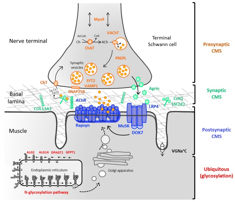
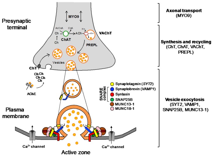
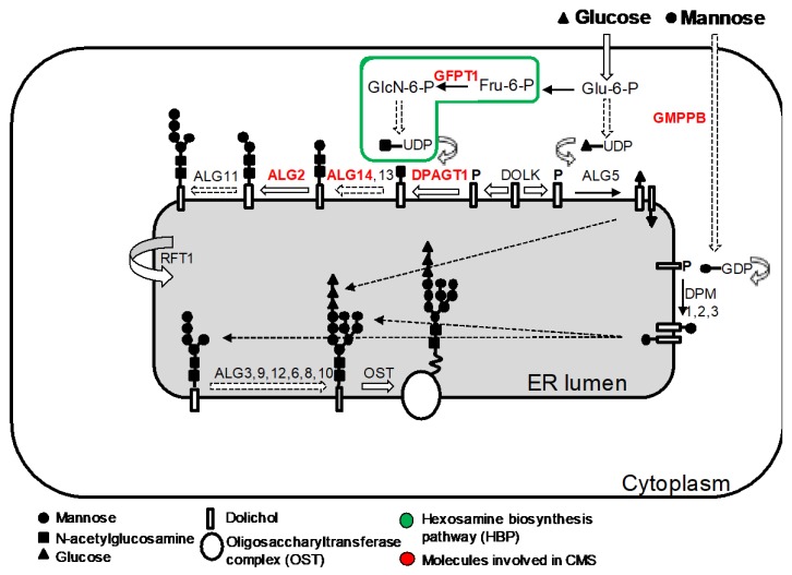
Congenital myasthenic syndromes (CMS) are genetic disorders characterised by impaired neuromuscular transmission. This review provides an overview on CMS and highlights recent advances in the field, including novel CMS causative genes and improved therapeutic strategies. CMS due to mutations in SLC5A7 and SLC18A3, impairing the synthesis and recycling of acetylcholine, have recently been described. In addition, a novel group of CMS due to mutations in SNAP25B, SYT2, VAMP1, and UNC13A1 encoding molecules implicated in synaptic vesicles exocytosis has been characterised. The increasing number of presynaptic CMS exhibiting CNS manifestations along with neuromuscular weakness demonstrate that the myasthenia can be only a small part of a much more extensive disease phenotype. Moreover, the spectrum of glycosylation abnormalities has been increased with the report that GMPPB mutations can cause CMS, thus bridging myasthenic disorders with dystroglycanopathies. Finally, the discovery of COL13A1 mutations and laminin α5 deficiency has helped to draw attention to the role of extracellular matrix proteins for the formation and maintenance of muscle endplates. The benefit of β2-adrenergic agonists alone or combined with pyridostigmine or 3,4-Dyaminopiridine is increasingly being reported for different subtypes of CMS including AChR-deficiency and glycosylation abnormalities, thus expanding the therapeutic repertoire available.

Keywords: COL13A1; GMPPB; N-glycosylation pathway; SNARE complex; congenital myasthenic syndromes; neuromuscular junction; neuromuscular transmission; presynaptic CMS; β2-adrenergic agonists.


Conflict of interest statement

The authors declare no conflict of interest.

Figures

Figure 1
Figure 1
Schematic of the neuromuscular junction (NMJ) and the main molecules involved in congenital myasthenic syndromes. CMS result from presynaptic (ChAT, ChT, MUNC13-1, MYO9, PREPL, SYT2, VAChT, and VAMP1), synaptic basal lamina (COLQ and COL13A1), and postsynaptic defects (AChR subunits: α, β, δ and ε, AGRN, DOK7, MUSK, LRP4, and rapsyn). An increasing number of presynaptic CMS is being reported due to abnormalities in the synthesis, recycling or release of acetylcholine (normal arrows). The Agrin-LRP4-MuSK signaling pathway (bold arrows) is crucial for the clustering of the AChRs at the postsynaptic muscle membrane. Novel genes encoding for ubiquitous molecules (GFPT1, DPAGT1, ALG2, ALG14, and GMPPB) are represented in the endoplasmic reticulum (ER) in a simplified view of the N-glycosylation pathway. Post-translational modifications of the saccharide structure of the AChR and other NMJ proteins take place at the ER and Golgi apparatus (dashed arrows), before reaching the muscle cell surface as mature proteins. ACh, acetylcholine; AChE, acetylcholinesterase; AcCoA, acetyl coenzyme A; Ch, choline; VGNa + C, voltage-gated sodium channel.
Figure 2
Figure 2
Proposed algorithm for targeted genetic screening of suspected CMS cases. Clinical evaluation should start by exploring age at onset and presence of manifestations beyond the neuromuscular boundaries. Ophthalmoplegia and limb-girdle weakness are clinically useful to guide genetic screening. Key diagnostic features are provided outside the boxes. Most frequent subtypes of CMS include AChR-deficiency, DOK7 CMS, and rapsyn CMS which stand for approximately 70% of all cases in the UK. (*) Slow channel syndrome, SYT2 CMS, and SNAP25B CMS are dominantly inherited. CNS, central nervous system; TA, tubular aggregates.
Figure 3
Figure 3
Schematic representation of the nerve terminal and the main molecules involved in presynaptic CMS. In the synaptic cleft, acetylcholinesterase (AChE) breaks down acetylcholine (ACh) into acetate and choline (Ch), which is uptaken by the sodium-dependent high-affinity choline transporter 1 (ChT) to the presynaptic terminal. The enzyme choline acetyltransferase (ChAT) catalyses the synthesis of ACh from acetyl coenzyme A (AcCoA) and choline, and the vesicular acetylcholine transporter (VAChT) loads ACh into synaptic vesicles. PREPL encodes a protein that is meant to act as an effector of the clathrin-associated adaptor protein 1 in the trafficking of VAChT [7]. The synaptic vesicles accumulate adjacent to the nerve terminal ready for exocytosis. Upon the arrival of an action potential, voltage-dependent Ca2+ channels open and the influx of Ca2+ cause the fusion of vesicles to the plasma membrane through the soluble N-ethylmaleimide-sensitive factor attachment protein receptor (SNARE) complex (synaptobrevin, syntaxin, and SNAP25B) and the Ca2+ sensor, synaptotagmin. Additionally, MUNC 13-1 and MUNC 18-1 (syntaxin-binding protein 1) take part in the assembly and disassembly of the complex through mechanisms still not fully understood [8]. Myosin-IX A is believed to be involved in axonal transport (two directions arrow). Adapted from [9].
Figure 4
Figure 4
Schematic representation of the synaptic basal lamina and its main components. Laminins are heterotrimeric proteins of high molecular weight formed by the incorporation of α, β, and γ chains. Laminins self-assemble, but also interact with integrins and α-dystroglycan [41]. Collagen IV, which is the most abundant protein at the basal lamina, self-assembles into dimers and hexamers thanks to its globular domains [41]. Nidogen-2 are non-collagenous glycoproteins responsible for linking collagen IV and laminin networks [42,43]. Additional collagens include COL13A1 [44] and COLQ, a collagen like tail responsible for anchoring AChE to the synaptic cleft. Muscle agrin binds to the basal lamina via laminin [45] and α-dystroglycan [46], and this is important for maintenance of the NMJ [47]. This differs from the role of neuronal agrin as a key organiser of the postsynaptic apparatus via the AChR clustering pathway [48]. Perlecan, another synaptic heparan sulphate proteoglycan is linked to both ColQ [49] and α-dystroglycan [50].
Figure 5
Figure 5
The adult AChR and the genetics of AChR-deficiency and kinetics abnormalities of the AChR. (A) The AChR is made up of five subunits organised around a central pore. Each subunit is composed of an extracellular domain, four transmembrane domains (M1–M4), and a large cytoplasmic loop that links M3 and M4; (B,C) Relative proportion of genetic defects in patients with AChR deficiency and kinetic abnormalities of the AChR within the Oxford CMS cohort. AChR deficiency is mainly caused by mutations in CHRNE encoding the ε-subunit of the AChR. SCS is often caused by mutations in CHRNA1 encoding the AChR α-subunit, while FCS is most commonly due to mutations in CHRNE.
Figure 6
Figure 6
The agrin-induced AChR clustering and ACh-CdK5 dispersal pathways. The AChR clustering pathway is shown blue and the dispersal pathway in red. Main molecules with a role in synapse formation and maintenance are represented although additional, still unknown, positive and negative factors are very likely to be involved. CMS patients harboring mutations within the AChR clustering pathway (excluding RAPSN) present common clinical features such as relative sparing of eye muscles, predominant limb girdle weakness, worsening of symptoms with drugs increasing ACh levels, and improvement on long-term therapy with β2-adrenergic agonists. DOK7 CMS represents the most frequent subtype in this category with other syndromes being rather infrequent.
Figure 7
Figure 7
Simplified representation of the N-glycosylation pathway of proteins and the molecules involved in CMS. The N-linked glycosylation of proteins takes place in the ER. It starts with the assembly of the core glycan (N-acetylglucosamine, glucose and mannose) on the lipid dolichol. A series of cytosolic glycosyltransferases proceed to dolichol glycosylation on the cytoplasmic face of the ER: GFPT1 synthesizes UDP-GlcNAc (Uridine diphosphate N-acetylglucosamine); DPAGT1 and the ALG13/14 complex are involved in adding the first and second N-acetylglucosamine to dolichol. Additional sugar residues are added by ALG2 and other enzymes until the resulting product is flipped into the ER lumen by RFT1. Inside the ER lumen, sugar moieties are incorporated until the glycan is transferred to asparagine residues of nascent proteins by the multimeric oligosaccharyl transferase complex (OST) that subsequently will be modified inside the ER and Golgi. DOLK, dolichol kinase; DPM, dolichol-phosphate mannose synthase; Fru-6-P, fructose-6-phosphate; GlcN-6-P, glucosamine-6-phosphate; Glu-6-P, glucose-6-phosphate; GMPPB, GDP-mannose pyrophosphrylase B.
Figure 8
Figure 8
Treatment strategy at the Oxford CMS Service for most common CMS subtypes. Whenever features of DOK7 and other members of the AGRN–MUSK pathway, COLQ, or SCS are present, first-line treatment with pyridostigmine should be avoided until confirmation of genetic diagnosis. 3,4-DAP can be added in with caution in certain CMS subtypes (* use with caution) [137]. Combined therapy with β2-adrenergic agonists may be tried on AChR deficiency, CMS due to glycosylation defects, FCS, and rapsyn CMS. In cases of SCS, quinidine may be used as an alternative to fluoxetine. FCS, Fast channel syndrome; N-glyc, N-glycosylation pathway; SCS, slow channel syndrome.

Similar articles

Cited by

References

    1. Palace J., Beeson D. The congenital myasthenic syndromes. J. Neuroimmunol. 2008;201–202:2–5. doi: 10.1016/j.jneuroim.2008.05.030. - DOI - PubMed
    1. Parr J.R., Andrew M.J., Finnis M., Beeson D., Vincent A., Jayawant S. How common is childhood myasthenia? The UK incidence and prevalence of autoimmune and congenital myasthenia. Arch. Dis. Child. 2014:5–9. doi: 10.1136/archdischild-2013-304788. - DOI - PubMed
    1. Zhang B., Shen C., Bealmear B., Ragheb S., Xiong W.-C., Lewis R.A., Lisak R.P., Mei L. Autoantibodies to agrin in myasthenia gravis patients. PLoS ONE. 2014;9:e91816. doi: 10.1371/journal.pone.0091816. - DOI - PMC - PubMed
    1. Pevzner A., Schoser B., Peters K., Cosma N.-C., Karakatsani A., Schalke B., Melms A., Kröger S. Anti-LRP4 autoantibodies in AChR- and MuSK-antibody-negative myasthenia gravis. J. Neurol. 2012;259:427–435. doi: 10.1007/s00415-011-6194-7. - DOI - PubMed
    1. Leite M.I., Jacob S., Viegas S., Cossins J., Clover L., Morgan B.P., Beeson D., Willcox N., Vincent A. IgG1 antibodies to acetylcholine receptors in “seronegative” myasthenia gravis. Brain. 2008:1940–1952. doi: 10.1093/brain/awn092. - DOI - PMC - PubMed

MeSH terms

LinkOut - more resources