

Neurodegeneration with brain iron accumulation
- PMID: 29325618
- PMCID: PMC8235601
- DOI: 10.1016/B978-0-444-63233-3.00019-1
Neurodegeneration with brain iron accumulation
Abstract
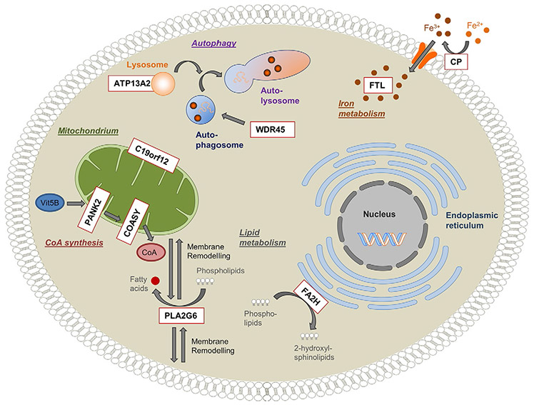
Neurodegeneration with brain iron accumulation (NBIA) comprises a clinically and genetically heterogeneous group of disorders affecting children and adults. These rare disorders are often first suspected when increased basal ganglia iron is observed on brain magnetic resonance imaging. For the majority of NBIA disorders the genetic basis has been delineated, and clinical testing is available. The four most common NBIA disorders include pantothenate kinase-associated neurodegeneration (PKAN) due to mutations in PANK2, phospholipase A2-associated neurodegeneration caused by mutation in PLA2G6, mitochondrial membrane protein-associated neurodegeneration from mutations in C19orf12, and beta-propeller protein-associated neurodegeneration due to mutations in WDR45. The ultrarare NBIA disorders are caused by mutations in CoASY, ATP13A2, and FA2H (causing CoA synthase protein-associated neurodegeneration, Kufor-Rakeb disease, and fatty acid hydroxylase-associated neurodegeneration, respectively). Together, these genes account for disease in approximately 85% of patients diagnosed with an NBIA disorder. New NBIA genes are being recognized with increasing frequency as a result of whole-exome sequencing, which is also facilitating early ascertainment of patients whose phenotype is often nonspecific.
Keywords: BPAN; INAD; MPAN; NBIA; PKAN; PLAN; infantile neuroaxonal dystrophy; neurodegeneration with brain iron accumulation; pantothenate kinase.
Copyright © 2018 Elsevier B.V. All rights reserved.
Figures
Similar articles
-
Analysis of the C19orf12 and WDR45 genes in patients with neurodegeneration with brain iron accumulation.J Neurol Sci. 2015 Feb 15;349(1-2):105-9. doi: 10.1016/j.jns.2014.12.036. Epub 2015 Jan 3. J Neurol Sci. 2015. PMID: 25592411
-
Mitochondria: A crossroads for lipid metabolism defect in neurodegeneration with brain iron accumulation diseases.Int J Biochem Cell Biol. 2015 Jun;63:25-31. doi: 10.1016/j.biocel.2015.01.018. Epub 2015 Feb 7. Int J Biochem Cell Biol. 2015. PMID: 25668476 Review.
-
Neurodegeneration with brain iron accumulation: Insights into the mitochondria dysregulation.Biomed Pharmacother. 2019 Oct;118:109068. doi: 10.1016/j.biopha.2019.109068. Epub 2019 Aug 9. Biomed Pharmacother. 2019. PMID: 31404774 Review.
-
Pantothenate kinase-associated neurodegeneration (PKAN) and PLA2G6-associated neurodegeneration (PLAN): review of two major neurodegeneration with brain iron accumulation (NBIA) phenotypes.Int Rev Neurobiol. 2013;110:49-71. doi: 10.1016/B978-0-12-410502-7.00003-X. Int Rev Neurobiol. 2013. PMID: 24209433 Free PMC article. Review.
-
Novel mutations in PANK2 and PLA2G6 genes in patients with neurodegenerative disorders: two case reports.BMC Med Genet. 2017 Aug 18;18(1):87. doi: 10.1186/s12881-017-0439-y. BMC Med Genet. 2017. PMID: 28821231 Free PMC article.
Cited by
-
Review of Hereditary and Acquired Rare Choreas.Tremor Other Hyperkinet Mov (N Y). 2020 Aug 6;10:24. doi: 10.5334/tohm.548. Tremor Other Hyperkinet Mov (N Y). 2020. PMID: 32832197 Free PMC article. Review.
-
Targeting Iron Dyshomeostasis for Treatment of Neurodegenerative Disorders.CNS Drugs. 2019 Nov;33(11):1073-1086. doi: 10.1007/s40263-019-00668-6. CNS Drugs. 2019. PMID: 31556017 Free PMC article. Review.
-
Pediatric-Onset Epilepsy and Developmental Epileptic Encephalopathies Followed by Early-Onset Parkinsonism.Int J Mol Sci. 2023 Feb 14;24(4):3796. doi: 10.3390/ijms24043796. Int J Mol Sci. 2023. PMID: 36835207 Free PMC article. Review.
-
Exploring the genetic and genomic connection underlying neurodegeneration with brain iron accumulation and the risk for Parkinson's disease.NPJ Parkinsons Dis. 2023 Apr 6;9(1):54. doi: 10.1038/s41531-023-00496-y. NPJ Parkinsons Dis. 2023. PMID: 37024536 Free PMC article.
-
Emerging Therapeutic Strategies for Parkinson's Disease and Future Prospects: A 2021 Update.Biomedicines. 2022 Feb 3;10(2):371. doi: 10.3390/biomedicines10020371. Biomedicines. 2022. PMID: 35203580 Free PMC article. Review.
References
-
- Delgado RF, Sanchez PR, Speckter H et al. (2012). Missense PANK2 mutation without “eye of the tiger” sign: MR findings in a large group of patients with pantothenate kinase-associated neurodegeneration (PKAN). J Magn Reson Imaging 35: 788–794. - PubMed
-
- Dogu O, Krebs CE, Kaleagasi H et al. (2013). Rapid disease progression in adult-onset mitochondrial membrane protein-associated neurodegeneration. Clin Genet 84: 350–355. - PubMed
Publication types
MeSH terms
Substances
Grants and funding
LinkOut - more resources
Full Text Sources
Other Literature Sources
Medical
Molecular Biology Databases
