

Deficiency of the sphingosine-1-phosphate lyase SGPL1 is associated with congenital nephrotic syndrome and congenital adrenal calcifications
- PMID: 28181337
- PMCID: PMC5384969
- DOI: 10.1002/humu.23192
Deficiency of the sphingosine-1-phosphate lyase SGPL1 is associated with congenital nephrotic syndrome and congenital adrenal calcifications
Abstract
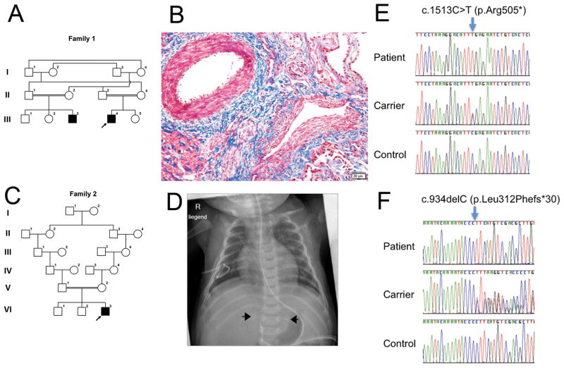
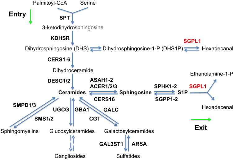
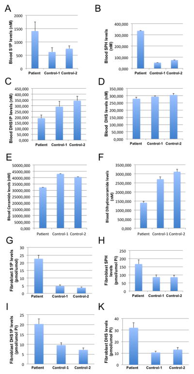
We identified two unrelated consanguineous families with three children affected by the rare association of congenital nephrotic syndrome (CNS) diagnosed in the first days of life, of hypogonadism, and of prenatally detected adrenal calcifications, associated with congenital adrenal insufficiency in one case. Using exome sequencing and targeted Sanger sequencing, two homozygous truncating mutations, c.1513C>T (p.Arg505*) and c.934delC (p.Leu312Phefs*30), were identified in SGPL1-encoding sphingosine-1-phosphate (S1P) lyase 1. SGPL1 catalyzes the irreversible degradation of endogenous and dietary S1P, the final step of sphingolipid catabolism, and of other phosphorylated long-chain bases. S1P is an intracellular and extracellular signaling molecule involved in angiogenesis, vascular maturation, and immunity. The levels of SGPL1 substrates, S1P, and sphingosine were markedly increased in the patients' blood and fibroblasts, as determined by liquid chromatography-tandem mass spectrometry. Vascular alterations were present in a patient's renal biopsy, in line with changes seen in Sgpl1 knockout mice that are compatible with a developmental defect in vascular maturation. In conclusion, loss of SGPL1 function is associated with CNS, adrenal calcifications, and hypogonadism.
Keywords: adrenal calcification; congenital adrenal insufficiency; congenital nephrotic syndrome; developmental; hypergonadotropic hypogonadism; hypogonadism; sphingolipids; sphingosine-1-phosphate; vascular.
© 2017 Wiley Periodicals, Inc.
Conflict of interest statement
The authors declare no conflict of interest.
Figures
Similar articles
-
Congenital adrenal calcifications as the first clinical indication of sphingosine lyase insufficiency syndrome: A case report and review of the literature.Am J Med Genet A. 2022 Nov;188(11):3312-3317. doi: 10.1002/ajmg.a.62956. Epub 2022 Aug 16. Am J Med Genet A. 2022. PMID: 35972040 Free PMC article. Review.
-
Sphingosine-1-phosphate lyase mutations cause primary adrenal insufficiency and steroid-resistant nephrotic syndrome.J Clin Invest. 2017 Mar 1;127(3):942-953. doi: 10.1172/JCI90171. Epub 2017 Feb 6. J Clin Invest. 2017. PMID: 28165343 Free PMC article.
-
A novel mutation in sphingosine-1-phosphate lyase causing congenital brain malformation.Brain Dev. 2018 Jun;40(6):480-483. doi: 10.1016/j.braindev.2018.02.008. Epub 2018 Mar 2. Brain Dev. 2018. PMID: 29501407
-
Sphingosine phosphate lyase insufficiency syndrome (SPLIS): A novel inborn error of sphingolipid metabolism.Adv Biol Regul. 2019 Jan;71:128-140. doi: 10.1016/j.jbior.2018.09.004. Epub 2018 Sep 25. Adv Biol Regul. 2019. PMID: 30274713 Free PMC article. Review.
-
Mutations in sphingosine-1-phosphate lyase cause nephrosis with ichthyosis and adrenal insufficiency.J Clin Invest. 2017 Mar 1;127(3):912-928. doi: 10.1172/JCI89626. Epub 2017 Feb 6. J Clin Invest. 2017. PMID: 28165339 Free PMC article.
Cited by
-
Druggable Sphingolipid Pathways: Experimental Models and Clinical Opportunities.Adv Exp Med Biol. 2020;1274:101-135. doi: 10.1007/978-3-030-50621-6_6. Adv Exp Med Biol. 2020. PMID: 32894509 Review.
-
Primary adrenal insufficiency: New genetic causes and their long-term consequences.Clin Endocrinol (Oxf). 2020 Jan;92(1):11-20. doi: 10.1111/cen.14109. Epub 2019 Oct 30. Clin Endocrinol (Oxf). 2020. PMID: 31610036 Free PMC article. Review.
-
An overview of inborn errors of metabolism manifesting with primary adrenal insufficiency.Rev Endocr Metab Disord. 2018 Mar;19(1):53-67. doi: 10.1007/s11154-018-9447-2. Rev Endocr Metab Disord. 2018. PMID: 29956047 Free PMC article. Review.
-
Latest Insights on the Etiology and Management of Primary Adrenal Insufficiency in Children.J Clin Res Pediatr Endocrinol. 2017 Dec 30;9(Suppl 2):9-22. doi: 10.4274/jcrpe.2017.S002. Epub 2017 Dec 27. J Clin Res Pediatr Endocrinol. 2017. PMID: 29280740 Free PMC article. Review.
-
Disarranged Sphingolipid Metabolism From Sphingosine-1-Phosphate Lyase Deficiency Leads to Congenital Nephrotic Syndrome.Kidney Int Rep. 2019 Aug 7;4(12):1763-1769. doi: 10.1016/j.ekir.2019.07.018. eCollection 2019 Dec. Kidney Int Rep. 2019. PMID: 31844815 Free PMC article. No abstract available.
References
-
- Bielawski J, Szulc ZM, Hannun YA, Bielawska A. Simultaneous quantitative analysis of bioactive sphingolipids by high-performance liquid chromatography-tandem mass spectrometry. Methods. 2006;39:82–91. - PubMed
Publication types
MeSH terms
Substances
Grants and funding
LinkOut - more resources
Full Text Sources
Other Literature Sources
Medical
Molecular Biology Databases
