. 2016 May 15;25(10):2031-2044.
doi: 10.1093/hmg/ddw077. Epub 2016 Mar 2.

ARL3 regulates trafficking of prenylated phototransduction proteins to the rod outer segment

Affiliations

ARL3 regulates trafficking of prenylated phototransduction proteins to the rod outer segment

Zachary C Wright et al. Hum Mol Genet. .

Abstract

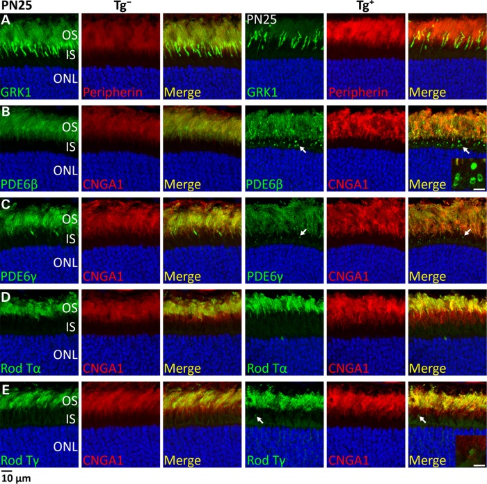
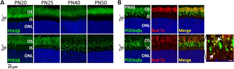
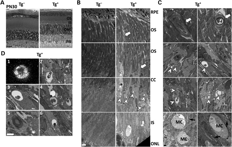
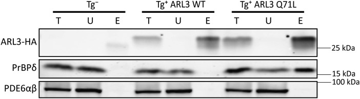
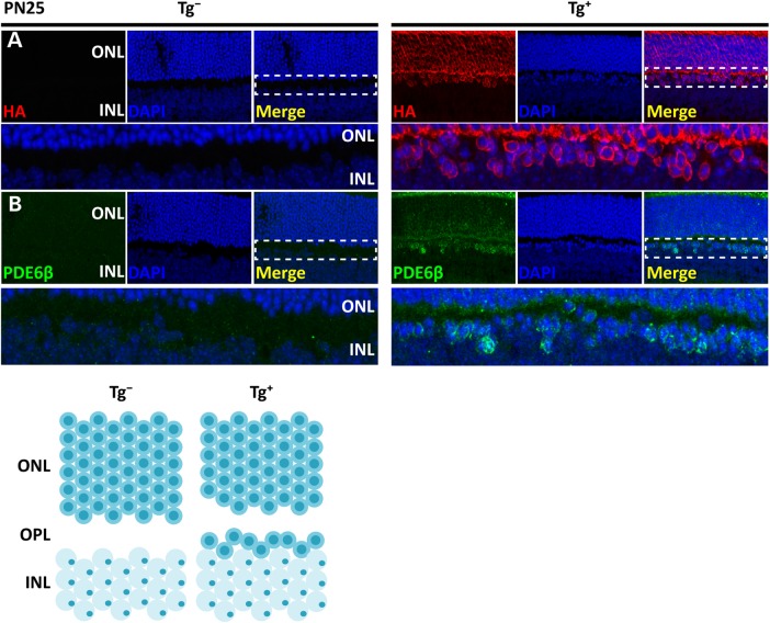
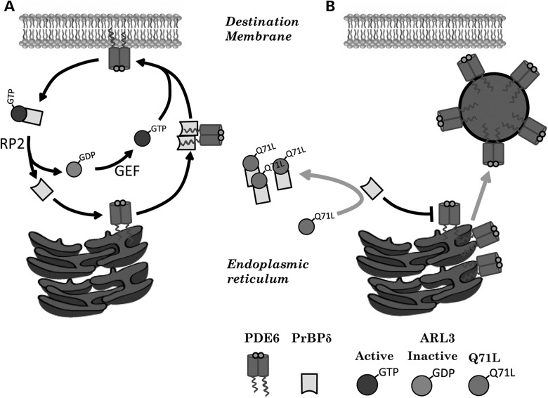
The small GTPase, ADP-ribosylation factor-like 3 (ARL3), has been proposed to participate in the transport of proteins in photoreceptor cells. Moreover, it has been implicated in the pathogenesis associated with X-linked retinitis pigmentosa (XLRP) resulting from mutations in the ARL3 GTPase activating protein, retinitis pigmentosa 2 (RP2). To determine the importance of ARL3 in rod photoreceptor cells, we generated transgenic mice expressing a dominant active form of ARL3 (ARL3-Q71L) under a rod-specific promoter. ARL3-Q71L animals exhibited extensive rod cell death after post-natal day 30 (PN30) and degeneration was complete by PN70. Prior to the onset of cell death, rod photoresponse was significantly reduced along with a robust decrease in rod phosphodiesterase 6 (PDE6) and G-protein receptor kinase-1 (GRK1) levels. Furthermore, assembled phosphodiesterase-6 (PDE6) subunits, rod transducin and G-protein receptor kinase-1 (GRK1) accumulated on large punctate structures within the inner segment in ARL3-Q71L retina. Defective trafficking of prenylated proteins is likely due to sequestration of prenyl binding protein δ (PrBPδ) by ARL3-Q71L as we demonstrate a specific interaction between these proteins in the retina. Unexpectedly, our studies also revealed a novel role for ARL3 in the migration of photoreceptor nuclei. In conclusion, this study identifies ARL3 as a key player in prenylated protein trafficking in rod photoreceptor cells and establishes the potential role for ARL3 dysregulation in the pathogenesis of RP2-related forms of XLRP.


Figures

Figure 1.
Figure 1.
ARL3-Q71L dominant active mutant transgenic model generation. (A) Scheme showing the construct used to express ARL3 in rod photoreceptors cells. The Arl3-Q71L (glutamine to leucine) dominant active mutant was expressed under a 4.4 kb rhodopsin promoter. This mutant protein was also C-terminally tagged with FLAG and Hemagglutinin (HA). (B) Western blot analysis of HEK 293 cells transfected with empty vector or wild-type ARL3 (lanes 1 and 2) and retinal lysate from Tg(−) and Tg(+) ARL3-Q71L animals (lanes 3 and 4). Staining was done with antibodies directed at ARL3 (red) and HA (green) to compare expression levels of transgenic versus endogenous ARL3 (refer to bar graph; P < 0.001). (C) Retinal sections from Tg(−) and Tg(+) animals stained for ARL3 (green) and CNGA1 (red) (top and middle panel) and ARL3 (green) and HA (red) (bottom panel). (Scale bar = 10 µm.) (D) Representative immunoblots showing the distribution of ARL3 and other proteins in a set of serial tangential 10 μm sections (1–10) of the mouse retina. RDS (perpherin) was used as a rod OS marker, COX I was used as a mitochondrial marker present in rod IS and synapse, the α subunit of rod transducin (rod Tα) was used as a general rod marker and β-tubulin was used as a general cellular marker. The inset on top, showing a mouse retinal section stained with toluidine blue, is provided to illustrate the origin of each section from various cellular compartments of rod photoreceptors.
Figure 2.
Figure 2.
Progressive loss of rod photoreceptor function and degeneration in ARL3-Q71L retina. (A) Representative scotopic (rod) and photopic (cone) electroretinograms (ERGs) comparing Tg(−) and Tg(+) animals at PN20 and PN70 across multiple light intensities. (B) Graph illustrating the scotopic a-wave amplitude measured at the light intensity of 0 Log Cd*s/m2 as a function of age. (C) Retinal sections showing nuclei from Tg(−) and Tg(+) animals stained with propidium iodide at PN30 and PN70. (D) Quantification of the ONL length (number of cell layers) at different locations within the retina from the inferior to superior portion between Tg(−) and Tg(+) animals at PN30 and PN70. *P < 0.01.
Figure 3.
Figure 3.
Normal elaboration of photoreceptor OS in ARL3-Q71L Mice. (A) Semi-thin sections of Tg(−) and Tg(+) retina from PN20 littermates stained with toluidine blue (scale bar = 20 µm). (B) Low magnification images from the OS and IS with endosomes indicated by white arrowheads (scale bar = 2 µm). (C) High magnification ultrastructure of the OS. White arrows point to membrane whorls in the rod OS from Tg(+) retina. Dilated endomembranes in the rod IS including Golgi apparatus indicated by black arrows and rough endoplasmic reticulum indicated by black arrowheads (scale bar = 0.5 µm).
Figure 4.
Figure 4.
Expression of ARL3-Q71L reduces the levels of prenylated phototransduction proteins. (A) Representative immunoblots of PN30 retinal protein samples showing levels of prenylated OS proteins and non-prenylated controls. (B) Quantification of protein levels from Tg(−) and Tg(+) retinal isolates (PDE6β : P = 0.0004, PDE6α : P = 0.009, GRK1 : P = 0.001, Tγ : P = 0.001). (C) Scotopic double flash (recovery) at PN20 shows the recovery of a-wave amplitude at different latencies between flashes from Tg(−) and Tg(+) animals (500 ms, P < 0.002; 750 ms, P < 0.001; 1000 ms, P < 0.002; 1500 ms, P = 0.32). Asterisk indicates statistically significant difference.
Figure 5.
Figure 5.
Loss of rod GRK1 and mislocalization of PDE6 in ARL3-Q71L mice. Immunolocalization of OS proteins in retinal cross-sections at PN25. (A) GRK1 (green) counterstained with peripherin/RDS (red) in Tg(−) and Tg(+) sections. (B) PDE6β (green) and (C) PDE6γ (green) each counterstained with CNGA1 (red) in Tg(−) and Tg(+) sections. Mislocalization of PDE6β or PDE6γ is indicated by white arrow. Inset in (B) shows a cropped and zoomed image (scale bar = 5 µm) of large punctate structures immunoreactive for PDE6 (ranging from 0.5 to 2 µm in diameter) within the inner segment. (D) Rod Tα (green) and (E) Rod Tγ (green) counterstained with CNGA1 (red) in Tg(−) and Tg(+) sections. White arrow indicates mislocalized transducin in the IS.
Figure 6.
Figure 6.
Progressive accumulation of rod PDE6 and colocalization of assembled rod PDE6 with GRK1 and transducin. (A) Immunolocalization of PDE6β in retinal cross-sections of Tg(−) and Tg(+) tissues from various ages (PN20–PN50). (B) PDE6αβγ (identified by ROS1 antibody), rod Tγ (Top Panel: red) immunolocalization in Tg(−) and Tg(+) retinal cross-sections at PN50. Zoomed image on right illustrates colocalization of PDE6αβγ with Tγ with PDE6αβγ in IS punctate structure indicated by white arrow (scale bar = 3 µm).
Figure 7.
Figure 7.
Accumulation of endosomal vesicles in the retina expressing ARL3-Q71L. (A) Semi-thin toluidine stained retinal sections from Tg(−) and Tg(+) animals at PN30 (scale bar = 20 µm). (B) Electron micrographs at low magnification illustrating the structural abnormalities of the photoreceptor OS and IS in Tg(+). Littermate Tg (−) serves as the control. Increased frequency of endosomes (white arrowhead), IS membrane whorls (*) and mis-oriented/dysmorphic OS discs (white arrow) (scale bar = 5 µm). (C) Electron micrographs from Tg(+) retina displaying high magnification images of the structures from (B) (top 3 rows) as well as dilated Golgi apparatus (black arrows) and Muller glial cells extending through the outer limiting membrane (MC) (scale bar = 0.5 µm). (D) Comparison of immunofluorescent punctate structure with PDE6β immunoreactivity on the same scale as the high magnification electron micrographs of the abnormal structures in the IS (scale bar = 1 µm).
Figure 8.
Figure 8.
PrBPδ interacts with ARL3-Q71L in vivo. Immunoprecipitation (IP) of retinal isolates from Tg(−), Tg(+) ARL3-WT (middle) and Tg(+) ARL3-Q71L (right) using rat monoclonal antibody against HA tag. Following IP, immunoblots were probed with indicated antibodies. T = Total, U = Unbound and E = Eluate.
Figure 9.
Figure 9.
Defective migration of rod photoreceptor cells in ARL3-Q71L retina. (A). Retinal cross-sections stained for HA (red) and DAPI (blue) comparing Tg(−) littermate and Tg(+) tissues at PN25. The zoomed image below was cropped from the white dashed box in the merge image above. (B). Retinal cross-sections stained for rod-specific PDE6β (green) and DAPI (blue) comparing Tg(−) and Tg(+) tissues at PN25. The diagram below illustrates the observed migration defect in transgenic animals expressing ARL3-Q71L.
Figure 10.
Figure 10.
Mechanistic model for mistrafficking of prenylated proteins in ARL3-Q71L mouse. (A) After prenylation and further processing, PDE6 and GRK1 are peripherally bound to the ER membrane. PrBPδ, or its homologs, extract prenylated proteins from the endomembrane and target them to the destination membrane. ARL3 is activated by its GEF (ARL13b) in the area of the destination membrane and binds to PrBPδ causing release of PDE6 and/or GRK1. RP2 accelerates the GTPase activity of ARL3 stimulating release of PrBPδ from the PrBPδ–ARL3–GTP complex. (B) Exogenously expressed active ARL3-Q71L sequesters PrBPδ and its homologs, thereby rendering it in capable of extracting PDE6 from the ER membrane. PDE6 and other prenylated proteins buildup in the ER membrane and are pushed out of the ER in endosomal vesicles which accumulate in the IS.

Similar articles

Cited by

References

    1. Sung C.H., Chuang J.Z. (2010) The cell biology of vision. J. Cell Biol., 190, 953–963. - PMC - PubMed
    1. Pearring J.N., Salinas R.Y., Baker S.A., Arshavsky V.Y. (2013) Protein sorting, targeting and trafficking in photoreceptor cells. Prog. Retin. Eye Res., 36, 24–51. - PMC - PubMed
    1. Schrick J.J., Vogel P., Abuin A., Hampton B., Rice D.S. (2006) ADP-ribosylation factor-like 3 is involved in kidney and photoreceptor development. Am. J. Pathol., 168, 1288–1298. - PMC - PubMed
    1. Gillingham A.K., Munro S. (2007) The small G proteins of the Arf family and their regulators. Annu. Rev. Cell Dev. Biol., 23, 579–611. - PubMed
    1. Veltel S., Gasper R., Eisenacher E., Wittinghofer A. (2008) The retinitis pigmentosa 2 gene product is a GTPase-activating protein for Arf-like 3. Nat. Struct. Mol. Biol., 15, 373–380. - PubMed

MeSH terms