

Abnormal centrosome and spindle morphology in a patient with autosomal recessive primary microcephaly type 2 due to compound heterozygous WDR62 gene mutation
- PMID: 24228726
- PMCID: PMC4225825
- DOI: 10.1186/1750-1172-8-178
Abnormal centrosome and spindle morphology in a patient with autosomal recessive primary microcephaly type 2 due to compound heterozygous WDR62 gene mutation
Abstract
Background: Autosomal recessive primary microcephaly (MCPH) is a rare neurodevelopmental disease with severe microcephaly at birth due to a pronounced reduction in brain volume and intellectual disability. Biallelic mutations in the WD repeat-containing protein 62 gene WDR62 are the genetic cause of MCPH2. However, the exact underlying pathomechanism of MCPH2 remains to be clarified.
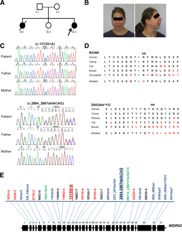
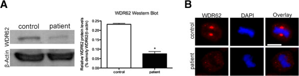
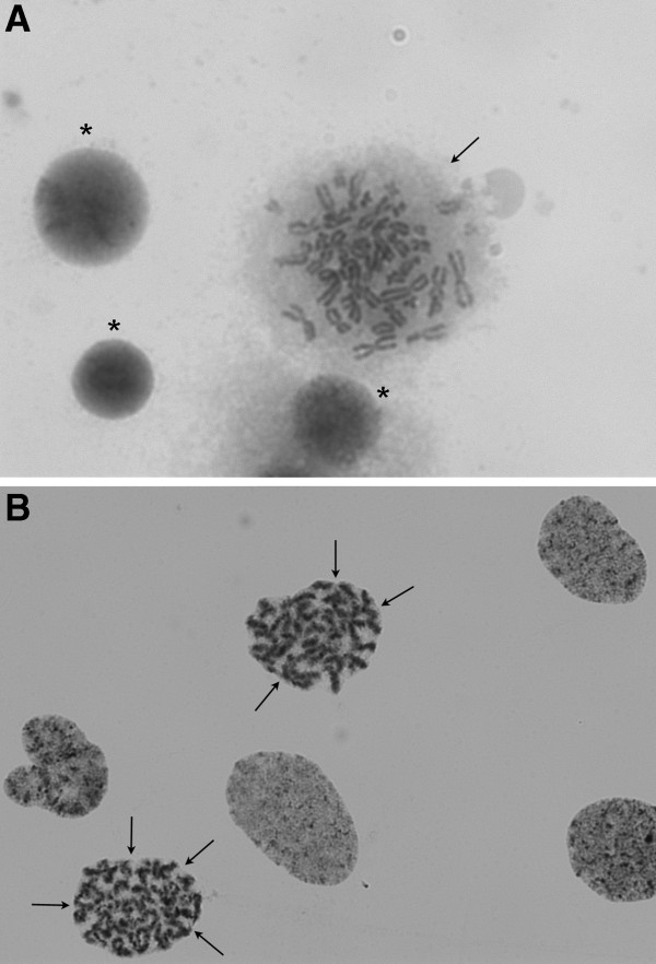
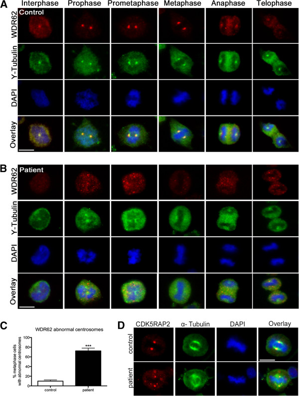
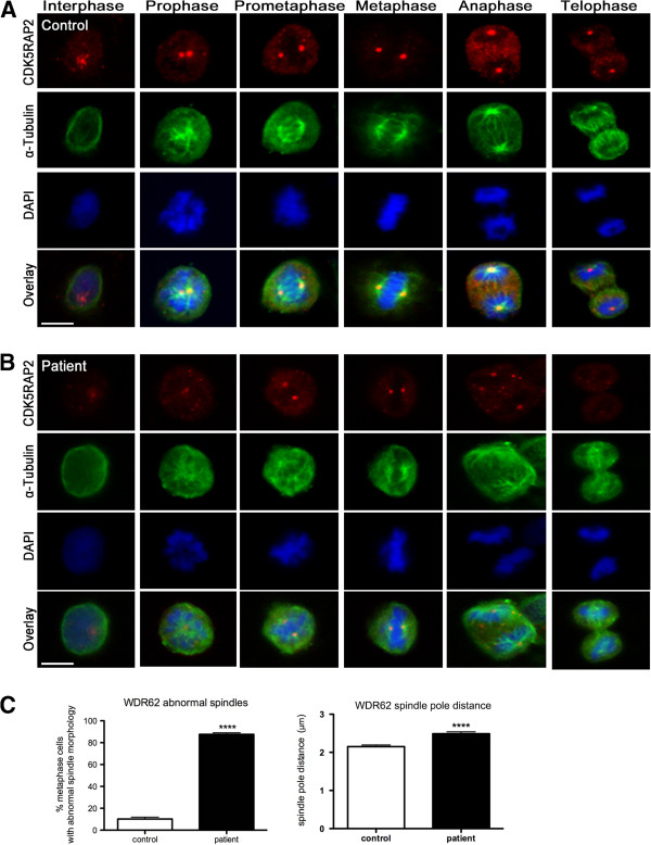
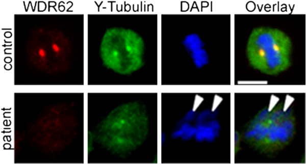
Methods/results: We characterized the clinical, radiological, and cellular features that add to the human MCPH2 phenotype. Exome sequencing followed by Sanger sequencing in a German family with two affected daughters with primary microcephaly revealed in the index patient the compound heterozygous mutations c.1313G>A (p.R438H) / c.2864-2867delACAG (p.D955Afs*112) of WDR62, the second of which is novel. Radiological examination displayed small frontal lobes, corpus callosum hypoplasia, simplified hippocampal gyration, and cerebellar hypoplasia. We investigated the cellular phenotype in patient-derived lymphoblastoid cells and compared it with that of healthy female controls. WDR62 expression in the patient's immortalized lymphocytes was deranged, and mitotic spindle defects as well as abnormal centrosomal protein localization were apparent.
Conclusion: We propose that a disruption of centrosome integrity and/or spindle organization may play an important role in the development of microcephaly in MCPH2.
Figures
Similar articles
-
PLK1-mediated phosphorylation of WDR62/MCPH2 ensures proper mitotic spindle orientation.Hum Mol Genet. 2017 Nov 15;26(22):4429-4440. doi: 10.1093/hmg/ddx330. Hum Mol Genet. 2017. PMID: 28973348
-
Clinical and cellular features in patients with primary autosomal recessive microcephaly and a novel CDK5RAP2 mutation.Orphanet J Rare Dis. 2013 Apr 15;8:59. doi: 10.1186/1750-1172-8-59. Orphanet J Rare Dis. 2013. PMID: 23587236 Free PMC article.
-
Mutations in WDR62, encoding a centrosomal and nuclear protein, in Indian primary microcephaly families with cortical malformations.Clin Genet. 2011 Dec;80(6):532-40. doi: 10.1111/j.1399-0004.2011.01686.x. Epub 2011 May 16. Clin Genet. 2011. PMID: 21496009
-
A novel WDR62 missense mutation in microcephaly with abnormal cortical architecture and review of the literature.J Appl Genet. 2019 May;60(2):151-162. doi: 10.1007/s13353-019-00486-y. Epub 2019 Feb 1. J Appl Genet. 2019. PMID: 30706430 Review.
-
The Role of WD40-Repeat Protein 62 (MCPH2) in Brain Growth: Diverse Molecular and Cellular Mechanisms Required for Cortical Development.Mol Neurobiol. 2018 Jul;55(7):5409-5424. doi: 10.1007/s12035-017-0778-x. Epub 2017 Sep 22. Mol Neurobiol. 2018. PMID: 28940170 Review.
Cited by
-
Two Novel Mutations (c.883-4_890del and c.1684C>G) of WDR62 Gene Associated With Autosomal Recessive Primary Microcephaly: A Case Report.Front Pediatr. 2019 Nov 7;7:457. doi: 10.3389/fped.2019.00457. eCollection 2019. Front Pediatr. 2019. PMID: 31788460 Free PMC article.
-
Molecular and cellular basis of autosomal recessive primary microcephaly.Biomed Res Int. 2014;2014:547986. doi: 10.1155/2014/547986. Epub 2014 Dec 8. Biomed Res Int. 2014. PMID: 25548773 Free PMC article. Review.
-
Nek5: a new regulator of centrosome integrity.Oncotarget. 2015 Sep 22;6(28):24594-5. doi: 10.18632/oncotarget.5139. Oncotarget. 2015. PMID: 26309075 Free PMC article. No abstract available.
-
Disorders of neurogenesis and cortical development.Dialogues Clin Neurosci. 2018 Dec;20(4):255-266. doi: 10.31887/DCNS.2018.20.4/ccardoso. Dialogues Clin Neurosci. 2018. PMID: 30936766 Free PMC article. Review.
-
Roots of the Malformations of Cortical Development in the Cell Biology of Neural Progenitor Cells.Front Neurosci. 2022 Jan 5;15:817218. doi: 10.3389/fnins.2021.817218. eCollection 2021. Front Neurosci. 2022. PMID: 35069108 Free PMC article. Review.
References
-
- Nicholas AK, Khurshid M, Desir J, Carvalho OP, Cox JJ, Thornton G, Kausar R, Ansar M, Ahmad W, Verloes A, Passemard S, Misson JP, Lindsay S, Gergely F, Dobyns WB, Roberts E, Abramowicz M, Woods CG. WDR62 is associated with the spindle pole and is mutated in human microcephaly. Nat Gen. 2010;42:1010–1014. doi: 10.1038/ng.682. DOI 10.1038/ng.682. - DOI - PMC - PubMed
Publication types
MeSH terms
Substances
LinkOut - more resources
Full Text Sources
Other Literature Sources
