

Global gene expression profiling in PAI-1 knockout murine heart and kidney: molecular basis of cardiac-selective fibrosis
- PMID: 23724005
- PMCID: PMC3665822
- DOI: 10.1371/journal.pone.0063825
Global gene expression profiling in PAI-1 knockout murine heart and kidney: molecular basis of cardiac-selective fibrosis
Abstract
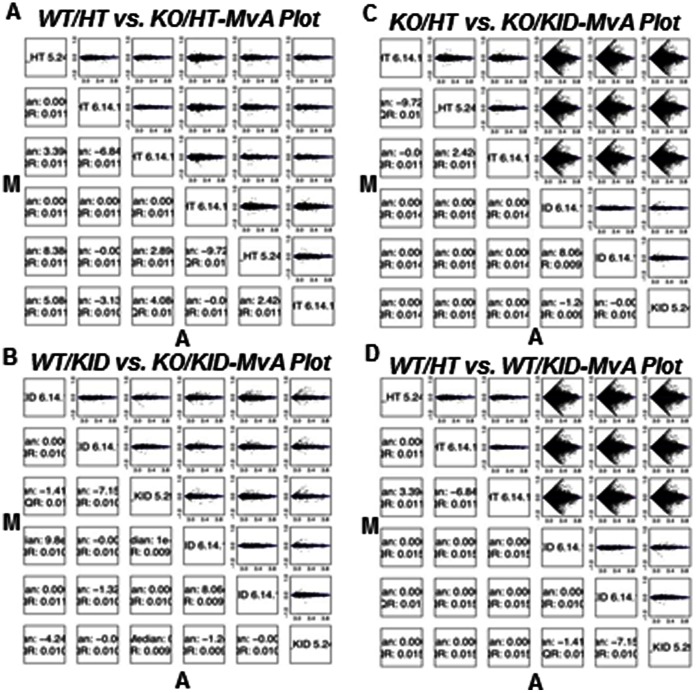
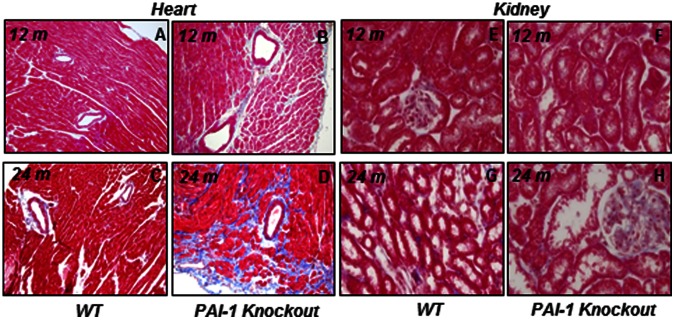
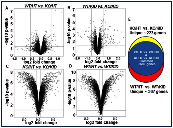
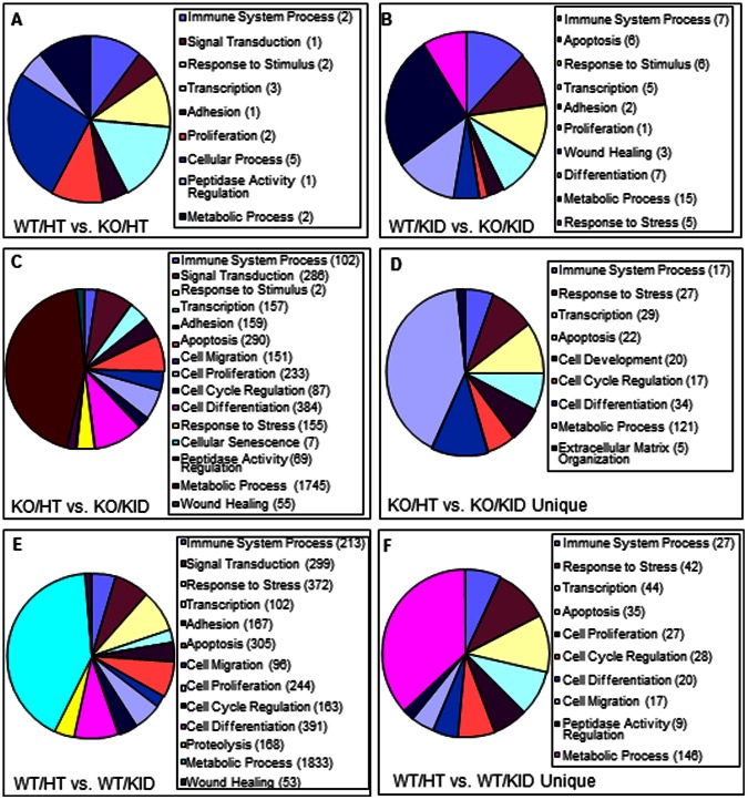
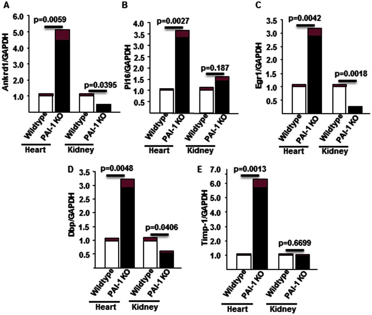
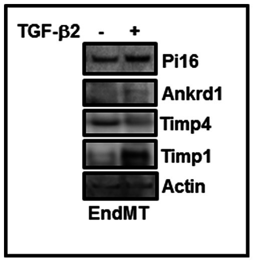
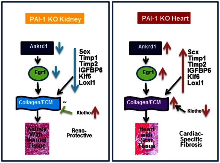
Fibrosis is defined as an abnormal matrix remodeling due to excessive synthesis and accumulation of extracellular matrix proteins in tissues during wound healing or in response to chemical, mechanical and immunological stresses. At present, there is no effective therapy for organ fibrosis. Previous studies demonstrated that aged plasminogen activator inhibitor-1 (PAI-1) knockout mice develop spontaneously cardiac-selective fibrosis without affecting any other organs. We hypothesized that differential expressions of profibrotic and antifibrotic genes in PAI-1 knockout hearts and unaffected organs lead to cardiac selective fibrosis. In order to address this prediction, we have used a genome-wide gene expression profiling of transcripts derived from aged PAI-1 knockout hearts and kidneys. The variations of global gene expression profiling were compared within four groups: wildtype heart vs. knockout heart; wildtype kidney vs. knockout kidney; knockout heart vs. knockout kidney and wildtype heart vs. wildtype kidney. Analysis of illumina-based microarray data revealed that several genes involved in different biological processes such as immune system processing, response to stress, cytokine signaling, cell proliferation, adhesion, migration, matrix organization and transcriptional regulation were affected in hearts and kidneys by the absence of PAI-1, a potent inhibitor of urokinase and tissue-type plasminogen activator. Importantly, the expressions of a number of genes, involved in profibrotic pathways including Ankrd1, Pi16, Egr1, Scx, Timp1, Timp2, Klf6, Loxl1 and Klotho, were deregulated in PAI-1 knockout hearts compared to wildtype hearts and PAI-1 knockout kidneys. While the levels of Ankrd1, Pi16 and Timp1 proteins were elevated during EndMT, the level of Timp4 protein was decreased. To our knowledge, this is the first comprehensive report on the influence of PAI-1 on global gene expression profiling in the heart and kidney and its implication in fibrogenesis and several other biological processes. The significance of these observations in the light of heart-specific profibrotic signaling and fibrogenesis are discussed.
Conflict of interest statement
Figures
Similar articles
-
Genetic deficiency of plasminogen activator inhibitor-1 promotes cardiac fibrosis in aged mice: involvement of constitutive transforming growth factor-beta signaling and endothelial-to-mesenchymal transition.Circulation. 2010 Sep 21;122(12):1200-9. doi: 10.1161/CIRCULATIONAHA.110.955245. Epub 2010 Sep 7. Circulation. 2010. PMID: 20823384
-
Plasminogen Activator Inhibitor Type I Controls Cardiomyocyte Transforming Growth Factor-β and Cardiac Fibrosis.Circulation. 2017 Aug 15;136(7):664-679. doi: 10.1161/CIRCULATIONAHA.117.028145. Epub 2017 Jun 6. Circulation. 2017. PMID: 28588076 Free PMC article.
-
Cardiomyocyte PAI-1 influences the cardiac transcriptome and limits the extent of cardiac fibrosis in response to left ventricular pressure overload.Cell Signal. 2023 Apr;104:110555. doi: 10.1016/j.cellsig.2022.110555. Epub 2022 Dec 28. Cell Signal. 2023. PMID: 36584735
-
PAI-1 in tissue fibrosis.J Cell Physiol. 2012 Feb;227(2):493-507. doi: 10.1002/jcp.22783. J Cell Physiol. 2012. PMID: 21465481 Free PMC article. Review.
-
The Role of Plasminogen Activator Inhibitor Type-1 in Fibrosis.Semin Thromb Hemost. 2017 Mar;43(2):169-177. doi: 10.1055/s-0036-1586228. Epub 2016 Aug 24. Semin Thromb Hemost. 2017. PMID: 27556351 Review.
Cited by
-
Molecular biomarkers of Graves' ophthalmopathy.Exp Mol Pathol. 2019 Feb;106:1-6. doi: 10.1016/j.yexmp.2018.11.004. Epub 2018 Nov 8. Exp Mol Pathol. 2019. PMID: 30414981 Free PMC article. Review.
-
HLA-B and TIMP1 as hub genes of the ventricular remodeling caused by hypertension.Aging (Albany NY). 2024 May 9;16(9):8260-8278. doi: 10.18632/aging.205816. Epub 2024 May 9. Aging (Albany NY). 2024. PMID: 38728374 Free PMC article.
-
Densification of Type I Collagen Matrices as a Model for Cardiac Fibrosis.Adv Healthc Mater. 2017 Nov;6(22):10.1002/adhm.201700114. doi: 10.1002/adhm.201700114. Epub 2017 Sep 7. Adv Healthc Mater. 2017. PMID: 28881428 Free PMC article.
-
Mechanisms in hypertension and target organ damage: Is the role of the thymus key? (Review).Int J Mol Med. 2018 Jul;42(1):3-12. doi: 10.3892/ijmm.2018.3605. Epub 2018 Mar 30. Int J Mol Med. 2018. PMID: 29620247 Free PMC article. Review.
-
A small molecule inhibitor of PAI-1 protects against doxorubicin-induced cellular senescence.Oncotarget. 2016 Nov 8;7(45):72443-72457. doi: 10.18632/oncotarget.12494. Oncotarget. 2016. PMID: 27736799 Free PMC article.
References
Publication types
MeSH terms
Substances
Grants and funding
- R01 HL091983/HL/NHLBI NIH HHS/United States
- HL091983/HL/NHLBI NIH HHS/United States
- R01 HL095874/HL/NHLBI NIH HHS/United States
- R01 HL053354/HL/NHLBI NIH HHS/United States
- HL105597/HL/NHLBI NIH HHS/United States
- HL053354/HL/NHLBI NIH HHS/United States
- HL095874/HL/NHLBI NIH HHS/United States
- P01 HL108795/HL/NHLBI NIH HHS/United States
- 1P01HL108795/HL/NHLBI NIH HHS/United States
- HL051387/HL/NHLBI NIH HHS/United States
- R01 HL051387/HL/NHLBI NIH HHS/United States
- R01 HL105597/HL/NHLBI NIH HHS/United States
- R37 HL053354/HL/NHLBI NIH HHS/United States
LinkOut - more resources
Full Text Sources
Other Literature Sources
Molecular Biology Databases
Research Materials
Miscellaneous
