Comparative Study
. 2012 Mar 14;32(11):3865-76.
doi: 10.1523/JNEUROSCI.3679-11.2012.

Loss of neuronal potassium/chloride cotransporter 3 (KCC3) is responsible for the degenerative phenotype in a conditional mouse model of hereditary motor and sensory neuropathy associated with agenesis of the corpus callosum

Affiliations
Comparative Study

Loss of neuronal potassium/chloride cotransporter 3 (KCC3) is responsible for the degenerative phenotype in a conditional mouse model of hereditary motor and sensory neuropathy associated with agenesis of the corpus callosum

Masoud Shekarabi et al. J Neurosci. .

Abstract

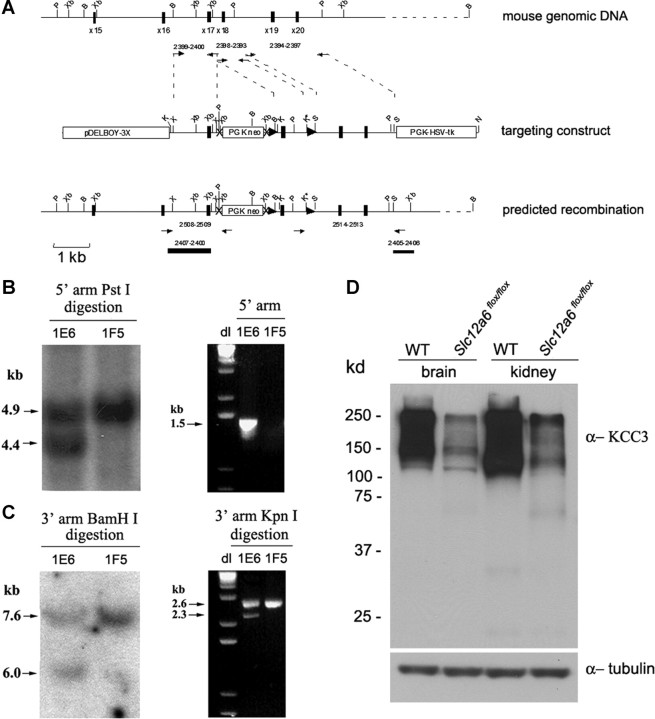
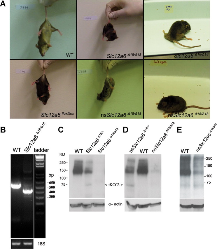
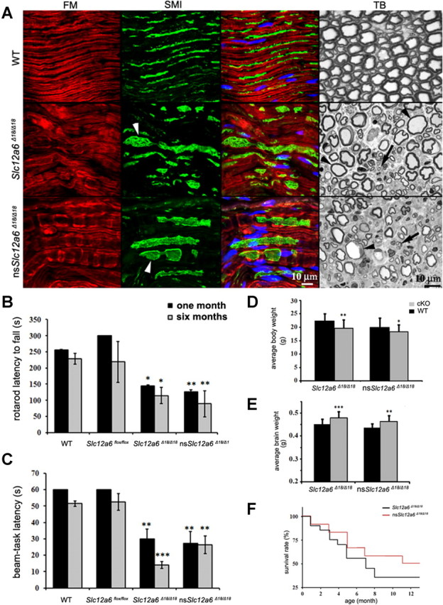
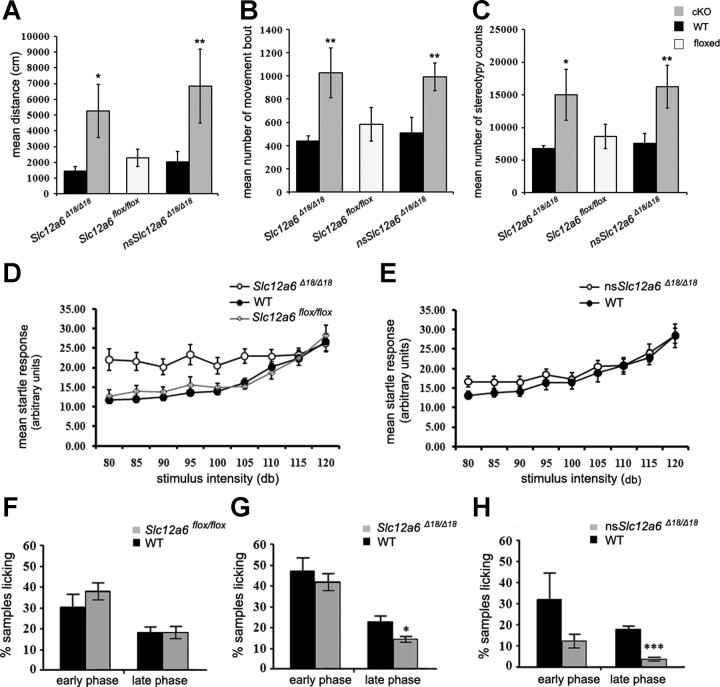
Disruption of the potassium/chloride cotransporter 3 (KCC3), encoded by the SLC12A6 gene, causes hereditary motor and sensory neuropathy associated with agenesis of the corpus callosum (HMSN/ACC), a neurodevelopmental and neurodegenerative disorder affecting both the peripheral nervous system and CNS. However, the precise role of KCC3 in the maintenance of ion homeostasis in the nervous system and the pathogenic mechanisms leading to HMSN/ACC remain unclear. We established two Slc12a6 Cre/LoxP transgenic mouse lines expressing C-terminal truncated KCC3 in either a neuron-specific or ubiquitous fashion. Our results suggest that neuronal KCC3 expression is crucial for axon volume control. We also demonstrate that the neuropathic features of HMSN/ACC are predominantly due to a neuronal KCC3 deficit, while the auditory impairment is due to loss of non-neuronal KCC3 expression. Furthermore, we demonstrate that KCC3 plays an essential role in inflammatory pain pathways. Finally, we observed hypoplasia of the corpus callosum in both mouse mutants and a marked decrease in axonal tracts serving the auditory cortex in only the general deletion mutant. Together, these results establish KCC3 as an important player in both central and peripheral nervous system maintenance.


Figures

Figure 1.
Figure 1.
Generation of Slc12a6 flox/flox transgenic mice. A, Schematic representation of mouse genomic DNA encompassing exons 15–20 of the Slc12a6 gene, the targeting construct and selection markers, and the predicted recombination between the 5′ and 3′ arms of the construct and the genomic Slc12a6 DNA. The modified allele contains LoxP sites (▶) in introns 17 and 18. The PGK-neo cassette was also flanked with Frt sequences (×) and resides in intron 17 in the same orientation. Arrows (→) indicate primers used for PCR or to generate probes for Southern blot screening of ES clones and animals. P, PstI; Xb, XbaI; B, BamHI; K, KpnI; S, SalI; X, XhoI. B, C, Southern blot analysis and PCR screening experiments used to identify 5′ and 3′ targeted recombination in positive clones using probes and specific primers (see Materials and Methods). D, A representative Western blot analysis of brain and kidney lysates from Slc12a6 flox/flox transgenic animals and WT littermates probed with anti-KCC3 antibody or anti-tubulin antibody (n = 3).
Figure 2.
Figure 2.
Characterization of Slc12a6 conditional KO mouse lines. A, Tail-clasping test done on Slc12a6Δ1818 and nsSlc12a6Δ1818 KO lines at 3–4 weeks of age, compared with Slc12a6flox/flox transgenic mice and WT littermates. Resting appearance of Slc12a6Δ1818 and nsSlc12a6Δ1818 mice at 12 months of age exhibiting extensive hindlimb weakness, movement disorganization, and kyphosis (curvature of the upper spine). B, A representative RT-PCR performed on brain mRNA isolated from WT and Slc12a6Δ1818 mice using primers designed from exon 17 (forward) and exon 20 (reverse). A band of ∼616 bp is seen in the WT, whereas a smaller fragment of ∼450 bp is seen in the Slc12a6Δ1818 mice (n = 3). C, D, A representative Western blot analysis of brain lysates isolated from Slc12a6Δ1818 (C) and nsSlc12a6Δ1818 (D) KO lines and their heterozygous and WT littermates (n = 3), and probed with anti-KCC3 and anti-actin antibodies. A truncated protein with a molecular weight of ∼75 kDa is weakly visible in the heterozygous and KO animals (arrowheads). E, A representative Western blot analysis of kidney lysates from nsSlc12a6Δ1818 and WT littermate mice using the same antibodies as in C and D, showing reduced KCC3 levels, but absence of the truncated KCC3 (n = 3).
Figure 3.
Figure 3.
Analysis of sciatic nerves, assessment of locomotor deficits, and changes in body and brain weights and survival rate of Slc12a6 cKO mice. A, Longitudinal sections of sciatic nerves from WT, Slc12a6Δ1818, and nsSlc12a6Δ1818 (n = 2) were analyzed using immunolabeling with an antibody against axons (SMI31 and 32; green) and a myelin marker (FlouroMyelin, red). Swollen axons were observed in both KOs (arrowheads). Semithin cross sections of sciatic nerves were also prepared for histological toluidine blue (TB) staining. Numerous swollen axons with thin myelin sheets (arrowheads) and axon degeneration (arrows) were observed in both lines when compared with a WT sciatic nerve section. B, C, Results of rotarod and beam-task tests performed on Slc12a6Δ1818 (n = 8) and nsSlc12a6Δ1818 (n = 10) mice at 1 and 6 months of age and compared with WT (n = 12) and Slc12a6 flox/flox (n = 8) littermates. D, Body weights of Slc12a6 conditional KO mice and their respective WT littermates were analyzed at 8 weeks of age (n = 35). E, The same animals were perfused, and their brains were weighed. A significant increase in the brain weights of both KO lines was observed, but their body weights were decreased. For both brain and body weight, the comparison was made using nontransgenic (WT) littermate animals from each line. F, Both KO lines were also monitored for 12 months, and their survival rates were calculated using the Kaplan–Meier method (n = 12). Student unpaired t test; *p < 0.05, **p < 0.005, ***p < 0.001. Data are reported as mean ± SE. Nuclei were stained with TOTO-3 iodide and are shown in blue.
Figure 4.
Figure 4.
Locomotor, startle response, and pain tests in Slc12a6flox/flox, Slc12a6 KO lines, and their WT littermates. A–C, Mean of the total traveled distance (A), number of discrete horizontal movement bouts separated by at least a 1 s rest period (B), and total number of times that a single infrared beam was broken repeatedly (C) were measured in Slc12a6flox/flox (n = 7), Slc12a6Δ1818 (n = 11), and nsSlc12a6Δ1818 (n = 8) mice (gray boxes), and their WT littermates (n = 8, 14, and 7, respectively, black boxes). D, E, Acoustic startle response was performed in Slc12a6flox/flox (n = 6), Slc12a6Δ1818 (n = 7), and nsSlc12a6Δ1818 (n = 8) mice, and compared with their WT littermates (n = 6, n = 10, and n = 10, respectively) with increasing stimulus intensities (in decibels). F–H, Formalin pain tests were performed on Slc12a6flox/flox (n = 6), Slc12a6Δ1818 (n = 15), and nsSlc12a6Δ1818(n = 14) mice (gray boxes), and their WT littermates (n = 6, 13, and 15, respectively, black boxes). Bars represent the percentage of 5 s samples featuring licking of the injected hindpaw 0–10 min (early phase) and 10–60 min (late phase) after application of formalin (all mice were tested at 5–6 months of age). *p < 0.05, **p < 0.005, ***p < 0.001. Data are reported as mean ± SE.
Figure 5.
Figure 5.
Immunodetection analysis of GABAergic neurons of Slc12a6Δ1818 and WT littermate mice. Brain sections of Slc12a6Δ1818 and WT littermates (5 months of age) were prepared and immunolabeled using anti-GAD67 antibodies to label GABAergic neurons (arrows, green) and presynaptic terminals (arrowheads, green) and anti-KCC3 antibodies to label KCC3 (red) (n = 3). Areas of motor cortex were analyzed using sequential scanning by confocal microscopy. No apparent difference was observed between the two groups of mice. However, KCC3 staining was reduced and punctated in neurons derived from Slc12a6Δ1818 mice, presumably because of the instability of the truncated protein. Nuclei were stained with TOTO-3 iodide and are shown in blue.
Figure 6.
Figure 6.
A, The corpus callosum analysis in Slc12a6 KO lines. Both Slc12a6 conditional KO lines were analyzed using gold chloride staining of mid-sagittal cut of the brains and compared with their respective wild-type littermates (only one shown) at 8 weeks of age. B, A representative WT brain that was stained by gold chloride is shown indicating the parameters to measure the CC and AC at midline sagittal plane. C, Cortical neurons were cultured from Slc12a6Δ1818 and WT embryos at E15.5, and the lengths of axons were compared.
Figure 7.
Figure 7.
dMRI and tractography reveal subtle hypoplasia of the corpus callosum. A, Representative sagittal view of color-coded dMRI and enlargement of a representative CC showing the regions of interest (ROIs) used for tractography: orange, splenium; yellow, rostrum; remaining green CC, body. The key to tissue color bidirectional orientations in the color-coded dMRI is as follows: blue, dorsal-ventral; green, medial-lateral; red, anterior-posterior. B, Representative dorsal three-dimensional views of CC tractography in WT, Slc12a6Δ1818, and nsSlc12a6Δ1818 brains. Each region of tractography has been color coded according to its seed ROI and overlaid on a two-dimensional color-coded dMRI horizontal plane at the level of the anterior commissure. A bilateral streamline deficit appears in the external capsule (ec) of the caudal cortex of the Slc12a6Δ1818 brain. These streamlines are adjacent to the auditory cortices. C, The number of voxels for each of the ROIs was quantified, giving a measure of volume, while slight, but insignificant, decreases in CC volume were observed in the rostrum and body. When calculated for the entire CC, volume was decreased by ∼12% and 11% of WTs, which were statistically significant, *p = 0.0019 and *p = 0.044, compared with WTs, respectively. D, Streamline number was unchanged in each of the ROIs. E, When streamlines were filtered to isolate those passing through the external capsule adjacent to the auditory cortex, significantly fewer streamlines were found in the Slc12a6Δ1818 brain (*p = 0.0325, n = 3, compared with WTs, t test). Values represent means ± SE. Scale bar, 1 mm in all images. post., Posterior; ant., anterior.
Figure 8.
Figure 8.
Immunostaining of axons and midline glial structures at E17.5 in Slc12a6Δ1818 KO and WT mice. A, C, E, The integrity of growing axons was assessed using immunohistochemistry for GAP-43 in brain coronal sections from both KO lines and WT mice. B, D, F, The three prominent midline glial populations were found to be present when visualized using immunohistochemistry for GFAP. These glial populations included the indusium griseum glia (IGG), the glial wedge (GW), and the midline zipper glia (MZ). Scale bar, 200 μm. n = 3.

Similar articles

Cited by

References

    1. Abramoff MD, Magelhaes PJ, Ram SJ. Image processing with ImageJ. Biophotonics Int. 2004;11:36–42.
    1. Alcamo EA, Chirivella L, Dautzenberg M, Dobreva G, Fariñas I, Grosschedl R, McConnell SK. Satb2 regulates callosal projection neuron identity in the developing cerebral cortex. Neuron. 2008;57:364–377. - PubMed
    1. Bishop KM, Wahlsten D. Sex and species differences in mouse and rat forebrain commissures depend on the method of adjusting for brain size. Brain Res. 1999;815:358–366. - PubMed
    1. Boettger T, Hübner CA, Maier H, Rust MB, Beck FX, Jentsch TJ. Deafness and renal tubular acidosis in mice lacking the K-Cl co-transporter Kcc4. Nature. 2002;416:874–878. - PubMed
    1. Boettger T, Rust MB, Maier H, Seidenbecher T, Schweizer M, Keating DJ, Faulhaber J, Ehmke H, Pfeffer C, Scheel O, Lemcke B, Horst J, Leuwer R, Pape HC, Völkl H, Hübner CA, Jentsch TJ. Loss of K-Cl co-transporter KCC3 causes deafness, neurodegeneration and reduced seizure threshold. EMBO J. 2003;22:5422–5434. - PMC - PubMed

Publication types

MeSH terms

LinkOut - more resources