. 2011 Jun;43(6):595-600.
doi: 10.1038/ng.830. Epub 2011 May 1.

Mutations in DNMT1 cause hereditary sensory neuropathy with dementia and hearing loss

Affiliations

Mutations in DNMT1 cause hereditary sensory neuropathy with dementia and hearing loss

Christopher J Klein et al. Nat Genet. 2011 Jun.

Abstract

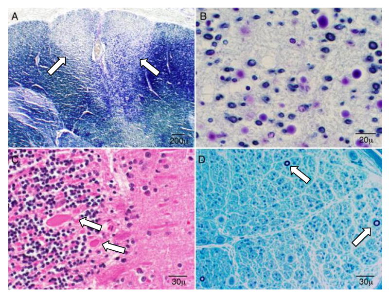
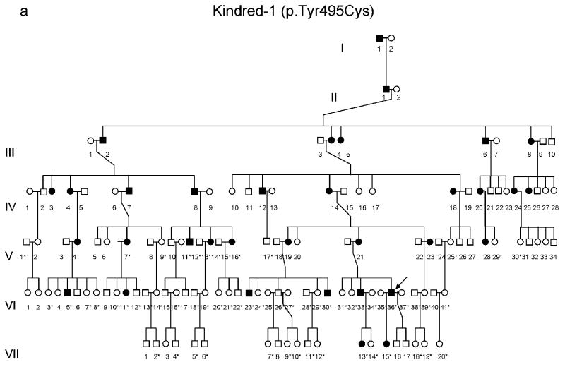
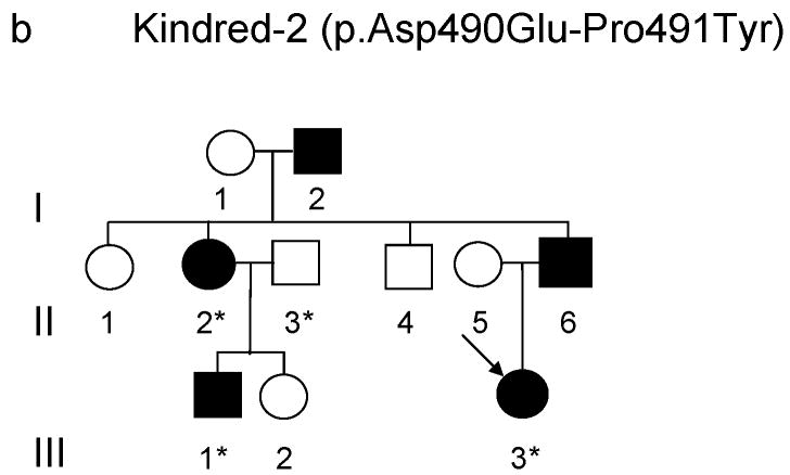
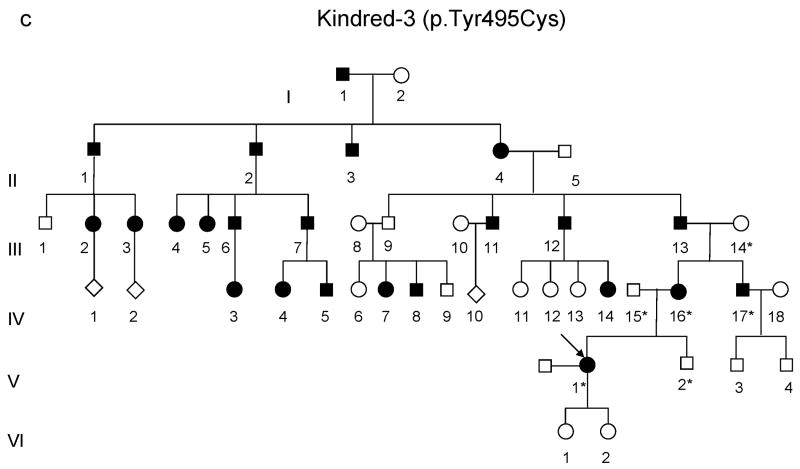
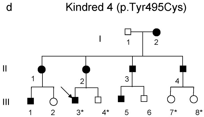
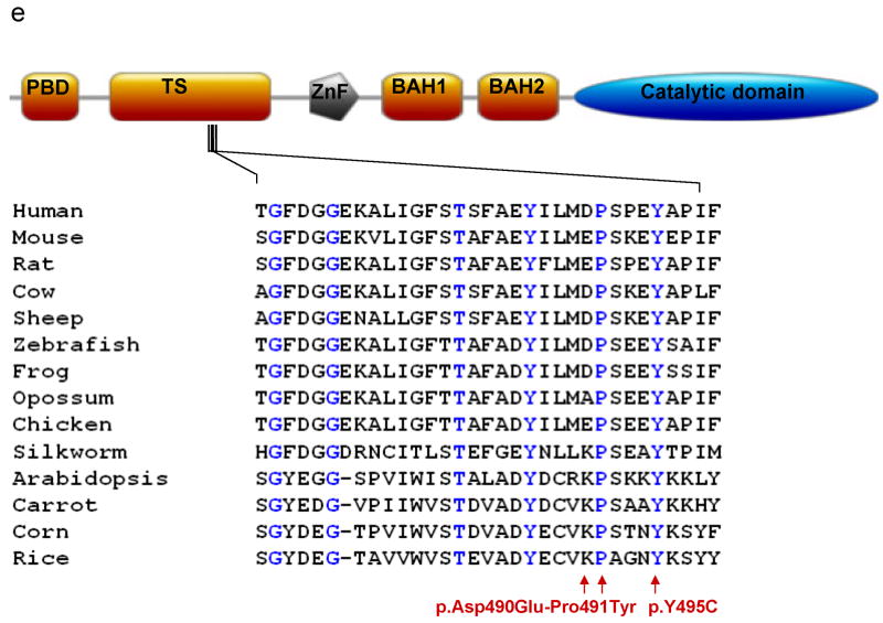
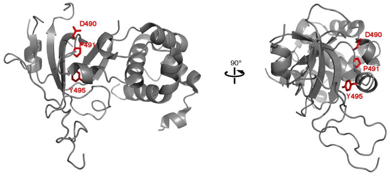
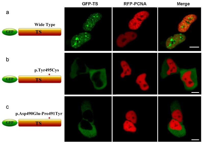
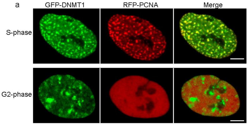
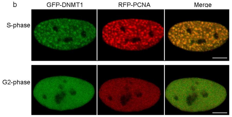
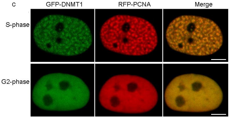
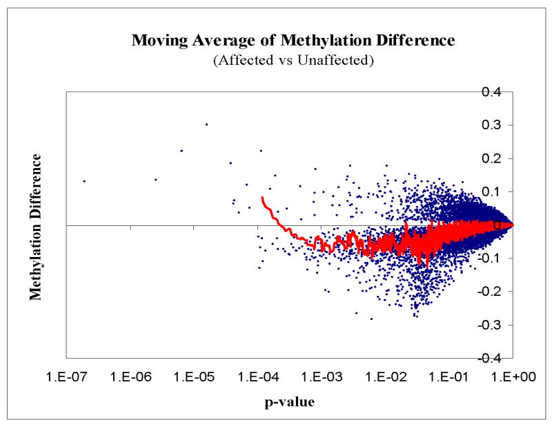
DNA methyltransferase 1 (DNMT1) is crucial for maintenance of methylation, gene regulation and chromatin stability. DNA mismatch repair, cell cycle regulation in post-mitotic neurons and neurogenesis are influenced by DNA methylation. Here we show that mutations in DNMT1 cause both central and peripheral neurodegeneration in one form of hereditary sensory and autonomic neuropathy with dementia and hearing loss. Exome sequencing led to the identification of DNMT1 mutation c.1484A>G (p.Tyr495Cys) in two American kindreds and one Japanese kindred and a triple nucleotide change, c.1470-1472TCC>ATA (p.Asp490Glu-Pro491Tyr), in one European kindred. All mutations are within the targeting-sequence domain of DNMT1. These mutations cause premature degradation of mutant proteins, reduced methyltransferase activity and impaired heterochromatin binding during the G2 cell cycle phase leading to global hypomethylation and site-specific hypermethylation. Our study shows that DNMT1 mutations cause the aberrant methylation implicated in complex pathogenesis. The discovered DNMT1 mutations provide a new framework for the study of neurodegenerative diseases.


Figures

Figure 1
Figure 1
Autopsy (a-c) and nerve biopsy (d) results of an affected person (VI-36, deceased at 48 y.o.) from Kindred 1. Similar results were also seen in two other affected and deceased persons from kindred 1. (a) Ascending spinal sensory tract degeneration with profound myelin and axonal loss (lighter staining, between arrowheads), involving the gracile fasciculus (medial posterior columns) at all spinal levels; (b). The neuronal loss described in panel a is shown at higher magnification; (c) Chronic cerebellar Purkinje cell swelling and axonal loss (arrowheads) with associated hyperplasia of the Bergman glia. There was also severe neuronal loss and gliosis of inferior olivary nucleus as well as generalized cerebral atrophy with brain weight of 1085 (normal 1300-1400) grams without distinct histopathologic alterations or inclusions which was determined by Bielschowsky silver and immunohistochemical stains for detection of beta-amyloid, tau, TDP-43, and alpha-synuclein; (d) Epoxy embedded sural nerve tissue showing severe loss of large and small myelinated fibers with only few fibers remaining (arrows) without distinctive interstitial infiltrative change.
Figure 2
Figure 2
(a-d) Pedigrees of four kindreds are shown. A heterozygous mutation c.A1484G, resulting in p.Tyr495Cys, was identified in exon 20 of DNMT1 in the HSAN kindred 1(a), 3(c) and 4(d). Three consecutive heterozygous mutations c.1470TCC-1472ATA, resulting in p.Asp490Glu-Pro491Tyr substitution, also in exon 20 of DNMT1 was identified in the HSAN kindred 2(b). We sequenced all available samples (with asterisk) from these four kindreds to confirm the mutation is segregated with the disease. (e) Schematic overview of DNMT1 and its multiple domains in the N-terminal region (PBD, PCNA binding domain; TS, targeting sequence; ZnF, zinc finger; BAH1 & 2, bromo adjacent homology domains1 & 2). Below is the ClustalW alignment of the part of TS domain where mutations occur from multiple DNMT1 homologs. Comparison of human DNMT1 (P26358) and its orthologs in mouse (P13864), rat (Q9Z330), cow (Q24K09), sheep (Q865V5), zebrafish (Q8QGB8), frog (Q6GQH0), opossum (Q8MJ28), chicken (Q92072), silkworm (Q5W7N6), arebiopsis (Q9SEG3), carrot (O48867), corn (Q8LPU6) and rice (A2XMY1). Conserved amino acids are colored in blue. Red arrow points to the mutations. (f). Location of mutated residues in the TS domain of human DNMT1. Shown in red are the side chains of Asp490, Pro491 and Tyr495 in the crystal structure of the TS domain (Protein Data Bank accession number 3EPZ). The image was generated using PyMOL (http://www.pymol.org/).
Figure 2
Figure 2
(a-d) Pedigrees of four kindreds are shown. A heterozygous mutation c.A1484G, resulting in p.Tyr495Cys, was identified in exon 20 of DNMT1 in the HSAN kindred 1(a), 3(c) and 4(d). Three consecutive heterozygous mutations c.1470TCC-1472ATA, resulting in p.Asp490Glu-Pro491Tyr substitution, also in exon 20 of DNMT1 was identified in the HSAN kindred 2(b). We sequenced all available samples (with asterisk) from these four kindreds to confirm the mutation is segregated with the disease. (e) Schematic overview of DNMT1 and its multiple domains in the N-terminal region (PBD, PCNA binding domain; TS, targeting sequence; ZnF, zinc finger; BAH1 & 2, bromo adjacent homology domains1 & 2). Below is the ClustalW alignment of the part of TS domain where mutations occur from multiple DNMT1 homologs. Comparison of human DNMT1 (P26358) and its orthologs in mouse (P13864), rat (Q9Z330), cow (Q24K09), sheep (Q865V5), zebrafish (Q8QGB8), frog (Q6GQH0), opossum (Q8MJ28), chicken (Q92072), silkworm (Q5W7N6), arebiopsis (Q9SEG3), carrot (O48867), corn (Q8LPU6) and rice (A2XMY1). Conserved amino acids are colored in blue. Red arrow points to the mutations. (f). Location of mutated residues in the TS domain of human DNMT1. Shown in red are the side chains of Asp490, Pro491 and Tyr495 in the crystal structure of the TS domain (Protein Data Bank accession number 3EPZ). The image was generated using PyMOL (http://www.pymol.org/).
Figure 3
Figure 3
Confocal microscopy was performed using HeLa cells co-transfected with plasmids containing RFP-PCNA and GFP-DNMT1 wild type TS domain (panel a), p.Tyr495Cys-TS domain (panel b) or p.Asp490Glu-Pro491Tyr-TS domain (panel c). Wild type and mutant TS domains appear green in the right panels, PCNA appears red in the middle panels and merged images are shown in the left panels. Scale bar, 5um. In panel a, wild type TS domain enters nucleus during S phase when PCNA localizes to the toroidal structures of replication foci, wild type TS also binds to heterochromatin during G2 phase when PCNA showed diffused pattern and toroidal structures are no longer visible. Panel b and panel c showed mutant TS domains was unable to enter into the nucleus and remained in the cytoplasm.
Figure 4
Figure 4
Confocal microscopy was performed using HeLa cells co-transfected with plasmids containing RFP-PCNA and full length (a) GFP-wild type DNMT1, (b) GFP-p.Tyr495Cys-DNMT1 or (c) GFP-p.Asp490Glu-Pro491Tyr-DNMT1. Wild type and mutant DNMT1 appear green in the right panels, PCNA appears red in the middle panels and merged images are shown in the left panels. Scale bar, 5um. Cell cycles are deciphered from the pattern of RFP-PCNA. In S phase, PCNA is present at the toroidal structures of the replication foci; in G2 phase, PCNA shows diffused pattern in the nucleus. In panel a, wild type DNMT1 co-localizes with PCNA at replication foci during both S phase and binds to heterochromatin during G2 phase. In panel b and c, p.Tyr495Cys-DNMT1 and p.Asp490GLu-Pro491Tyr-DNMT1 localize along with PCNA at the replication foci during S phase but did not show binding of heterochromatin during G2 phase.
Figure 4
Figure 4
Confocal microscopy was performed using HeLa cells co-transfected with plasmids containing RFP-PCNA and full length (a) GFP-wild type DNMT1, (b) GFP-p.Tyr495Cys-DNMT1 or (c) GFP-p.Asp490Glu-Pro491Tyr-DNMT1. Wild type and mutant DNMT1 appear green in the right panels, PCNA appears red in the middle panels and merged images are shown in the left panels. Scale bar, 5um. Cell cycles are deciphered from the pattern of RFP-PCNA. In S phase, PCNA is present at the toroidal structures of the replication foci; in G2 phase, PCNA shows diffused pattern in the nucleus. In panel a, wild type DNMT1 co-localizes with PCNA at replication foci during both S phase and binds to heterochromatin during G2 phase. In panel b and c, p.Tyr495Cys-DNMT1 and p.Asp490GLu-Pro491Tyr-DNMT1 localize along with PCNA at the replication foci during S phase but did not show binding of heterochromatin during G2 phase.
Figure 5
Figure 5
Moving average of methylation difference of ∼25000 CpG sites between affected and unaffected groups from kindred-1. Kindred 1 was chosen to optimize same number of gender and the first degree relation between affected and unaffected group. The methylation profile of affected and unaffected groups was compared. Y-axis represents the methylation difference between the two groups. X-axis represents p-value. Red colored line represents the moving average of methylation difference between affected vs. unaffected group. Each blue dot represents methylation difference for a CpG site. Blue dots below the 0 line represents reduced methylation in the affected group while blue dots above 0 line represents increased methylation in the affected group. The red moving average line suggests local hypermethylation and moderate global hypomethylation in the affected group, consistent with the 5-mdC content measurement by LC-ESI-MS/MS (supplementary fig. 5).

Comment in

Similar articles

Cited by

References

    1. Feng J, Fan G. The role of DNA methylation in the central nervous system and neuropsychiatric disorders. Int Rev Neurobiol. 2009;89:67–84. - PubMed
    1. Chen WG, et al. Derepression of BDNF transcription involves calcium-dependent phosphorylation of MeCP2. Science. 2003;302:885–9. - PubMed
    1. Tohgi H, et al. Reduction with age in methylcytosine in the promoter region -224 approximately -101 of the amyloid precursor protein gene in autopsy human cortex. Brain Res Mol Brain Res. 1999;70:288–92. - PubMed
    1. Herrup K, Yang Y. Cell cycle regulation in the postmitotic neuron: oxymoron or new biology? Nat Rev Neurosci. 2007;8:368–78. - PubMed
    1. Brooks PJ, Marietta C, Goldman D. DNA mismatch repair and DNA methylation in adult brain neurons. J Neurosci. 1996;16:939–45. - PMC - PubMed

Publication types

Substances

Associated data