

Protein profile of exosomes from trabecular meshwork cells
- PMID: 21362503
- PMCID: PMC3085584
- DOI: 10.1016/j.jprot.2011.02.024
Protein profile of exosomes from trabecular meshwork cells
Abstract
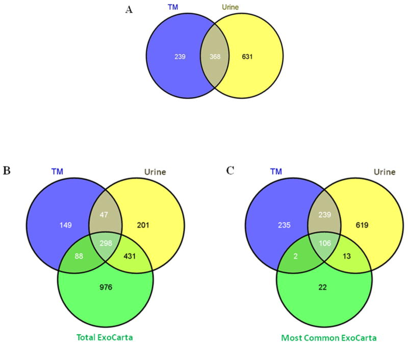
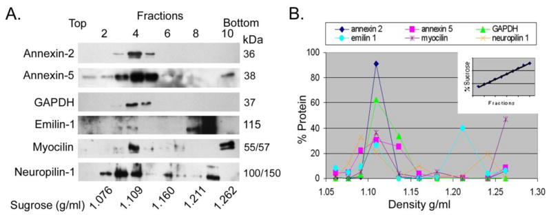
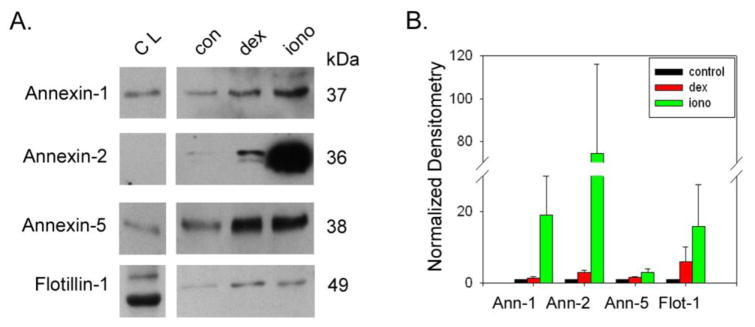
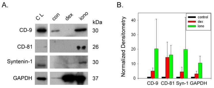
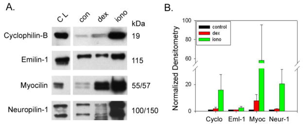
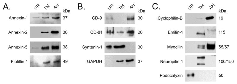
To better understand the role of exosomes in the trabecular meshwork (TM), the site of intraocular pressure control, the exosome proteome from primary cultures of human TM cell monolayers was analyzed. Exosomes were purified from urine and conditioned media from primary cultures of human TM cell monolayers and subjected to a two dimensional HPLC separation and MS/MS analyses using the MudPIT strategy. Spectra were searched against a human protein database using Sequest. Protein profiles were compared to each other and the Exocarta database and the presence of specific protein markers confirmed by Western blot analyses of exosomes from aqueous humor and human TM cell strains (n=5) that were untreated, or exposed to dexamethasone and/or ionomycin. TM cell exosomes contained 108 of the 143 most represented exosome proteins in ExoCarta, including previously characterized markers such as membrane organizing and tetraspanin proteins. Several cell-specific proteins in TM exosomes were identified including myocilin, emilin-1 and neuropilin-1. All TM exosome proteins had flotation densities on sucrose gradients and release responses to ionomycin typical for exosomes. Taken together, TM exosomes have a characteristic exosome protein profile plus contain unique proteins, including the glaucoma-causing protein, myocilin; suggesting a role for exosomes in the control of intraocular pressure.
Copyright © 2011 Elsevier B.V. All rights reserved.
Figures
Similar articles
-
Regulation of myocilin-associated exosome release from human trabecular meshwork cells.Invest Ophthalmol Vis Sci. 2009 Mar;50(3):1313-8. doi: 10.1167/iovs.08-2326. Epub 2008 Oct 24. Invest Ophthalmol Vis Sci. 2009. PMID: 18952916 Free PMC article.
-
Primary trabecular meshwork cells incubated in human aqueous humor differ from cells incubated in serum supplements.Invest Ophthalmol Vis Sci. 2005 Aug;46(8):2848-56. doi: 10.1167/iovs.05-0101. Invest Ophthalmol Vis Sci. 2005. PMID: 16043859
-
Characterizing the metabolic profile of dexamethasone treated human trabecular meshwork cells.Exp Eye Res. 2022 Jan;214:108888. doi: 10.1016/j.exer.2021.108888. Epub 2021 Dec 9. Exp Eye Res. 2022. PMID: 34896106 Free PMC article.
-
Targeting the ER-autophagy system in the trabecular meshwork to treat glaucoma.Exp Eye Res. 2016 Mar;144:38-45. doi: 10.1016/j.exer.2015.08.017. Epub 2015 Aug 22. Exp Eye Res. 2016. PMID: 26302411 Free PMC article. Review.
-
Effects of glucocorticoids on the trabecular meshwork: towards a better understanding of glaucoma.Prog Retin Eye Res. 1999 Sep;18(5):629-67. doi: 10.1016/s1350-9462(98)00035-4. Prog Retin Eye Res. 1999. PMID: 10438153 Review.
Cited by
-
Asymmetric depth-filtration: A versatile and scalable method for high-yield isolation of extracellular vesicles with low contamination.J Extracell Vesicles. 2022 Aug;11(8):e12256. doi: 10.1002/jev2.12256. J Extracell Vesicles. 2022. PMID: 35942823 Free PMC article.
-
Neuropilin-1 is present on Foxp3+ T regulatory cell-derived small extracellular vesicles and mediates immunity against skin transplantation.J Extracell Vesicles. 2022 Jun;11(6):e12237. doi: 10.1002/jev2.12237. J Extracell Vesicles. 2022. PMID: 35676234 Free PMC article.
-
Parallel Analysis of Exosomes and Cytokines in Aqueous Humor Samples to Evaluate Biomarkers for Glaucoma.Cells. 2024 Jun 13;13(12):1030. doi: 10.3390/cells13121030. Cells. 2024. PMID: 38920659 Free PMC article.
-
Extracellular Vesicles and Matrix Remodeling Enzymes: The Emerging Roles in Extracellular Matrix Remodeling, Progression of Diseases and Tissue Repair.Cells. 2018 Oct 13;7(10):167. doi: 10.3390/cells7100167. Cells. 2018. PMID: 30322133 Free PMC article. Review.
-
The emerging role of extracellular vesicles in retinal diseases.Am J Transl Res. 2021 Dec 15;13(12):13227-13245. eCollection 2021. Am J Transl Res. 2021. PMID: 35035672 Free PMC article. Review.
References
-
- AGIS-Investigators. The Advanced Glaucoma Intervention Study (AGIS): The relationship between control of intraocular pressure and visual field deterioration. Am J Ophthalmol. 2000;130:429–440. - PubMed
-
- Grant WM. Clinical tonography. Trans Am Acad Ophthalmol Otolaryngol. 1951;55:774–81. - PubMed
-
- Grant WM. Open-angle glaucoma. AMA Arch Ophthalmol. 1953;50:125–6. - PubMed
-
- Grant WM. Experimental aqueous perfusion in enucleated human eyes. Arch Ophthalmol. 1963;69:783. - PubMed
Publication types
MeSH terms
Substances
Grants and funding
LinkOut - more resources
Full Text Sources
Other Literature Sources
Molecular Biology Databases
