

Case Reports
doi: 10.1212/WNL.0b013e3181f4d7ac.
Evolution of a tumor-like lesion in cerebroretinal vasculopathy and TREX1 mutation
Affiliations
- PMID: 20876473
- PMCID: PMC3013489
- DOI: 10.1212/WNL.0b013e3181f4d7ac
Item in Clipboard
Case Reports
Evolution of a tumor-like lesion in cerebroretinal vasculopathy and TREX1 mutation
Neurology.
.
No abstract available
Figures
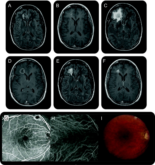
Figure Brain and retinal imaging in a patient with cerebroretinal vasculopathy Sequential axial MRI showing an ovoid T2-hyperintense (A) and gadolinium-enhancing (B) lesion (2.1 × 1 cm), abutting the frontal horn of the right lateral ventricle, bright on both diffusion-weighted imaging (DWI) and apparent diffusion coefficient (ADC) map. At 6 months, a larger, more aggressive-appearing lesion (2 × 2 × 3 cm) with surrounding edema occupied the right frontal lobe. There was a central zone of presumed necrosis and gadolinium enhancement of the lesion rim (C, D). Diffusion imaging showed more heterogeneous signal, without the characteristic bright DWI and dark ADC signal of acute infarction. At 12 months, the lesion approximated its size on initial imaging (1.4 × 1.2 × 0.9 cm) with a persistent rim pattern of enhancement (E, F). At 18 months, the lesion further decreased in size (1.1 × 0.9 × 0.3 cm) with near resolution of the surrounding edema. Fluorescein angiography and indocyanine green with corresponding color photographs of the retina show views of the macula of the right eye (G–I). Periarteriolar narrowing and sheathing, focal leakage, telangiectasias, and cotton wool spots are present.
Similar articles
-
A Case of TREX1-Associated Retinal Vasculopathy with Cerebral Leukodystrophy.Ophthalmol Retina. 2020 Jan;4(1):115-117. doi: 10.1016/j.oret.2019.07.014. Epub 2019 Jul 30. Ophthalmol Retina. 2020. PMID: 31540853 No abstract available.
-
Evolution of brain lesions in a patient with TREX1 cerebroretinal vasculopathy.Neurology. 2015 Nov 3;85(18):1633-4. doi: 10.1212/WNL.0000000000002092. Neurology. 2015. PMID: 26527794 No abstract available.
-
New roles for the major human 3'-5' exonuclease TREX1 in human disease.Cell Cycle. 2008 Jun 15;7(12):1718-25. doi: 10.4161/cc.7.12.6162. Epub 2008 Jun 16. Cell Cycle. 2008. PMID: 18583934 Free PMC article. Review.
-
A 31-Year-Old Man With a Ring-Enhancing Brain Lesion.J Neuroophthalmol. 2017 Jun;37(2):172-175. doi: 10.1097/WNO.0000000000000469. J Neuroophthalmol. 2017. PMID: 28079760 No abstract available.
-
Human disease phenotypes associated with mutations in TREX1.J Clin Immunol. 2015 Apr;35(3):235-43. doi: 10.1007/s10875-015-0147-3. Epub 2015 Mar 4. J Clin Immunol. 2015. PMID: 25731743 Review.
Cited by
-
Retinal vasculopathy with cerebral leukoencephalopathy and systemic manifestations in conjunction with systemic lupus erythematosus: Missed diagnosis or misdiagnosis?Immun Inflamm Dis. 2024 Aug;12(8):e1367. doi: 10.1002/iid3.1367. Immun Inflamm Dis. 2024. PMID: 39119967 Free PMC article.
-
Lesion evolution and neurodegeneration in RVCL-S: A monogenic microvasculopathy.Neurology. 2020 Oct 6;95(14):e1918-e1931. doi: 10.1212/WNL.0000000000010659. Epub 2020 Sep 4. Neurology. 2020. PMID: 32887784 Free PMC article.
-
Multiple sclerosis-like lesions and type I interferon signature in a patient with RVCL.Neurol Neuroimmunol Neuroinflamm. 2014 Dec 23;2(1):e55. doi: 10.1212/NXI.0000000000000055. eCollection 2015 Feb. Neurol Neuroimmunol Neuroinflamm. 2014. PMID: 25566545 Free PMC article. No abstract available.
-
A 44-year-old man with eye, kidney, and brain dysfunction.Ann Neurol. 2016 Apr;79(4):507-19. doi: 10.1002/ana.24583. Epub 2016 Mar 7. Ann Neurol. 2016. PMID: 26691497 Free PMC article.
-
Type I interferon dysregulation and neurological disease.Nat Rev Neurol. 2015 Sep;11(9):515-23. doi: 10.1038/nrneurol.2015.143. Epub 2015 Aug 25. Nat Rev Neurol. 2015. PMID: 26303851 Review.
References
-
- Grand MG, Kaine J, Fulling K, et al. Cerebroretinal vasculopathy: a new hereditary syndrome. Ophthalmology 1988;95:649–659. - PubMed
-
- Richards A, van den Maagdenberg AM, Jen JC, et al. C-terminal truncations in human 3′-5′ DNA exonuclease TREX1 cause autosomal dominant retinal vasculopathy with cerebral leukodystrophy. Nat Genet 2007; 39:1068–1070. - PubMed
-
- Entrez-Pubmed Gene References into Function. Available at: http://www.ncbi.nlm.nih.gov/sites/entrez?cmd_current=&db=gene&orig_db=ge.... Accessed August 30, 2009.
-
- Mazur DJ, Perrino FW. Identification and expression of the TREX1 and TREX2 cDNA sequences encoding mammalian 3′->5′ exonucleases. J Biol Chem 1999;274:19655–19660. - PubMed
-
- Chowdhury D, Beresford PJ, Zhu P, et al. The exonuclease TREX1 is in the SET complex and acts in concert with NM23-H1 to degrade DNA during granzyme A-mediated cell death. Mol Cell 2006;23:133–142. - PubMed
Publication types
MeSH terms
Substances
Grants and funding
LinkOut - more resources
Full Text Sources
Medical