. 2010 Sep 22;30(38):12777-86.
doi: 10.1523/JNEUROSCI.1102-10.2010.

CDKL5, a protein associated with rett syndrome, regulates neuronal morphogenesis via Rac1 signaling

Affiliations

CDKL5, a protein associated with rett syndrome, regulates neuronal morphogenesis via Rac1 signaling

Qian Chen et al. J Neurosci. .

Abstract

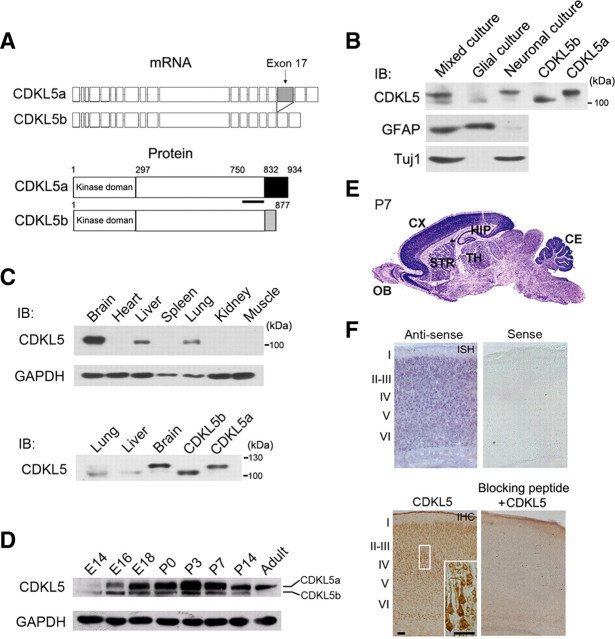
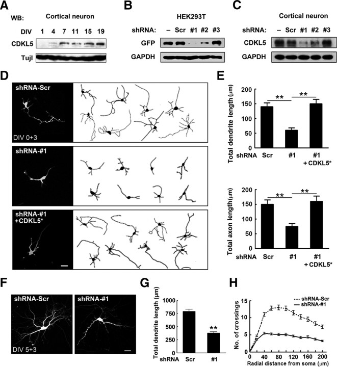
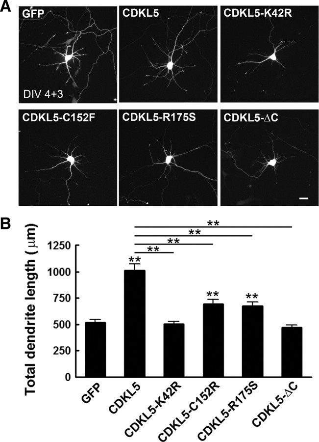
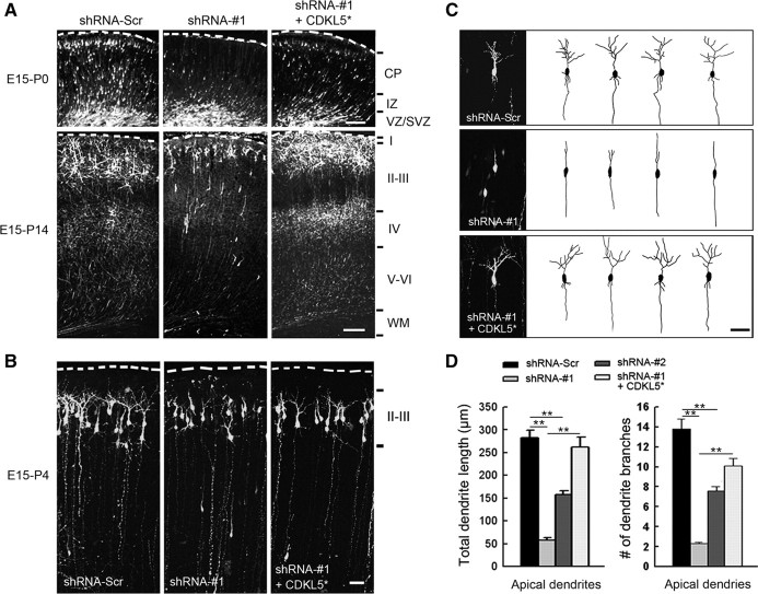
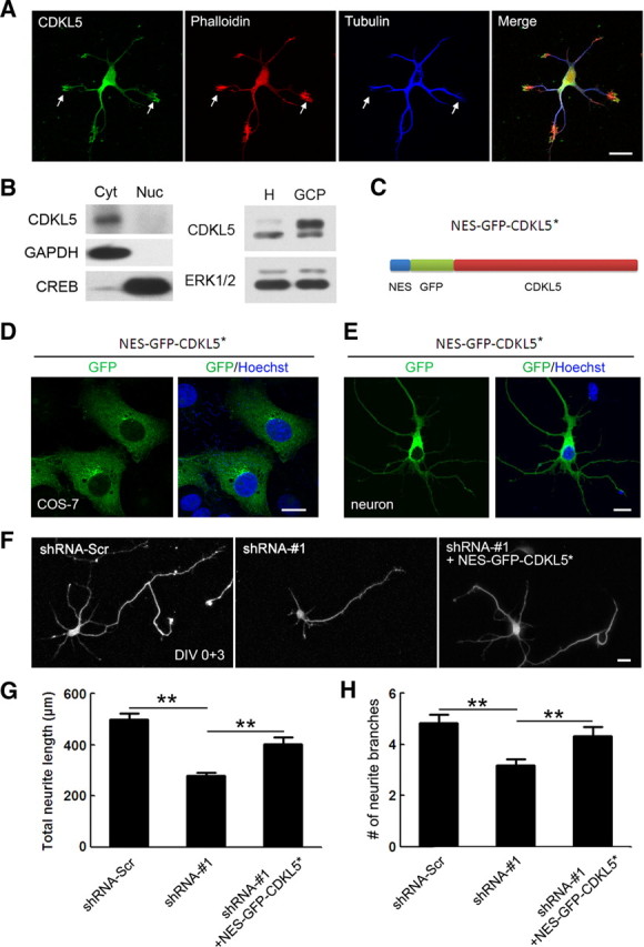
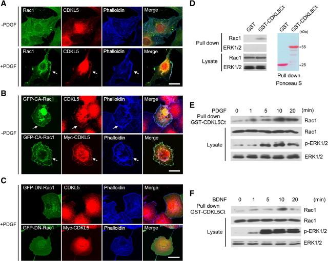
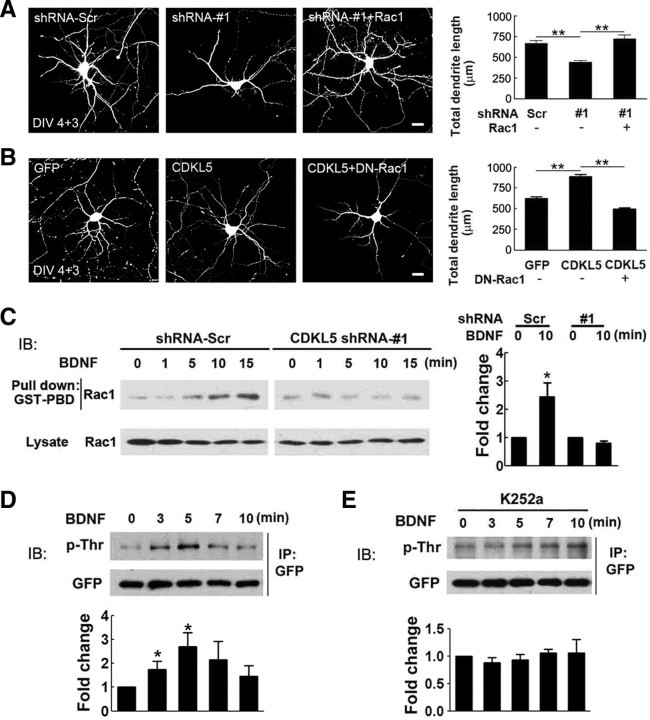
Mutations in cyclin-dependent kinase-like 5 (CDKL5), also known as serine/threonine kinase 9 (STK9), have been identified in patients with Rett syndrome (RTT) and X-linked infantile spasm. However, the function of CDKL5 in the brain remains unknown. Here, we report that CDKL5 is a critical regulator of neuronal morphogenesis. We identified a neuron-specific splicing variant of CDKL5 whose expression was markedly induced during postnatal development of the rat brain. Downregulating CDKL5 by RNA interference (RNAi) in cultured cortical neurons inhibited neurite growth and dendritic arborization, whereas overexpressing CDKL5 had opposite effects. Furthermore, knocking down CDKL5 in the rat brain by in utero electroporation resulted in delayed neuronal migration, and severely impaired dendritic arborization. In contrast to its proposed function in the nucleus, we found that CDKL5 regulated dendrite development through a cytoplasmic mechanism. In fibroblasts and in neurons, CDKL5 colocalized and formed a protein complex with Rac1, a critical regulator of actin remodeling and neuronal morphogenesis. Overexpression of Rac1 prevented the inhibition of dendrite growth caused by CDKL5 knockdown, and the growth-promoting effect of ectopically expressed CDKL5 on dendrites was abolished by coexpressing a dominant-negative form of Rac1. Moreover, CDKL5 was required for brain-derived neurotrophic factor (BDNF)-induced activation of Rac1. Together, these results demonstrate a critical role of CDKL5 in neuronal morphogenesis and identify a Rho GTPase signaling pathway which may contribute to CDKL5-related disorders.


Figures

Figure 1.
Figure 1.
Molecular cloning of rat CDKL5 and expression of CDKL5 in the developing rat brain. A, Schematic diagrams showing the mRNA and protein structures of CDKL5a and CDKL5b. The immunogen region for the CDKL5 antibody is marked by a thick black line. B, Western blot analysis of lysates extracted from neuronal, glial, or mixed neuron-glia cultures, as well as 293T cells transfected with plasmids expressing CDKL5a and CDKL5b; using antibodies against CDKL5, Tuj1, and GFAP. C, Western blot analysis of CDKL5 protein levels in brain, heart, liver, spleen, lung, kidney, and muscle lysates (top); and in lung, liver, brain and HEK293T cells expressing CDKL5a or CDKL5b (bottom). D, Western blot analysis of CDKL5 protein levels during cortical development at the indicated time points. E, In situ hybridization of CDKL5 mRNA in the P7 rat brain. CX, cortex; OB, olfactory bulb; STR, striatum; CE, cerebellum; TH, thalamus; HIP, hippocampus. F, In situ hybridization (ISH) and immunohistochemistry (IHC) of CDKL5 mRNA and protein in adjacent sections of P7 rat cerebral cortex. The inset (bottom right) shows a higher-magnification view of the boxed region of the cortex. ISH with Sense probe and IHC with blocking peptide to neutralize CDKL5 antibody are negative controls. Scale bars, 50 μm.
Figure 2.
Figure 2.
CDKL5 regulates neuronal morphogenesis in vitro. A, Western blot analysis of CDKL5 expression in cultured cortical neurons at the indicated time points. B, C, Downregulation of CDKL5 in HEK293T cells and cultured cortical neurons. Extracts from HEK293T cells (B) or cortical neurons (C) transfected with (B) or without (C) GFP-CDKL5 together with shRNA-Scr, shRNA-#1, shRNA-#2, or shRNA-#3 were immunoblotted with GFP antibody (B) or CDKL5 antibody (C). − indicates lysates of untransfected cells. D, E, Effects of CDKL5 knockdown on neurite growth. D, Representative images of neurons transfected with GFP together with indicated constructs. CDKL5* is an RNAi resistant form of CDKL5. Scale bar, 20 μm. E, Quantitative analysis of total dendritic length and total axon length of neurons treated as in D. Data represent mean ± SEM, n = 50–70 in each group; **p < 0.001; t test. F–H, Effects of CDKL5 knockdown on dendritic arborization. F, Representative images of neurons transfected with GFP together with indicated constructs. Scale bar, 20 μm. G, Quantitative analysis of total dendritic length of neurons treated as in F. Data represent mean ± SEM; n = 50–53; **p < 0.001; t test. H, Sholl analysis of dendritic arborization of neurons in G.
Figure 3.
Figure 3.
Effects of CDKL5 and mutated CDKL5 variants on dendrite growth. A, Representative images of neurons transfected with plasmids expressing wild-type or the indicated variants of CDKL5 together with GFP. B, Quantitative analysis of total dendrite length of neurons treated as in A. Data represent mean ± SEM; n = 80–100 in each group; **p < 0.001 relative to the GFP or CDKL5 group, as indicated on the graph; t test.
Figure 4.
Figure 4.
CDKL5 is required for dendritic arborization in vivo. A, Representative images of coronal slices of P0 and P14 rat brains that were transfected with GFP together with indicated constructs by in utero electroporation at E15. Transfected cells were visualized by staining coronal slices with GFP antibody. Scale bar, 100 μm. B, Representative images of P4 slices of rat brains transfected with GFP together with indicated constructs. Scale bar, 50 μm. C, Representative high-magnification images and Neurolucida tracings of layer II/III pyramidal neurons in B. Scale bar, 50 μm. D, Quantitative analysis of total dendritic length and number of dendritic branches in neurons treated as in B. Data represent mean ± SEM; n = 30–40 in each group; **p < 0.001; t test.
Figure 5.
Figure 5.
Subcellular localization of CDKL5. A, Representative images of DIV 2 neurons costained with CDKL5 and tubulin antibody. F-actin was visualized by phalloidin staining. Arrows indicate growth cones with abundant CDKL5. Scale bar, 20 μm. B, Left, Western blot showing CDKL5 enrichment in the cytoplasmic fraction, but not the nucleus, of DIV 14 cultured cortical neurons. Cytoplasmic and nuclear lysate were immunoblotted with antibodies against CDKL5, the cytoplasmic factor GAPDH and the nuclear factor CREB. Right, Anti-CDKL5 Western blot analysis of whole-cell homogenate (H) and GCPs isolated from E18 rat cortex. ERK1/2 was used as the loading control. C, Schematic illustration of the protein structure of NES-GFP-CDKL5*. D, E, Representative images showing the localization of NES-GFP-CDKL5* in COS-7 cells (D) and in neurons (E). Scale bar, 10 μm. F, Representative images of neurons transfected with GFP together with the indicated constructs. Scale bar, 20 μm. G, H, Quantitative analysis of total neurite length and total number of dendrite branches in neurons treated as in F. Data represent mean ± SEM; n = 70–80 in each group; **p < 0.001; t test.
Figure 6.
Figure 6.
CDKL5 forms a complex with Rac1. A, Colocalization of CDKL5 and Rac1 in membrane ruffles. To detect endogenous CDKL5, NIH3T3 fibroblasts were serum starved for 48 h, and then left unstimulated or stimulated with PDGF (25 ng/ml) for 5 min. Cells were fixed and costained with CDKL5, Rac1, and phalloidin. Arrows indicate membrane ruffles induced by PDGF. Scale bar, 20 μm. B, Colocalization of CDKL5 and CA-Rac1 in membranes of serum-starved COS-7 cells. Cells transfected with GFP-CA-Rac1 either with or without Myc-CDKL5 were serum starved and stained for CDKL5 or Myc. Arrows indicate membranes where CDKL5 and CA-Rac1 colocalize. Scale bar, 20 μm. C, DN-Rac1 does not colocalize with CDKL5 in membrane regions in response to PDGF. COS-7 cells transfected with GFP-DN-Rac1 either with or without Myc-CDKL5 were serum starved and stimulated with PDGF (25 ng/ml) for 5 min. Cells were fixed and stained for CDKL5 or Myc. Scale bar, 20 μm. D, GST pull-down assay showing the interaction of CDKL5 and Rac1. COS-7 cell lysates were incubated with agarose bead embedded GST or GST-CDKL5Ct. Proteins eluted from the beads and in the lysates were immunoblotted with GFP antibody. GST and GST fusion proteins were visualized by staining the same membrane with Ponceau S. E, F, Western blots showing that the association of CDKL5 with Rac1 is induced by growth factor stimulation. COS-7 cells (E) or cultured cortical neurons (F) were serum starved and stimulated with PDGF (25 ng/ml) or BDNF (25 ng/ml), respectively, for 0, 1, 5, 10, and 20 min. Lysates from the cells were then incubated with GST-CDKL5Ct, and proteins bound to the beads and in the lysates were immunoblotted with Rac1, pERK1/2, and ERK1/2 antibodies.
Figure 7.
Figure 7.
CDKL5 regulates neuronal morphogenesis through Rac1. A, Left, Representative images of neurons transfected with GFP together with shRNA-Scr, shRNA-#1, or shRNA-#1 plus wild-type Rac1. Scale bar, 20 μm. Right, Quantitative analysis of total dendritic length of transfected neurons. Data represent mean ± SEM; n = 30 in each group; **p < 0.001; t test. B, Left, Representative images of neurons transfected with GFP alone, GFP together with CDKL5, or GFP with CDKL5 plus DN-Rac1. Scale bar, 20 μm. Right, Quantitative analysis of total dendritic length of transfected neurons. Data represent mean ± SEM; n = 132, 110, and 109 from left to right; **p < 0.001; t test. C, Western blot analysis showing the effects of CDKL5 knockdown on BDNF-induced Rac1 activation. DIV 1 cultured cortical neurons were infected with lentivirus expressing shRNA-Scr or shRNA-#1. Four days after infection, cells were stimulated with BDNF (10 ng/ml) for the indicated length of time, and active Rac1 was pulled down by GST-PBD. The activation of Rac1 was determined by normalizing the amount of pulled down Rac1 to total Rac1 in the lysate. Data represent mean ± SEM; n = 4; *p < 0.05 relative to the unstimulated group. D, E, Western blot analysis showing the effects of BDNF on CDKL5 phosphorylation. Neurons infected with SFV-expressing GFP-CDKL5 were serum starved and subsequently stimulated with BDNF (10 ng/ml) for the indicated length of time (D). To inhibit Trk activity, neurons were preincubated with K252a 1 h before BDNF application (E). After BDNF stimulation, GFP-CDKL5 was immunoprecipitated with GFP antibody and probed with phospho-threonine antibody. The same membrane was stripped and reprobed with GFP antibody to confirm equal protein loading. Data represent mean ± SEM; n = 5; *p < 0.05 relative to the unstimulated group; t test.

Similar articles

Cited by

References

    1. Archer HL, Evans J, Edwards S, Colley J, Newbury-Ecob R, O'Callaghan F, Huyton M, O'Regan M, Tolmie J, Sampson J, Clarke A, Osborne J. CDKL5 mutations cause infantile spasms, early onset seizures, and severe mental retardation in female patients. J Med Genet. 2006;43:729–734. - PMC - PubMed
    1. Armstrong D, Dunn JK, Antalffy B, Trivedi R. Selective dendritic alterations in the cortex of Rett syndrome. J Neuropathol Exp Neurol. 1995;54:195–201. - PubMed
    1. Bertani I, Rusconi L, Bolognese F, Forlani G, Conca B, De Monte L, Badaracco G, Landsberger N, Kilstrup-Nielsen C. Functional consequences of mutations in CDKL5, an X-linked gene involved in infantile spasms and mental retardation. J Biol Chem. 2006;281:32048–32056. - PubMed
    1. Buyse IM, Fang P, Hoon KT, Amir RE, Zoghbi HY, Roa BB. Diagnostic testing for Rett syndrome by DHPLC and direct sequencing analysis of the MECP2 gene: identification of several novel mutations and polymorphisms. Am J Hum Genet. 2000;67:1428–1436. - PMC - PubMed
    1. Chang Q, Khare G, Dani V, Nelson S, Jaenisch R. The disease progression mutant mice is affected of Mecp2 by the level of BDNF expression. Neuron. 2006;49:341–348. - PubMed

Publication types

MeSH terms

Substances