

Characterization of the chromosome 1q41q42.12 region, and the candidate gene DISP1, in patients with CDH
- PMID: 20799323
- PMCID: PMC3797530
- DOI: 10.1002/ajmg.a.33618
Characterization of the chromosome 1q41q42.12 region, and the candidate gene DISP1, in patients with CDH
Abstract
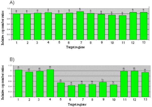
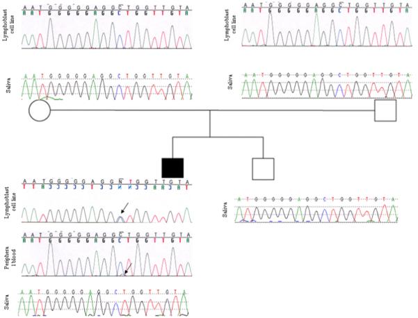
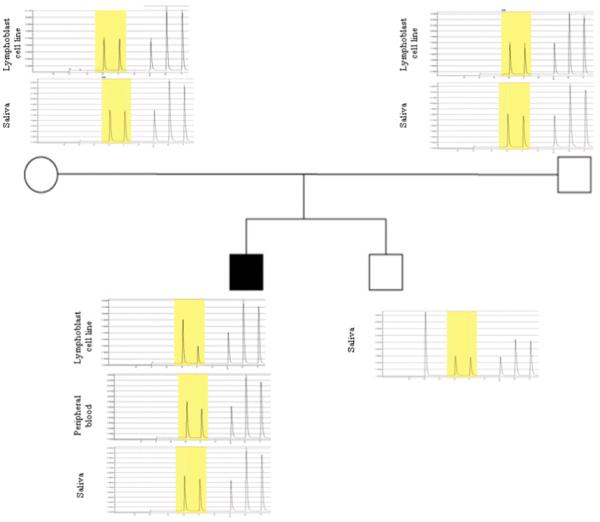
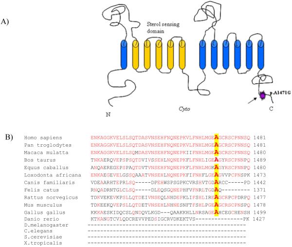
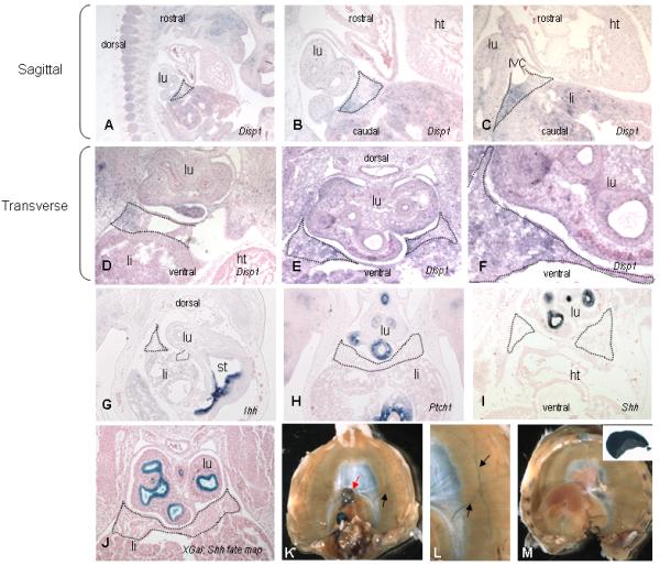
Cytogenetic and molecular cytogenetic studies demonstrate association between congenital diaphragmatic hernia (CDH) and chromosome 1q41q42 deletions. In this study, we screened a large CDH cohort (N=179) for microdeletions in this interval by the multiplex ligation-dependent probe amplification (MLPA) technique, and also sequenced two candidate genes located therein, dispatched 1 (DISP1) and homo sapiens H2.0-like homeobox (HLX). MLPA analysis verified deletions of this region in two cases, an unreported patient with a 46,XY,del(1)(q41q42.13) karyotype and a previously reported patient with a Fryns syndrome phenotype [Kantarci et al., 2006]. HLX sequencing showed a novel but maternally inherited single nucleotide variant (c.27C>G) in a patient with isolated CDH, while DISP1 sequencing revealed a mosaic de novo heterozygous substitution (c.4412C>G; p.Ala1471Gly) in a male with a left-sided Bochdalek hernia plus multiple other anomalies. Pyrosequencing demonstrated the mutant allele was present in 43%, 12%, and 4.5% of the patient's lymphoblastoid, peripheral blood lymphocytes, and saliva cells, respectively. We examined Disp1 expression at day E11.5 of mouse diaphragm formation and confirmed its presence in the pleuroperitoneal fold, as well as the nearby lung which also expresses Sonic hedgehog (Shh). Our report describes the first de novo DISP1 point mutation in a patient with complex CDH. Combining this finding with Disp1 embryonic mouse diaphragm and lung tissue expression, as well as previously reported human chromosome 1q41q42 aberrations in patients with CDH, suggests that DISP1 may warrant further consideration as a CDH candidate gene.
Copyright © 2010 Wiley-Liss, Inc.
Figures
Similar articles
-
New cases and refinement of the critical region in the 1q41q42 microdeletion syndrome.Eur J Med Genet. 2011 Jan-Feb;54(1):42-9. doi: 10.1016/j.ejmg.2010.10.002. Epub 2010 Oct 15. Eur J Med Genet. 2011. PMID: 20951845
-
Genomic alterations that contribute to the development of isolated and non-isolated congenital diaphragmatic hernia.J Med Genet. 2011 May;48(5):299-307. doi: 10.1136/jmg.2011.089680. J Med Genet. 2011. PMID: 21525063 Free PMC article.
-
Congenital diaphragmatic hernia (CDH) etiology as revealed by pathway genetics.Am J Med Genet C Semin Med Genet. 2007 May 15;145C(2):217-26. doi: 10.1002/ajmg.c.30132. Am J Med Genet C Semin Med Genet. 2007. PMID: 17436295 Review.
-
The discovery of microdeletion syndromes in the post-genomic era: review of the methodology and characterization of a new 1q41q42 microdeletion syndrome.Genet Med. 2007 Sep;9(9):607-16. doi: 10.1097/gim.0b013e3181484b49. Genet Med. 2007. PMID: 17873649 Review.
-
Sequence variants in the HLX gene at chromosome 1q41-1q42 in patients with diaphragmatic hernia.Clin Genet. 2009 May;75(5):429-39. doi: 10.1111/j.1399-0004.2009.01182.x. Clin Genet. 2009. PMID: 19459883 Free PMC article.
Cited by
-
The Complement Regulator Susd4 Influences Nervous-System Function and Neuronal Morphology in Mice.iScience. 2020 Mar 27;23(3):100957. doi: 10.1016/j.isci.2020.100957. Epub 2020 Feb 28. iScience. 2020. PMID: 32179479 Free PMC article.
-
Kif7 is required for the patterning and differentiation of the diaphragm in a model of syndromic congenital diaphragmatic hernia.Proc Natl Acad Sci U S A. 2013 May 21;110(21):E1898-905. doi: 10.1073/pnas.1222797110. Epub 2013 May 6. Proc Natl Acad Sci U S A. 2013. PMID: 23650387 Free PMC article.
-
Polygenic Causes of Congenital Diaphragmatic Hernia Produce Common Lung Pathologies.Am J Pathol. 2016 Oct;186(10):2532-43. doi: 10.1016/j.ajpath.2016.07.006. Epub 2016 Aug 24. Am J Pathol. 2016. PMID: 27565037 Free PMC article. Review.
-
Computational Reconstruction of NFκB Pathway Interaction Mechanisms during Prostate Cancer.PLoS Comput Biol. 2016 Apr 14;12(4):e1004820. doi: 10.1371/journal.pcbi.1004820. eCollection 2016 Apr. PLoS Comput Biol. 2016. PMID: 27078000 Free PMC article.
-
Muscle connective tissue controls development of the diaphragm and is a source of congenital diaphragmatic hernias.Nat Genet. 2015 May;47(5):496-504. doi: 10.1038/ng.3250. Epub 2015 Mar 25. Nat Genet. 2015. PMID: 25807280 Free PMC article.
References
-
- Al-Salem AH, Alkhuwaher H. Coexisting congenital diaphragmatic hernia, esophageal atresia, and tracheoesophageal fistula: a case report and review of the literature. Int Surg. 2008;93:141–144. - PubMed
-
- Allan DW, Greer JJ. Pathogenesis of nitrofen-induced congenital diaphragmatic hernia in fetal rats. J Appl Physiol. 1997;83:338–347. - PubMed
-
- Babiuk RP, Zhang W, Clugston R, Allan DW, Greer JJ. Embryological origins and development of the rat diaphragm. J Comp Neurol. 2003;455:477–487. - PubMed
-
- Bates MD, Schatzman LC, Lints T, Hamlin PE, Harvey RP, Potter SS. Structural and functional characterization of the mouse Hlx homeobox gene. Mamm Genome. 2000;11:836–842. - PubMed
Publication types
MeSH terms
Substances
Grants and funding
LinkOut - more resources
Full Text Sources
Medical
Molecular Biology Databases
Research Materials
