

Planar cell polarity acts through septins to control collective cell movement and ciliogenesis
- PMID: 20671153
- PMCID: PMC3509789
- DOI: 10.1126/science.1191184
Planar cell polarity acts through septins to control collective cell movement and ciliogenesis
Abstract
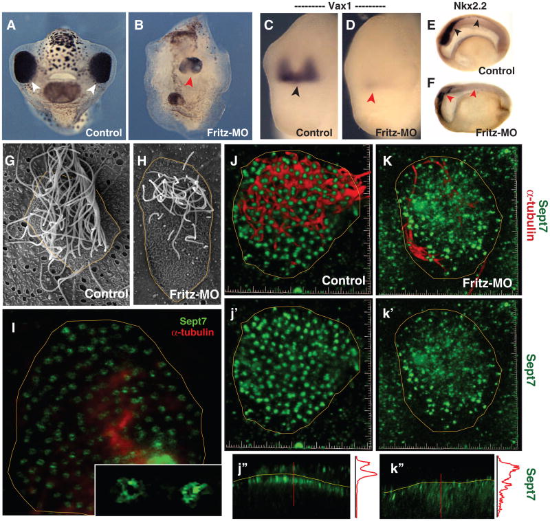
The planar cell polarity (PCP) signaling pathway governs collective cell movements during vertebrate embryogenesis, and certain PCP proteins are also implicated in the assembly of cilia. The septins are cytoskeletal proteins controlling behaviors such as cell division and migration. Here, we identified control of septin localization by the PCP protein Fritz as a crucial control point for both collective cell movement and ciliogenesis in Xenopus embryos. We also linked mutations in human Fritz to Bardet-Biedl and Meckel-Gruber syndromes, a notable link given that other genes mutated in these syndromes also influence collective cell movement and ciliogenesis. These findings shed light on the mechanisms by which fundamental cellular machinery, such as the cytoskeleton, is regulated during embryonic development and human disease.
Figures


Comment in
-
Cell biology. Septins at the nexus.Science. 2010 Sep 10;329(5997):1289-90. doi: 10.1126/science.1195445. Science. 2010. PMID: 20829470 No abstract available.
Similar articles
-
Cell biology. Septins at the nexus.Science. 2010 Sep 10;329(5997):1289-90. doi: 10.1126/science.1195445. Science. 2010. PMID: 20829470 No abstract available.
-
PCP and septins compartmentalize cortical actomyosin to direct collective cell movement.Science. 2014 Feb 7;343(6171):649-52. doi: 10.1126/science.1243126. Science. 2014. PMID: 24503851 Free PMC article.
-
Wdpcp, a PCP protein required for ciliogenesis, regulates directional cell migration and cell polarity by direct modulation of the actin cytoskeleton.PLoS Biol. 2013 Nov;11(11):e1001720. doi: 10.1371/journal.pbio.1001720. Epub 2013 Nov 26. PLoS Biol. 2013. PMID: 24302887 Free PMC article.
-
Cell and molecular biology of septins.Int Rev Cell Mol Biol. 2014;310:289-339. doi: 10.1016/B978-0-12-800180-6.00007-4. Int Rev Cell Mol Biol. 2014. PMID: 24725429 Review.
-
Here come the septins: novel polymers that coordinate intracellular functions and organization.J Cell Sci. 2006 Jan 1;119(Pt 1):4-10. doi: 10.1242/jcs.02746. J Cell Sci. 2006. PMID: 16371649 Free PMC article. Review.
Cited by
-
Substitution of a single non-coding nucleotide upstream of TMEM216 causes non-syndromic retinitis pigmentosa and is associated with reduced TMEM216 expression.Am J Hum Genet. 2024 Sep 5;111(9):2012-2030. doi: 10.1016/j.ajhg.2024.07.020. Epub 2024 Aug 26. Am J Hum Genet. 2024. PMID: 39191256 Free PMC article.
-
Septin dynamics are essential for exocytosis.J Biol Chem. 2015 Feb 27;290(9):5280-97. doi: 10.1074/jbc.M114.616201. Epub 2015 Jan 9. J Biol Chem. 2015. PMID: 25575596 Free PMC article.
-
Direct role of Bardet-Biedl syndrome proteins in transcriptional regulation.J Cell Sci. 2012 Jan 15;125(Pt 2):362-75. doi: 10.1242/jcs.089375. Epub 2012 Feb 2. J Cell Sci. 2012. PMID: 22302990 Free PMC article.
-
Mutations in SDCCAG8/NPHP10 Cause Bardet-Biedl Syndrome and Are Associated with Penetrant Renal Disease and Absent Polydactyly.Mol Syndromol. 2011 Sep;1(6):273-281. doi: 10.1159/000331268. Epub 2011 Sep 14. Mol Syndromol. 2011. PMID: 22190896 Free PMC article.
-
Planar cell polarity: coordinating morphogenetic cell behaviors with embryonic polarity.Dev Cell. 2011 Jul 19;21(1):120-33. doi: 10.1016/j.devcel.2011.06.011. Dev Cell. 2011. PMID: 21763613 Free PMC article. Review.
References
-
- Trinkaus JP. Cell Into Organs—The Forces That Shape the Embryo. Prentice-Hall; Englewood Cliffs, NJ: 1969.
-
- Shih J, Keller R. Development. 1992;116:901. - PubMed
-
- Keller R, Shih J, Sater A. Dev Dyn. 1992;193:199. - PubMed
-
- Keller R. Science. 2002;298:1950. - PubMed
-
- Wallingford JB, Harland RM. Development. 2002;129:5815. - PubMed
Publication types
MeSH terms
Substances
Grants and funding
LinkOut - more resources
Full Text Sources
Other Literature Sources
Molecular Biology Databases
