

Review
doi: 10.1038/ni.1887.
Epub 2010 Jul 20.
Decision checkpoints in the thymus
Affiliations
- PMID: 20644572
- PMCID: PMC3388799
- DOI: 10.1038/ni.1887
Item in Clipboard
Review
Decision checkpoints in the thymus
Nat Immunol.
2010 Aug.
Erratum in
- Nat Immunol. 2011 Mar;12(3):271
Abstract
The development of T cells in the thymus involves several differentiation and proliferation events, during which hematopoietic precursors give rise to T cells ready to respond to antigen stimulation and undergo effector differentiation. This review addresses signaling and transcriptional checkpoints that control the intrathymic journey of T cell precursors. We focus on the divergence of alphabeta and gammadelta lineage cells and the elaboration of the alphabeta T cell repertoire, with special emphasis on the emergence of transcriptional programs that direct lineage decisions.
Figures
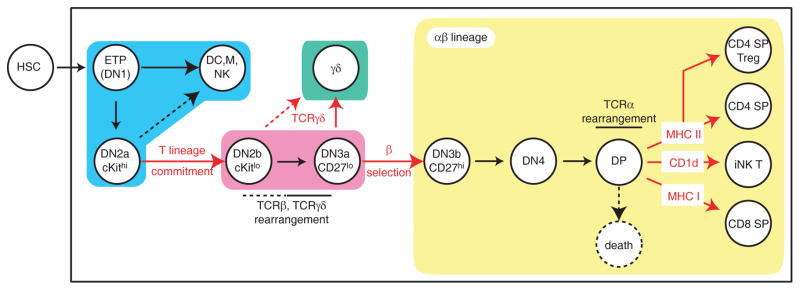
Thymic developmental stages are depicted. Expression of CD4 and CD8 separates CD4−CD8− double negative (DN), CD4+CD8+ double positive (DP) and cells expressing either coreceptor (single positive, SP), whereas the expression of CD44 and CD25 defines four DN subsets: CD44+CD25− [DN1], CD44+CD25+ [DN2], CD44−CD25+ [DN3], and CD44−CD25− [DN4]. The earliest precursors, known as ETP, that enter the thymus from the bone marrow are part of an heterogeneous DN1 subset that includes both subsequent intermediates in the T differentiation pathway and cells belonging to other lineages. The DN2 and DN3 subsets are themselves divided into two stages based on the expression of the receptor cKit and of CD27, respectively. Critical checkpoints addressed in the text are shown in red. Rounded rectangles group cell subsets according to the key developmental step they belong to: early uncommitted progenitors (blue), T committed progenitors before the separation of αβ and γδ lineages (purple) and committed αβ (yellow) or γδ (green) lineage cells.

In pre-selection DP thymocytes (left), E2A is thought to promote expression of Rorc and Rag genes, thereby ensuring TCR gene receptor expression and TCRα locus accessibility. In addition, E2A restrains expression of IL-7Rα, Bcl-2 and CCR7, although it is not clear whether such effects are direct or indirect (e.g. through effects on Foxo1 expression). Positive selection signals (right) reduce E2A activity by increasing Id3 expression, indirectly through Erk-dependent up-regulation of Egr proteins. Positively selected thymocytes have ceased expression of DP-stage genes and up-regulated Bcl-2, CCR7 and IL-7Rα, presumably as a result of the termination of E2A activity and of the induction of activators that may include Foxo1. Arrows or block signs do not necessarily indicate direct effects.
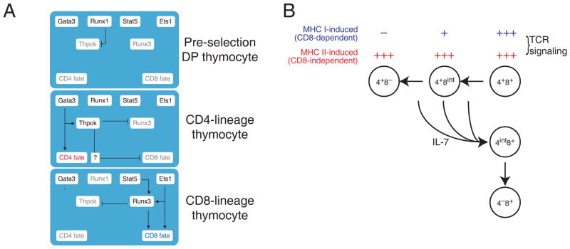
(A) Components of the transcriptional circuitry that promotes CD4-CD8 differentiation are schematically depicted and interconnected at three stages of T cell development. In preselection cells, Runx1-nucleated activities repress Thpok expression. In CD4-differentiating cells, Runx1-mediated Thpok expression is relieved, although Runx1 is still expressed in CD4 cells in which it binds the Thpok gene. Gata3 promotes both Thpok expression and additional developmental events required for CD4 cell differentiation. Thpok prevents Runx3 up-regulation and CD8 differentiation. In CD8-differentiating cells, Thpok repression is maintained, presumably through Runx3. Ets1 promotes Runx3 expression, and binds the Runx3 locus, whereas Stat5 has been reported to relay IL-7 signaling to Runx3. Grey lettering indicates factors not expressed at a particular stage. Other factors (including Tox) are omitted for clarity. Arrows or block signs do not imply direct effects. (B) The ‘kinetic signaling’ model of lineage differentiation posits that intrathymic TCR signaling, regardless of MHC specificity, represses Cd8 expression, causing thymocytes to adopt a CD4+CD8int surface phenotype. TCR signaling in MHC II-restricted thymocytes is not affected by CD8 down-regulation, and its persistence eventually seals CD4 commitment. In contrast, TCR signaling in MHC I-restricted thymocytes is impaired by CD8 down-regulation, and its cessation causes ‘coreceptor reversal’ i.e. the cessation of Cd4 expression and the resumption of Cd8 expression.
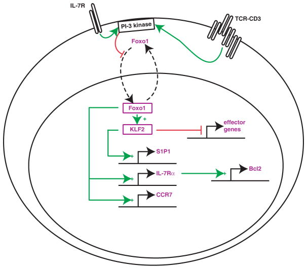
Based on analyses in mature T cells, a transcriptional circuitry is proposed in mature thymocytes, that enables expression of IL-7Rα, CCR7 and Klf12, which itself controls thymic egress by increasing expression of the receptor for sphingosine 1-phophate (S1P1), T cell trafficking and quiescence. Foxo1 activity is inhibited by PI-3 kinase-dependent phosphorylation, that promotes its sequestration in the cytosol, contributing to the self-limiting IL-7Rα expression characteristic of mature T cells. It may also act as a ‘licensing’ factor in the thymus to prevent the release of self reactive thymocytes due to their persistent TCR signaling.
Similar articles
-
Specific Notch receptor-ligand interactions control human TCR-αβ/γδ development by inducing differential Notch signal strength.J Exp Med. 2013 Apr 8;210(4):683-97. doi: 10.1084/jem.20121798. Epub 2013 Mar 25. J Exp Med. 2013. PMID: 23530123 Free PMC article. Clinical Trial.
-
Delta-like 4-mediated Notch signaling is required for early T-cell development in a three-dimensional thymic structure.Eur J Immunol. 2015 Aug;45(8):2252-62. doi: 10.1002/eji.201445123. Epub 2015 Jun 3. Eur J Immunol. 2015. PMID: 25976373
-
A comparative view on vitamin C effects on αβ- versus γδ T-cell activation and differentiation.J Leukoc Biol. 2020 Jun;107(6):1009-1022. doi: 10.1002/JLB.1MR1219-245R. Epub 2020 Feb 7. J Leukoc Biol. 2020. PMID: 32034803 Review.
-
αβ and γδ T cell receptors: Similar but different.J Leukoc Biol. 2020 Jun;107(6):1045-1055. doi: 10.1002/JLB.2MR1219-233R. Epub 2020 Jan 29. J Leukoc Biol. 2020. PMID: 31994778 Review.
-
Lineage divergence at the first TCR-dependent checkpoint: preferential γδ and impaired αβ T cell development in nonobese diabetic mice.J Immunol. 2011 Jan 15;186(2):826-37. doi: 10.4049/jimmunol.1002630. Epub 2010 Dec 10. J Immunol. 2011. PMID: 21148803 Free PMC article.
Cited by
-
Late stages of T cell maturation in the thymus involve NF-κB and tonic type I interferon signaling.Nat Immunol. 2016 May;17(5):565-73. doi: 10.1038/ni.3419. Epub 2016 Apr 4. Nat Immunol. 2016. PMID: 27043411 Free PMC article.
-
Synergistic effects of interleukin-7 and pre-T cell receptor signaling in human T cell development.J Biol Chem. 2012 Sep 28;287(40):33826-35. doi: 10.1074/jbc.M112.380113. Epub 2012 Aug 2. J Biol Chem. 2012. PMID: 22859301 Free PMC article.
-
TET Proteins in the Spotlight: Emerging Concepts of Epigenetic Regulation in T Cell Biology.Immunohorizons. 2023 Jan 1;7(1):106-115. doi: 10.4049/immunohorizons.2200067. Immunohorizons. 2023. PMID: 36645853 Free PMC article.
-
Neutrophil and T Cell Functions in Patients with Congenital Heart Diseases: A Review.Pediatr Cardiol. 2021 Oct;42(7):1478-1482. doi: 10.1007/s00246-021-02681-3. Epub 2021 Jul 20. Pediatr Cardiol. 2021. PMID: 34282478 Free PMC article. Review.
-
IL-7-Induced Proliferation of Human Naive CD4 T-Cells Relies on Continued Thymic Activity.Front Immunol. 2017 Jan 19;8:20. doi: 10.3389/fimmu.2017.00020. eCollection 2017. Front Immunol. 2017. PMID: 28154568 Free PMC article.
References
-
- Hayday AC. Gammadelta T cells and the lymphoid stress-surveillance response. Immunity. 2009;31:184–196. - PubMed
-
- Bhandoola A, von Boehmer H, Petrie HT, Zuniga-Pflucker JC. Commitment and developmental potential of extrathymic and intrathymic T cell precursors: plenty to choose from. Immunity. 2007;26:678–689. - PubMed
-
- Feyerabend TB, et al. Deletion of Notch1 converts pro-T cells to dendritic cells and promotes thymic B cells by cell-extrinsic and cell-intrinsic mechanisms. Immunity. 2009;30:67–79. - PubMed
Publication types
MeSH terms
Substances
Grants and funding
LinkOut - more resources
Full Text Sources
Other Literature Sources
