

Neuroradiologic features of CASK mutations
- PMID: 20595373
- PMCID: PMC3756090
- DOI: 10.3174/ajnr.A2173
Neuroradiologic features of CASK mutations
Abstract
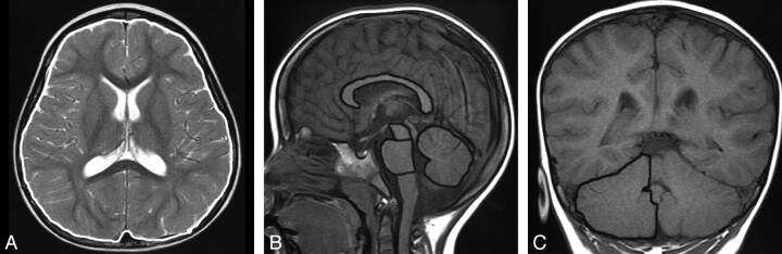
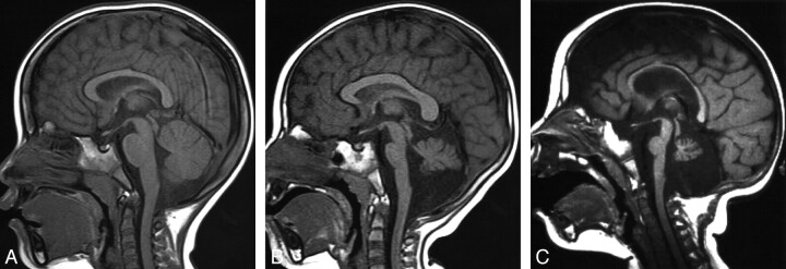
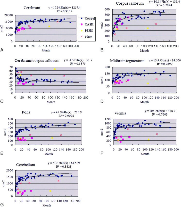
Mutations of the CASK gene are associated with X-linked mental retardation with microcephaly and disproportionate brain stem and cerebellar hypoplasia in females. The areas of the cerebrum, corpus callosum, pons, midbrain, and cerebellar vermis and hemisphere and a ratio of cerebrum/corpus callosum areas were measured in 5 female patients with CASK mutations, 67 female controls, and 5 patients with pontine hypoplasia. MR imaging in patients with CASK mutations revealed a normal size of the corpus callosum and a low ratio of the cerebrum/corpus callosum with a reduced area of the cerebrum, pons, midbrain, and cerebellar vermis and hemispheres. The 5 patients with pontine hypoplasia showed thinning of the corpus callosum and a high ratio of the cerebrum/corpus callosum, irrespective of the size of the cerebrum. The normal size of the corpus callosum, which gives an impression of callosal thickening at first glance, may be an imaging clue to detect patients with CASK mutations.
Figures
Similar articles
-
Novel intragenic duplications and mutations of CASK in patients with mental retardation and microcephaly with pontine and cerebellar hypoplasia (MICPCH).Hum Genet. 2012 Jan;131(1):99-110. doi: 10.1007/s00439-011-1047-0. Epub 2011 Jul 7. Hum Genet. 2012. PMID: 21735175
-
Survival of a male patient harboring CASK Arg27Ter mutation to adolescence.Mol Genet Genomic Med. 2020 Oct;8(10):e1426. doi: 10.1002/mgg3.1426. Epub 2020 Jul 21. Mol Genet Genomic Med. 2020. PMID: 32696595 Free PMC article.
-
Clinical and radiological features of Japanese patients with a severe phenotype due to CASK mutations.Am J Med Genet A. 2012 Dec;158A(12):3112-8. doi: 10.1002/ajmg.a.35640. Epub 2012 Nov 19. Am J Med Genet A. 2012. PMID: 23165780
-
Associated brain abnormalities in patients with corpus callosum anomalies.Turk J Pediatr. 1999 Apr-Jun;41(2):173-80. Turk J Pediatr. 1999. PMID: 10770655 Review.
-
Possible new autosomal recessive syndrome of partial agenesis of the corpus callosum, pontine hypoplasia, focal white matter changes, hypotonia, mental retardation, and minor anomalies.Am J Med Genet. 1997 Dec 12;73(2):184-8. Am J Med Genet. 1997. PMID: 9409870 Review.
Cited by
-
Case report: A novel CASK mutation in a Chinese female child with microcephaly with pontine and cerebellar hypoplasia.Front Genet. 2022 Sep 7;13:856636. doi: 10.3389/fgene.2022.856636. eCollection 2022. Front Genet. 2022. PMID: 36159992 Free PMC article.
-
Two heterozygous mutations in the calcium/calmodulin-dependent serine protein kinase gene (CASK) in cases with developmental disorders.Mol Genet Genomic Med. 2022 Nov;10(11):e2065. doi: 10.1002/mgg3.2065. Epub 2022 Sep 28. Mol Genet Genomic Med. 2022. PMID: 36168867 Free PMC article.
-
Spectrum of pontocerebellar hypoplasia in 13 girls and boys with CASK mutations: confirmation of a recognizable phenotype and first description of a male mosaic patient.Orphanet J Rare Dis. 2012 Mar 27;7:18. doi: 10.1186/1750-1172-7-18. Orphanet J Rare Dis. 2012. PMID: 22452838 Free PMC article.
-
A developmental and genetic classification for malformations of cortical development: update 2012.Brain. 2012 May;135(Pt 5):1348-69. doi: 10.1093/brain/aws019. Epub 2012 Mar 16. Brain. 2012. PMID: 22427329 Free PMC article. Review.
-
Drosophila CASK regulates brain size and neuronal morphogenesis, providing a genetic model of postnatal microcephaly suitable for drug discovery.Neural Dev. 2023 Oct 7;18(1):6. doi: 10.1186/s13064-023-00174-y. Neural Dev. 2023. PMID: 37805506 Free PMC article.
References
-
- Najm J, Horn D, Wimplinger I, et al. . Mutations of CASK cause an X-linked brain malformation phenotype with microcephaly and hypoplasia of the brainstem and cerebellum. Nat Genet 2008; 40: 1065– 67 - PubMed
-
- Hayashi S, Mizuno S, Migata O, et al. . The CASK gene harbored in a deletion detected by array-CGH as a potential candidate for a gene causative of X-linked dominant mental retardation. Am J Med Genet A 2008; 146A: 2145– 51 - PubMed
-
- Tanaka M, Tanaka Y, Hamano S, et al. . A case of PEHO (progressive encephalopathy with edema, hypsarrhythmia and optic atrophy) syndrome: changes in clinical and neuroradiological findings [in Japanese]. No To Hattatsu 1997; 29: 488– 93 - PubMed
Publication types
MeSH terms
Substances
Grants and funding
LinkOut - more resources
Full Text Sources
Medical