. 2010 May 14;141(4):632-44.
doi: 10.1016/j.cell.2010.04.008. Epub 2010 Apr 29.

Transmembrane receptor DCC associates with protein synthesis machinery and regulates translation

Affiliations

Transmembrane receptor DCC associates with protein synthesis machinery and regulates translation

Joseph Tcherkezian et al. Cell. .

Erratum in

Abstract

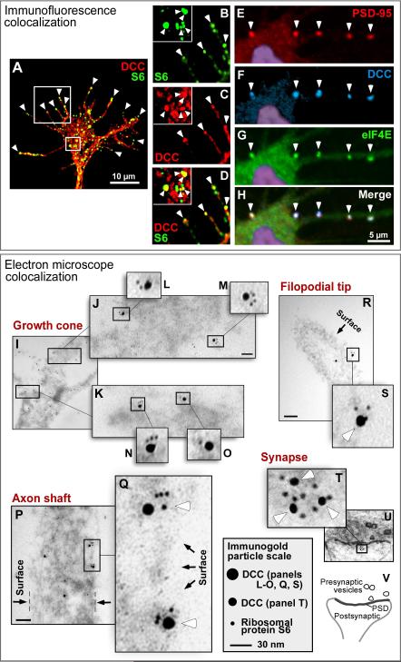
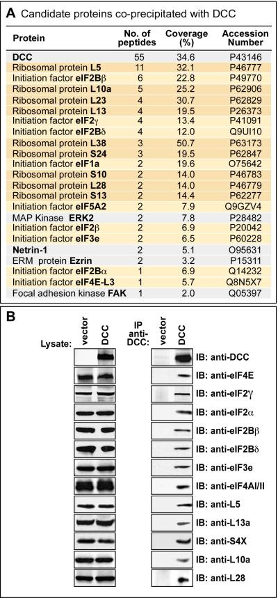
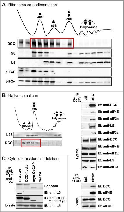
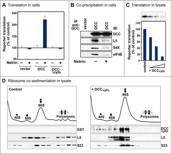
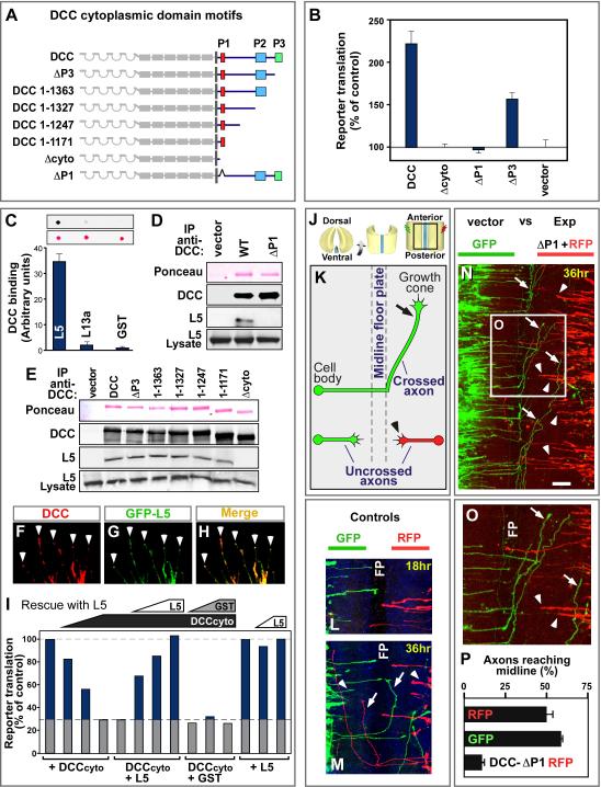
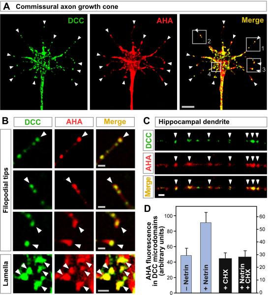
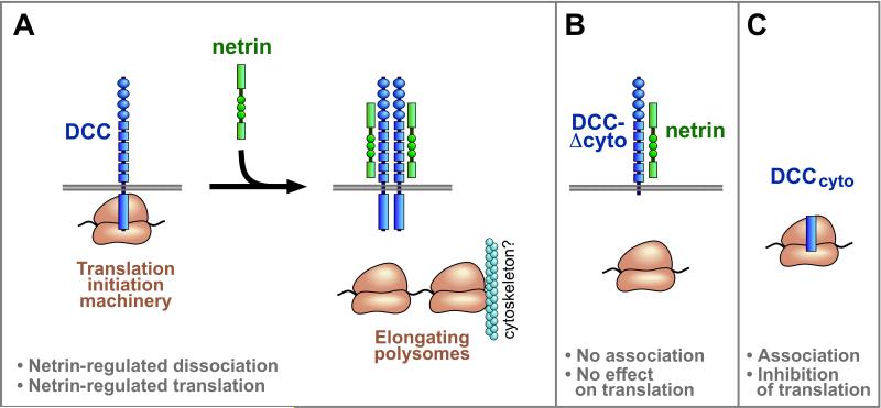
Extracellular signals regulate protein translation in many cell functions. A key advantage of control at the translational level is the opportunity to regulate protein synthesis within specific cellular subregions. However, little is known about mechanisms that may link extracellular cues to translation with spatial precision. Here, we show that a transmembrane receptor, DCC, forms a binding complex containing multiple translation components, including eukaryotic initiation factors, ribosomal large and small subunits, and monosomes. In neuronal axons and dendrites DCC colocalizes in particles with translation machinery, and newly synthesized protein. The extracellular ligand netrin promoted DCC-mediated translation and disassociation of translation components. The functional and physical association of a cell surface receptor with the translation machinery leads to a generalizable model for localization and extracellular regulation of protein synthesis, based on a transmembrane translation regulation complex.


Figures

Figure 1
Figure 1. DCC colocalizes with translational components
(A-H) Immunofluorescence. (A-D) Cultured commissural axons. DCC (red) and ribosomal protein S6 (green) appeared in puncta within the growth cone, with a subset of puncta overlapping (yellow). Panels B-D are enlargement of boxes from A, showing filopodial tips, with a central growth cone area in the inset. Arrowheads: examples of colocalized puncta, notably at filopodial tips. (E-H) Cultured hippocampal dendrites. Labeling for DCC (blue), eIF4E (green), and the postsynaptic marker PSD-95 was seen in puncta at synapses (arrowheads). Nuclear DAPI staining is shown in purple. (I-V) Electron microscopy, showing double immunogold labeling for endogenous DCC (15 nm gold particles in panels I-S; 10 nm particles in T-V) and ribosomal protein S6 (5 nm particles). Some variation in particle size is caused by silver enhancement. (I-S) Cultured commissural axons. Panels I-O show distal part of growth cone near the base of the filopodia; P and Q show axon shaft, oriented vertically; R shows filopodium, with distal end toward upper left. (T-V) Cultured hippocampal neuron synapse, diagrammed in panel V. White arrowheads: examples of clustered particles. Black arrows: outer boundary of the cell. Boxes show enlargement of clustered particles. Scale bar: 70 nm in J, R and P; 15 nm in T. See also Figure S1.
Figure 2
Figure 2. Tandem mass spectrometry screen identifying translational components coprecipitated with DCC
(A) Tandem mass spectrometry identification of proteins following immunoprecipitation with anti-DCC antibody. Proteins listed showed specific coprecipitation from 293-net cells transfected with DCC, versus vector control. Ribosomal proteins are highlighted in dark orange, and initiation factors in light orange. Grey indicates proteins previously shown to interact directly with DCC (although other known interaction partners such as Nck, Src and Fyn were not seen here, presumably reflecting detection limits in this screen). Accession numbers: UniProtKB/Swiss-Prot data base. (B) Most of the candidate translational components from the mass spectrometry screen were tested by Western blot, and in every case tested were confirmed to show co-immunoprecipitation with DCC. See also Figure S2.
Figure 3
Figure 3. DCC receptor physically associates with translation machinery
(A) Ribosome sedimentation from netrin-expressing 293-net cells transfected with full length DCC. DCC cosedimented in 40S, 60S and 80S fractions (red box) but not prominently in the polysome fraction. Control proteins were distributed as expected from previous studies: S6 in 40S, 80S and polysomal fractions; L5 in 60S, 80S and polysomal fractions; eIF4E primarily in the 40S fraction; and eIF2α in all four fractions. (B) DCC association with translation machinery, with all proteins expressed at endogenous levels in developing spinal cord. Left panel: DCC cosedimented in 40S, 60S and 80S fractions. Other markers such as eIF4E distributed as expected in spinal cord (not shown). Upper right panel: translational components co-immunoprecipitated with DCC. Lower right panel: DCC co-immunoprecipitated with eIF4E. (C) DCC-Δcyto mutant lacking a cytoplasmic domain did not co-precipitate ribosomal marker L5. myc-tagged mCdGAP was a negative control. See also Figure S3.
Figure 4
Figure 4. Effects of DCC and netrin on translation
(A) Full length DCC mediated netrin promotion of translation. 293 cells were transfected with DCC constructs, together with a reporter plasmid for cap-dependent translation (Figure S4A). Reporter translation was normalized to a cap-independent internal control, and is compared in netrin-expressing 293-net cells (+) versus 100% in control cells (–). DCC lacking a cytoplasmic domain (DCC-Δcyto) showed no effect on translation. n=3 experiments; error bars, SEM. Without netrin, full length DCC promoted translation slightly in some experiments, although to a much lesser degree than with netrin (data not shown). (B) Netrin reduced the association of DCC with translation components. Western analysis showed reduced coprecipitation of markers of the ribosomal large subunit (L5), small subunit (S4X) and initiation factor eIF4E with exogenous DCC in netrin-expressing 293-net cells (+), compared with control 293 cells (–). An association above background remained, however, in the presence of netrin (see Figure S4H). (C) DCC cytoplasmic domain (DCCcyto) inhibited translation. Purified recombinant DCCcyto (100 ng, 200 ng, and 500 ng) or GST control protein was added to a rabbit reticulocyte lysate translation system. (D) Ribosome sedimentation profiles of reticulocyte lysates following incubation with purified recombinant DCCcyto (right) or GST control (left). DCCcyto cosedimented with 40S, 60S and 80S fractions. DCCcyto decreased the polysome fraction and increased the 80S fraction, implying that a step after 80S ribosome assembly was inhibited and became rate limiting in this assay, although earlier steps may also be affected. Ribosomal proteins distributed as expected: S23 in 40S, 80S and polysome fractions; L5 in 60S, 80S and polysome fractions. See also Figure S4.
Figure 5
Figure 5. Involvement of the DCC P1 domain and ribosomal protein L5
(A) DCC cytoplasmic domain contains three conserved motifs: P1, P2 and P3. A nested series of deletions were made, and an internal deletion of the P1 motif. (B) DCC promotion of translation was prevented by P1 deletion (DCC-ΔP1), and partially reduced by P3 deletion (DCC-ΔP3). The assay was as in Figure 4A, with cap-dependent reporter translation normalized to a cap-independent control. n=3 experiments; error bars, SEM. (C-I) Physical and functional interactions with ribosomal protein L5. (C) Far Western dot blot quantitation of direct binding of purified recombinant DCC cytoplasmic domain to purified recombinant L5. Ribosomal protein L13a and GST provide controls. Ponceau stain shows similar loading. n=4 experiments; error bars, SEM. (D, E) Western analysis of endogenous L5 co-immunopreciptation with DCC, showing L5 associated with all mutants in the nested deletion series except DCC-Δcyto, and not with DCC-ΔP1. (F-H) Immunofluorescence of DCC (red) and GFP-L5 (green) co-transfected into 293 cells, showing colocalization in neurite-like extensions, notably at the tips (arrowheads). (I) Functional interaction between ribosomal protein L5 and DCCcyto. The inhibitory effect of DCCcyto in a reticulocyte lysate (see Figure 4C) was rescued in a dose-dependent manner by recombinant L5 (100 ng, 200 ng, and 500 ng). GST control at the same concentrations did not rescue. L5 alone (200 ng and 500 ng) had no noticeable effect. Column 5 is a duplicate of column 4 to clarify control value. (J-P) Effect of DCC-ΔP1 as a dominant inhibitory mutant on commissural axon pathfinding in developing spinal cord. Chick spinal cords at E3.5 (HH 23) were electroporated and analyzed 36 hr later, compared with controls electroporated on the contralateral side of the same spinal cord. (J) Illustration of spinal cord open book preparations, with imaged area in box. (K) Diagram of axon trajectories seen in panel N: axons grow toward the midline floor plate (arrowheads), cross it, then turn and grow anteriorly (arrow). (L, M) Control plasmids expressing GFP and RFP; equivalent axon numbers, growth rates and patterns were seen for both tracers after 18 or 36 hours. (N, O) Axons electroporated with DCC-ΔP1 (right side, red) compared to GFP control (left side, green) showed greater numbers remaining at locations midway toward the floorplate (red; examples indicated by arrowheads), and fewer had reached or crossed the floor plate compared with control (green; examples indicated by arrows). Panel O shows enlargement of boxed area. (P) Quantitation of axons reaching the midline after electroporation with control or DCC-ΔP1 mutant construct. A total of 530 axons were analyzed in seven experiments; error bars, SEM. Scale bar in N, 100 μm. FP, floor plate. See also Figure S5.
Figure 6
Figure 6. DCC colocalizes with newly synthesized protein
(A, B) Localization of DCC (green) and AHA labeled sites of newly synthesized protein (red) in cultured commissural axon growth cone. Panel B shows enlargement of boxes in A. Arrowheads show examples of overlapping puncta (yellow) notably at filopodial tips. (C) DCC (green) and AHA (red) labeling in cultured hippocampal neuron dendrite. Arrowheads indicate overlap. (D) Quantitation of AHA labeling in DCC puncta at filopodial tips. AHA was added for 20 min; then cycloheximide (CHX) for 20 min; then 200 ng/ml netrin-1 for 10 min. Netrin induced an increase in labeling that was blocked by cycloheximide. Panels A-C were in the presence of netrin-1. Scale bars: 5 μm in panel A; 1 μm in B and C. See also Figure S6.
Figure 7
Figure 7. Models for interaction of DCC with translational machinery
(A) In the model illustrated, full length DCC associates with translation initiation machinery, including 40S, 60S and 80S ribosomes and eIFs. Signaling by DCC within this complex is enhanced by netrin, and promotes formation of actively elongating polysomes which are no longer associated with DCC. Polysomes may nevertheless tend to remain locally within micron scale structures such as a filopodial tip or synaptic spine, and this may be facilitated by their known interaction with the cytoskeleton. Transduction of a signal across the membrane by DCC and other receptors is believed to be mediated by dimerization or higher-order clustering. In the absence of netrin, spontaneous dimerization and signaling by DCC may promote some translation, although at a lower level than with netrin. Signaling by some DCC molecules in the cell would not preclude the coexistence of other DCC molecules that remain in a complex with translation initiation machinery. In addition to DCC and translation initiation machinery, the complex is proposed also to include signal transduction proteins, and DCC may regulate more than one step of initiation. (B) No prominent association with translation components or functional effect on translation was seen with DCC mutants lacking the cytoplasmic domain (DCC-Δcyto) or lacking the 20 amino acid P1 motif (DCC-ΔP1), showing a requirement of these regions for physical and functional interactions with translation machinery. (C) The DCC cytoplasmic domain by itself, DCCcyto, cosedimented with 40S, 60S and 80S ribosomes, and inhibited protein synthesis in a cell-free system, showing that this region is sufficient for physical and functional interactions with translational machinery. DCCcyto is proposed to act here as an interfering mutant, occupying downstream components but lacking an extracellular domain necessary for positive signaling. Although the diagrams are not to a precise scale, receptor length and ribosome diameter are approximately in proportion: the eukaryotic ribosome is ~20-30 nm across (Spahn et al., 2001); each Ig domain (oval) and FNIII domain (small square) is ~4 nm long; the intracellular domain of DCC (rectangle) is approximately 25% of the protein sequence as illustrated here in proportion with the extracellular length to account for potential folding, or in more extended conformation it could be up to 100 nm long.

Comment in

Similar articles

Cited by

References

    1. Amrani N, Ghosh S, Mangus DA, Jacobson A. Translation factors promote the formation of two states of the closed-loop mRNP. Nature. 2008;453:1276–1280. - PMC - PubMed
    1. Beatty KE, Tirrell DA. Two-color labeling of temporally defined protein populations in mammalian cells. Bioorg Med Chem Lett. 2008;18:5995–5999. - PMC - PubMed
    1. Bilic J, Huang YL, Davidson G, Zimmermann T, Cruciat CM, Bienz M, Niehrs C. Wnt induces LRP6 signalosomes and promotes dishevelled-dependent LRP6 phosphorylation. Science. 2007;316:1619–1622. - PubMed
    1. Brittis PA, Lu Q, Flanagan JG. Axonal protein synthesis provides a mechanism for localized regulation at an intermediate target. Cell. 2002;110:223–235. - PubMed
    1. Campbell DS, Holt CE. Chemotropic responses of retinal growth cones mediated by rapid local protein synthesis and degradation. Neuron. 2001;32:1013–1026. - PubMed

Publication types

MeSH terms