. 2010 Aug;38(14):4586-98.
doi: 10.1093/nar/gkq214. Epub 2010 Apr 5.

CdnL, a member of the large CarD-like family of bacterial proteins, is vital for Myxococcus xanthus and differs functionally from the global transcriptional regulator CarD

Affiliations

CdnL, a member of the large CarD-like family of bacterial proteins, is vital for Myxococcus xanthus and differs functionally from the global transcriptional regulator CarD

Diana García-Moreno et al. Nucleic Acids Res. 2010 Aug.

Abstract

CarD, a global transcriptional regulator in Myxococcus xanthus, interacts with CarG via CarDNter, its N-terminal domain, and with DNA via a eukaryotic HMGA-type C-terminal domain. Genomic analysis reveals a large number of standalone proteins resembling CarDNter. These constitute, together with the RNA polymerase (RNAP) interacting domain, RID, of transcription-repair coupling factors, the CarD_TRCF protein family. We show that one such CarDNter-like protein, M. xanthus CdnL, cannot functionally substitute CarDNter (or vice versa) nor interact with CarG. Unlike CarD, CdnL is vital for growth, and lethality due to its absence is not rescued by homologs from various other bacteria. In mycobacteria, with no endogenous DksA, the function of the CdnL homolog mirrors that of Escherichia coli DksA. Our finding that CdnL, like DksA, is indispensable in M. xanthus implies that they are not functionally redundant. Cells are normal on CdnL overexpression, but divide aberrantly on CdnL depletion. CdnL localizes to the nucleoid, suggesting piggyback recruitment by factors such as RNAP, which we show interacts with CdnL, CarDNter and RID. Our study highlights a complex network of interactions involving these factors and RNAP, and points to a vital role for M. xanthus CdnL in an essential DNA transaction that affects cell division.


Figures

Figure 1.
Figure 1.
M. xanthus CarD and the PF02559 protein family. (A) Schematic showing CarD domain architecture. Residue numbers (on top) demarcate the distinct regions. The CarD N-terminal domain, CarDNter, defines the PF02559 family that includes a number of CarDNter-like proteins and TRCF-RID. CarDCter denotes the HMGA-like CarD C-terminal domain, with human HMGA1a shown for comparison (‘+’ indicate the basic AT-hooks and ‘−’ the acidic region). (B) Sequence alignment of representative PF02559 family members. NCBI accession codes and the corresponding bacterium are indicated in brackets: CarDNter (CAA91224; M. xanthus); CdnL (YP_630846; M. xanthus); TRCF-RID, the segment corresponding to residues 514–587 of TRCF (YP_629274; M. xanthus); BbCdnL (NP_969149; B. bacteriovorus); CgCdnL (YP_001139479; C. glutamicum); ScCdnL (NP_628406; S. coelicolor); TtCdnL (YP_005787; T. thermophilus). Residues are shaded black (with an asterisk in the consensus line below) when identical in the majority of the aligned sequences, or grey when similar.
Figure 2.
Figure 2.
CdnL cannot replace CarDNter in CarD function. (A) Schematic of the CarD chimera with CarDNter (residues 1–178) replaced by CdnL. (B) Western blot of pure CarD (lane 1) and cell extracts of strains expressing CarD (‘WT’; lane 2), the CdnL-CarDCter chimera (‘Chimera’, lane 4), or no CarD (‘ΔcarD’; lane 3) probed with monoclonal anti-CarD antibodies. (C) Top: colour phenotypes of cell spots after growth for 1 day on CTT plates in the dark or in the light. Bottom: expression levels of the reporter PQRS::lacZ probe in strains bearing the indicated carD allele. Cell cultures were grown to early exponential phase in the dark, divided into two, grown for a further 8 h, one in the dark (filled bars) and the other in the light (empty bars). The specific β-galactosidase activities (in nanomoles of o-nitrophenyl β-d-galactoside hydrolyzed/min/mg protein) are from two or more independent measurements. (D) Bacterial two-hybrid analysis of cell spots expressing the protein pair indicated on plates containing X-gal. Cells expressing only one fusion protein served as the negative control.
Figure 3.
Figure 3.
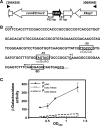
Genome context, promoter elements and expression of cdnL. (A) cdnL genome context. Genes are represented by the thick arrows. Those unlabeled encode hypothetical proteins of unknown functions. comEC/rec2 encodes a predicted DNA-internalization competence protein and k9ap1 is Pk9 associate protein 1, a predicted FHA (forkhead associated) domain signaling protein. (B) DNA sequence of the cdnL promoter region. The –10 and –35 promoter elements and the Shine-Dalgarno ribosome binding site are boxed; ‘+1’ is the transcription start site determined by 5′-RACE. The two arrows bracket the DNA segment fused to a reporter lacZ gene used for analyzing promoter activity. Lowercase letters in the sequences below the boxed –10 and –35 regions indicate bases that were mutated in the corresponding hexamers. (C) Specific β-galactosidase activities in nmol of o-nitrophenyl β-d-galactoside hydrolyzed/min/mg protein (mean of three independent measurements) of reporter lacZ probe with native (filled circle) or mutated PcdnL in the –35 (open triangle) or –10 (open square) promoter regions for different cell densities during vegetative growth.
Figure 4.
Figure 4.
Conditional expression of cdnL shows it is essential. (Top) Schematic for M. xanthus strains conditionally expressing cdnL. (Bottom) Cells grown on CTT plates in the light were streaked on CTT plates ± B12, then incubated at 33°C for 2 days under permissive (plates without B12 incubated in the light) or restrictive (plates with B12 incubated in the dark) conditions. Note the lack of growth of MR1467 under restrictive conditions. The red colour in the light is due to carotenogenesis.
Figure 5.
Figure 5.
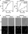
Growth rate and cellular morphology of cells conditionally expressing cdnL. (A) Growth curves for MR1466 and MR1467 under permissive (left) or restrictive (right) conditions. Cells were grown in the light at 33°C, 300 rpm, and aliquots of ∼106 cells were innoculated into 10 ml CTT in separate flasks. One was grown in the light (permissive conditions) and one in the dark with 0.75 µM B12 (restrictive conditions) at 33°C, 300 rpm; the abscissa indicates time (in hours) after the shift in growth conditions. (B) Cellular morphology of cell samples from cultures grown as in A, examined at an OD550 ∼0.8 after DAPI-staining by DIC and fluorescence microscopy. Note the unusually elongated cells indicative of aberrant cell division on shifting to restrictive conditions for the strain conditionally expressing cdnL and the extended span of the pole regions lacking DAPI-stained foci (scale marker: 5 μm).
Figure 6.
Figure 6.
CdnL subcellular localization. (A) Scheme of cdnL alleles in MR1488 and MR1489 (left). Western blots of the corresponding cell extracts probed with polyclonal anti-CdnL antibodies are shown on the right. (B) Subcellular localization of CdnL-eGFP in MR1488 and MR1489. Cells were grown to mid-log phase and examined by DIC (left panels) and fluorescence microscopy for CdnL-eGFP (middle panels) or DAPI-stained nucleoid (right panels). Arrows point to the more intense CdnL-eGFP foci in MR1489, which coincide with the nucleoid (scale marker: 5 μm).
Figure 7.
Figure 7.
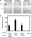
CdnL and CarDNter physically interact with M. xanthus RNAP. (A) Analysis of RNAP retention by CdnL-His6 immobilized on TALON metal affinity resin. Shown are silver-stained 10% SDS–PAGE gels of the flowthrough (FT), low salt (50 mM) and high salt (500 mM) eluates on passing M. xanthus core RNAP through a column of TALON resin alone (‘No His6-protein’), TALON bound to His6-DksA as the positive control, or TALON bound to CdnL-His6. The α, β and β′ RNAP subunits are indicated. (B) Bacterial two-hybrid analysis of the interaction of M. xanthus RNAP β19–148 region (in plasmid pUT18C) with the TRCF segment (514–645), CdnL, or CarDNter (in pKT25) as indicated. Cells expressing only one fusion protein are negative controls.
Figure 8.
Figure 8.
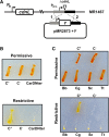
M. xanthus CdnL cannot be replaced by CarDNter or by CdnL homologs from other bacteria. (A) Scheme showing the strategy used for complementation analysis. A plasmid derived from pMR2873, with ‘Y’ (CarDNter, CdnL, or a given CdnL homolog) under PcdnL control and DNA segments flanking cdnL upstream (grey) and downstream (black) in the genome, was introduced into the MR1467 strain conditionally expressing cdnL. Merodiploids resulting from plasmid integration by recombination at either ‘1’ or ‘2’ would exhibit constitutive expression of the inserted variant and conditional expression of the cdnL allele at the heterologous site. (B) Complementation analysis with CarDNter. The test strain ‘CarDNter’ resulted from using pMR2873 with ‘Y’ = CarDNter coding sequence; C+, is the positive control derived from using pMR2873 with ‘Y’ = cdnL, and the negative control C− is the recipient strain MR1467. (C) Complementation analysis with BbCdnL, CgCdnl, ScCdnL, and TtCdnL. Test strains were generated using the pMR2873 with ‘Y’ = cdnLBb (‘Bb’), cdnLCg (‘Cg’), cdnLSc (‘Sc’) or cdnLTt (‘Tt’). C+ and C− are as in B. In B and C, cells grown on CTT plates in the light were streaked on CTT plates ± B12, then incubated at 33°C for 2 days under permissive (plates without B12 incubated in the light) or restrictive conditions (plates with B12 incubated in the dark). The red colour in the light is due to carotenogenesis.

Similar articles

Cited by

References

    1. Dawid W. Biology and global distribution of myxobacteria in soils. FEMS Microbiol. Rev. 2000;24:403–427. - PubMed
    1. Kaiser D. Signaling in myxobacteria. Annu. Rev. Microbiol. 2004;58:75–98. - PubMed
    1. Zusman DR, Scott AE, Yang Z, Kirby JR. Chemosensory pathways, motility and development in Myxococcus xanthus. Nat. Rev. Microbiol. 2007;5:862–72. - PubMed
    1. Kroos L. Eukaryotic-like signaling and gene regulation in a prokaryote that undergoes multicellular development. Proc. Natl Acad. Sci. USA. 2005;102:2681–2682. - PMC - PubMed
    1. Goldman BS, Nierman WC, Kaiser D, Slater SC, Durkin AS, Eisen JA, Ronning CM, Barbazuk WB, Blanchard M, Field C, et al. Evolution of sensory complexity recorded in a myxobacterial genome. Proc. Natl Acad. Sci. USA. 2006;103:15200–15205. - PMC - PubMed

Publication types

MeSH terms