. 2009 Nov 11;4(11):e7786.
doi: 10.1371/journal.pone.0007786.

Disorganized innervation and neuronal loss in the inner ear of Slitrk6-deficient mice

Affiliations

Disorganized innervation and neuronal loss in the inner ear of Slitrk6-deficient mice

Kei-ichi Katayama et al. PLoS One. .

Abstract

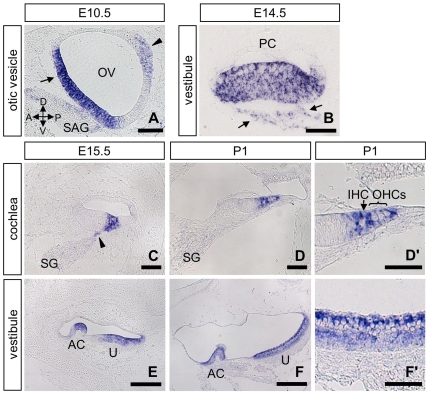
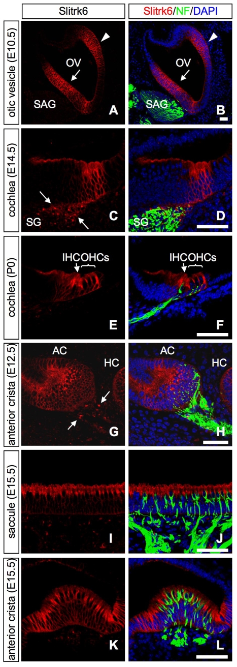
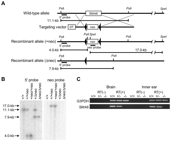
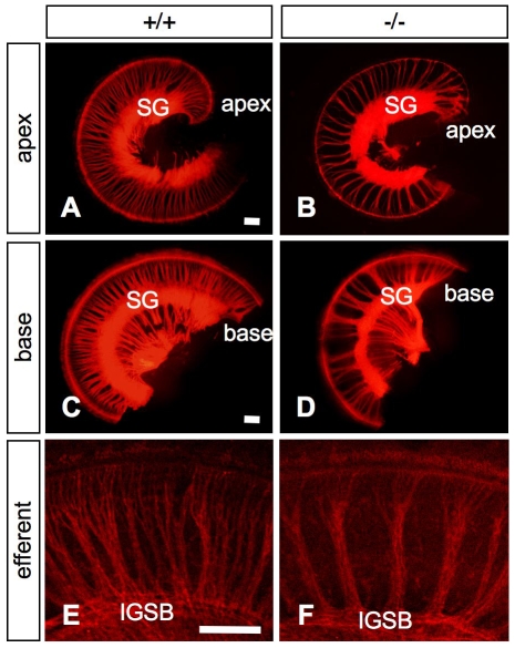
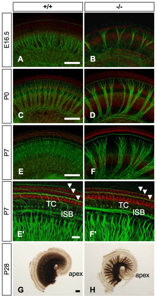
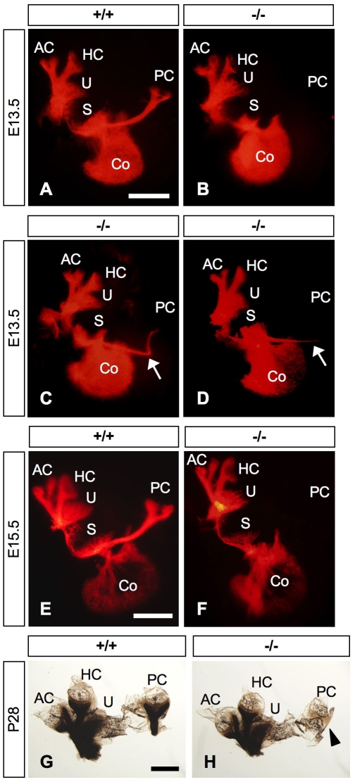
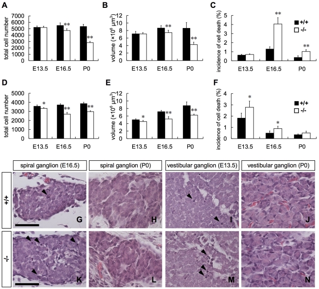
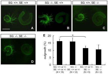
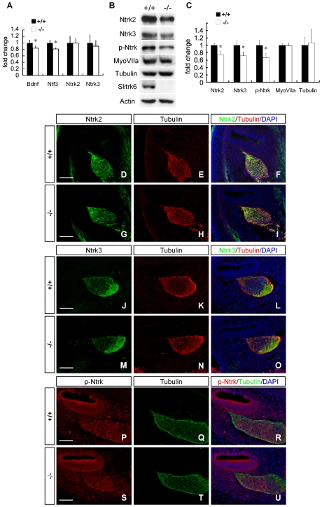
Slitrks are type I transmembrane proteins that share conserved leucine-rich repeat domains similar to those in the secreted axonal guidance molecule Slit. They also show similarities to Ntrk neurotrophin receptors in their carboxy-termini, sharing a conserved tyrosine residue. Among 6 Slitrk family genes in mammals, Slitrk6 has a unique expression pattern, with strong expression in the sensory epithelia of the inner ear. We generated Slitrk6-knockout mice and investigated the development of their auditory and vestibular sensory organs. Slitrk6-deficient mice showed pronounced reduction in the cochlear innervation. In the vestibule, the innervation to the posterior crista was often lost, reduced, or sometimes misguided. These defects were accompanied by the loss of neurons in the spiral and vestibular ganglia. Cochlear sensory epithelia from Slitrk6-knockout mice have reduced ability in promoting neurite outgrowth of spiral ganglion neurons. Indeed the Slitrk6-deficient inner ear showed a mild but significant decrease in the expression of Bdnf and Ntf3, both of which are essential for the innervation and survival of sensory neurons. In addition, the expression of Ntrk receptors, including their phosphorylated forms was decreased in Slitrk6-knockout cochlea. These results suggest that Slitrk6 promotes innervation and survival of inner ear sensory neurons by regulating the expression of trophic and/or tropic factors including neurotrophins from sensory epithelia.


Conflict of interest statement

Competing Interests: The authors have declared that no competing interests exist.

Figures

Figure 1
Figure 1. Expression of Slitrk6 mRNA during inner ear development.
In situ hybridization of Slitrk6 at E10.5 (A), E14.5 (B), E15.5 (C, E), and P1 (D, D', F, F') in the otic vesicle (A), cochlea (C, D, D'), and vestibule (B, E, F, F'). Slitrk6 transcripts are found in the ventromedial (arrow) and laterodorsal (arrowhead) regions of the otic vesicle at E10.5 (A). At E15.5, the expression of Slitrk6 mRNA marks the region of the developing organ of Corti (C). In addition, a faint positive signal is seen in the nascent spiral ganglion neurons adjacent to the sensory epithelium (arrowhead in C). Slitrk6 mRNA is still expressed in the organ of Corti at P1 (D), and higher magnification of the organ of Corti reveals that Slitrk6 transcripts are localized densely in supporting cells and weakly in inner and outer hair cells (D'). Slitrk6 mRNA is also detected in vestibular sensory epithelia, including ampullary cristae and utricular macula (B, E, F). Higher magnification of the utricular epithelium reveals that Slitrk6 transcripts are densely located at the lumenal layer of the sensory epithelium (F'). A faint positive signal is also observed in the nascent vestibular ganglion neurons adjacent to the sensory epithelium (arrows in B). Anterior, posterior, dorsal and ventral directions are indicated by arrows in (A). AC, anterior crista; IHC, inner hair cell; OHC, outer hair cell; OV, otic vesicle; PC, posterior crista; SAG, statoacoustic ganglion; SG, spiral ganglion; U, utricle. Scale bars: A, B, C, D, D', F' 50 µm; E, F, 100 µm.
Figure 2
Figure 2. Expression of Slitrk6 protein during inner ear morphogenesis.
Double immunohistochemistry for Slitrk6 (red; A–L), and neurofilament (NF, green; B, D, F, H, J, L) on transverse sections of the E10.5 otic vesicle (A, B), developing cochlea (C–F), and vestibular (G–L) sensory epithelia. Slitrk6 protein is found in the ventromedial (arrow) and laterodorsal (arrowhead) regions of the otic vesicle at E10.5 (A, B). At E14.5, Slitrk6 is strongly expressed in the area of the presumptive organ of Corti (C, D). The spiral ganglion neurons adjacent to the sensory epithelium are immunolabeled with Slitrk6 in a pattern that partly overlaps with neurofilament (arrows in C). At P0, Slitrk6 expression is confined to the supporting cells within the area of the organ of Corti (E, F). The neurofilament-labeled fibers project below the hair cells. In the macula of the sensory epithelium from an E15.5 saccule, Slitrk6 is uniformly and heavily distributed in the lumenal layer of the epithelium (I, J). In the sensory epithelium from E15.5 anterior crista, Slitrk6 expression is highly observed at the lumenal layer of the striolar region of the crista (K, L). At E12.5, faint Slitrk6-positive signals are also observed in the vestibular ganglion neurons adjacent to the sensory epithelium (arrows in G). DAPI-stained nuclei (blue) are seen in all panels of the merged images (B, D, F, H, J, L). AC, anterior crista; HC, horizontal crista; IHC, inner hair cell; OHC, outer hair cell; OV, otic vesicle; SAG, statoacoustic ganglion; SG, spiral ganglion. Scale bars, 50 µm.
Figure 3
Figure 3. Targeted disruption of the Slitrk6 gene.
(A) Schematic structures of the Slitrk6 genomic locus, targeting vector, and a mutated allele. The locations of the probes for Southern blotting (5′ and neo probes) are shown. DT, diphtheria toxin A; neo, neomycin-resistance gene. (B) Confirmation of homologous recombination of the mutant alleles by Southern blot. (C) RT-PCR performed on cDNAs prepared from brain stem and inner ear of Slitrk6+/+, Slitrk6+/−, and Slitrk6−/− mice at E16.5.
Figure 4
Figure 4. Cochlear innervation defects in Slitrk6-deficient mice.
DiI-traced afferent innervation of apical (A, B) and basal (C, D) turns of the neonatal (P0) cochlea in Slitrk6+/+ (A, C) and Slitrk6−/− (B, D) mice. Slitrk6-deficient inner ear displays a marked alteration in the spacing of radial fibers in the cochlear innervation (B, D). Innervation defects are distributed evenly throughout the cochlea with no basal-to-apical gradient. DiI-labeled efferent innervation of the cochlear middle turn of Slitrk6+/+ (E) and Slitrk6−/− (F) neonatal mice. Efferent fibers show abnormalities similar to those noted for the afferent innervation. IGSB, intraganglionic spiral bundle; SG, spiral ganglion. Scale bars, 100 µm.
Figure 5
Figure 5. Patterns of cochlear innervation in whole-mount surface preparations of the cochlear sensory epithelia of wild-type and Slitrk6-deficient mice.
Immunohistochemistry for neurofilament (green) in the mid-cochlear turn of Slitrk6+/+ (A, C, E) and Slitrk6−/− (B, D, F) mice at E16.5 (A, B), P0 (C, D), and P7 (E, F). Specimens were stained with phalloidin-TRITC (red) to visualize the hair cells. Mutant mice show a marked reduction in the cochlear innervation as compared to wild-type mice. The density of radial fibers is reduced in the absence of Slitrk6. A higher magnification of the P7 cochlea indicates that the spiral fibers navigating in the outer hair cell area are also reduced in knockout mice (arrowheads in E', F'). Myelin-stained flat mounts of Slitrk6+/+ (G) and Slitrk6−/− (H) cochleae at P28. The innervation defect of reduced radial fiber density is prominent in Slitrk6-deficient cochlea. ISB, inner spiral bundle; TC, tunnel of Corti. Scale bars: A, C, E, G, 100 µm; E', 20 µm.
Figure 6
Figure 6. Vestibular innervation defects in Slitrk6-deficient mice.
DiI-labeled flat mounts of Slitrk6+/+ (A, E) and Slitrk6−/− (B–D, F) inner ears at E13.5 (A–D) and E15.5 (E, F). Slitrk6-deficient mice frequently lack innervation to the posterior crista (B, F), although, in some cases, the fiber projections to the posterior crista are misrouted (arrows in C, D). Myelin-stained flat mounts of Slitrk6+/+ (G) and Slitrk6−/− (H) vestibule at P28; the whole innervation to the posterior crista is lacking (arrowhead). AC, anterior crista; Co, cochlea; HC, horizontal crista; PC, posterior crista; S, saccule; U, utricle. Scale bars, 500 µm.
Figure 7
Figure 7. Neuronal loss in the spiral and vestibular ganglia in Slitrk6-deficient mice.
Number of neurons (A, D), volume (B, E), and incidence of cell death (C, F) in the spiral (A–C) and vestibular (D–F) ganglia. Both the number of neurons and volume of the spiral ganglion of Slitrk6-knockout mice were significantly lower than those of wild-type mice at E16.5, and they decreased to about a half of those of wild-type mice at P0 (A, B). Cell death in the spiral ganglion significantly increased in Slitrk6-deficient mice at E16.5 and P0 (C). Both the number of neurons and the volume of the vestibular ganglion were significantly lower in the knockout than in the wild-type from E13.5, and both were about 75% of those of wild-type mice at E16.5 and P0 (D, E). Cell death in the vestibular ganglion was significantly increased in knockout mice at E13.5 and E16.5 (F). Measurements of Slitrk6-deficient vestibular ganglia were made in inner ears without posterior crista innervation. n = 5 to 7 in the spiral ganglion and n = 3 to 5 in the vestibular ganglion. Mean + SD, * p<0.05, ** p<0.01, Student's t-test. H&E-stained spiral ganglia at E16.5 (G, K) and P0 (H, L) and vestibular ganglia at E13.5 (I, M) and P0 (J, N) of Slitrk6+/+ (G–J) and Slitrk6−/− mice (K–N). Pyknotic cells are frequently observed in the spiral ganglion of Slitrk6-deficient mice at E16.5 (arrowheads in G, K). In the vestibular ganglion, the number of pyknotic cells is significantly increased at E13.5 in mutant mice (arrowheads in I, M). At P0, there are no apparent differences between wild-type and knockout mice in cell size and organization in either the spiral or vestibular ganglia (H, J, L, N). Scale bars: 50 µm.
Figure 8
Figure 8. Cochlear sensory epithelia of Slitrk6-knockout mice have less activity in promoting neurite outgrowth of spiral ganglion neurons.
Spiral ganglia (SG) and sensory epithelia (SE) were dissected from E14.5 wild-type and Slitrk6-deficient mice and the explants were cultured. Neurites were visualized by neurofilament immunostaining (green). There were hardly any detectable processes at the beginning of the explant culture. Spiral ganglia of wild-type (A) and Slitrk6-deficient (B) mice can strongly extend their neurites to sensory epithelia obtained from wild-type mice, whereas sensory epithelia of Slitrk6-knockout mice weakly attract neurites of spiral ganglia obtained from both wild-type (C) and knockout mice (D). Scale bar, 400 µm. (E) Measurement of neurite outgrowth (%) of spiral ganglia. The graphs represent mean + SEM. Neurite outgrowth of spiral ganglia was reduced in the presence of Slitrk6-deficient sensory epithelia compared to the presence of wild-type or heterozygous sensory epithelia. * p<0.05, Mann-Whitney's U-test.
Figure 9
Figure 9. Expression of neurotrophins and their receptors was decreased in Slitrk6-deficient inner ear.
(A) Real-time PCR analysis of Bdnf, Ntf3, Ntrk2, and Ntrk3 in E14.5 inner ear (includes cochlear and vestibular sensory epithelia, and spiral and vestibular ganglia). The graphs depict mean + SD of 3 independent analyses. The mRNAs of Bdnf and Ntf3 were significantly decreased in Slitrk6-deficient inner ear, whereas those of Ntrk2 and Ntrk3 were not. * p<0.05, Student's t-test. (B) Western blot of protein samples extracted from E14.5 cochlea (includes cochlear sensory epithelium and spiral ganglion). (C) Both Ntrk2 and Ntrk3 proteins in the cochlea were significantly decreased in Slitrk6-knockout mice. Furthermore, phosphorylated form of Ntrk (p-Ntrk) protein was decreased in the knockout mice. Amounts of a neuronal marker (βIII tubulin) and a sensory epithelium marker (Myosin VIIa) were comparable between the wild-type and knockout mice. The graphs represent mean + SD of 3 independent analyses. * p<0.05, Student's t-test. Immunohistochemistry for Ntrk2 (green; D, G), Ntrk3 (green; J, M), p-Ntrk (red; P, S), and βIII tubulin (red; E, H, K, N and green; Q, T) in mid-turn cochlea of Slitrk6+/+ (D–F, J–L, P–R) and Slitrk6−/− (G–I, M–O, S–U) mice. Specimens were counterstained with DAPI (blue) and their merged images are shown in F, I, L, O, R, U. The spatial localizations of Ntrk2, Ntrk3 and p-Ntrk are not altered in knockout mice. Scale bars: 50 µm.

Similar articles

Cited by

References

    1. Aruga J, Mikoshiba K. Identification and characterization of Slitrk, a novel neuronal transmembrane protein family controlling neurite outgrowth. Mol Cell Neurosci. 2003;24:117–129. - PubMed
    1. Aruga J, Yokota N, Mikoshiba K. Human SLITRK family genes: genomic organization and expression profiling in normal brain and brain tumor tissue. Gene. 2003;315:87–94. - PubMed
    1. Kobe B, Kajava AV. The leucine-rich repeat as a protein recognition motif. Curr Opin Struct Biol. 2001;11:725–732. - PubMed
    1. Brose K, Tessier-Lavigne M. Slit proteins: key regulators of axon guidance, axonal branching, and cell migration. Curr Opin Neurobiol. 2000;10:95–102. - PubMed
    1. Huang EJ, Reichardt LF. Trk receptors: roles in neuronal signal transduction. Annu Rev Biochem. 2003;72:609–642. - PubMed

Publication types