

Additional sex combs-like 1 belongs to the enhancer of trithorax and polycomb group and genetically interacts with Cbx2 in mice
- PMID: 19833123
- PMCID: PMC2807749
- DOI: 10.1016/j.ydbio.2009.10.004
Additional sex combs-like 1 belongs to the enhancer of trithorax and polycomb group and genetically interacts with Cbx2 in mice
Abstract
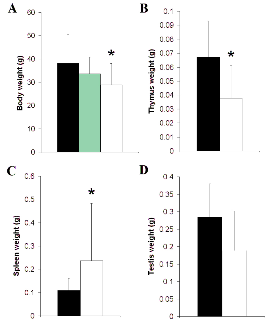
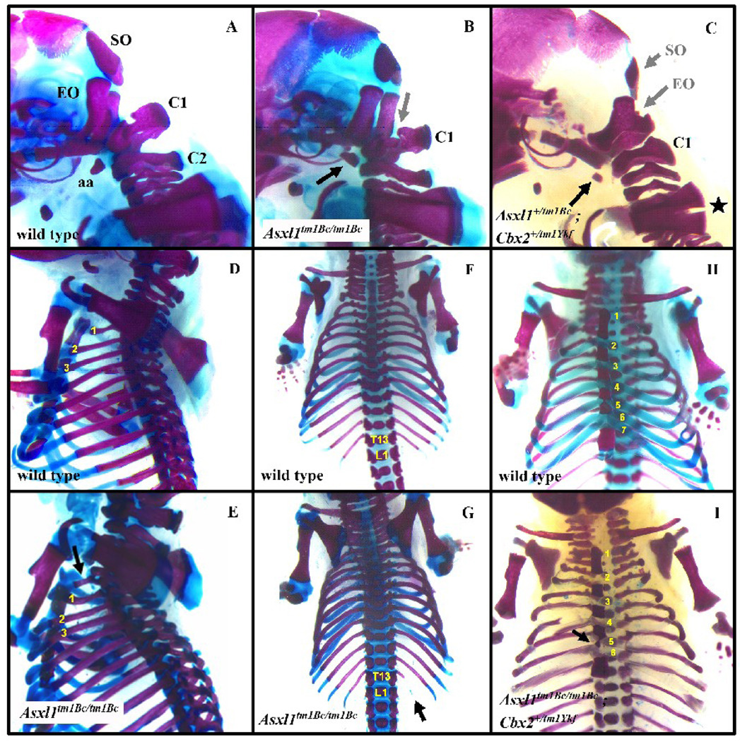
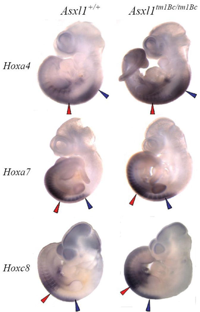
The Additional sex combs (Asx) gene of Drosophila behaves genetically as an enhancer of trithorax and polycomb (ETP) in displaying bidirectional homeotic phenotypes, suggesting that is required for maintenance of both activation and silencing of Hox genes. There are three murine homologs of Asx called Additional sex combs-like1, 2, and 3. Asxl1 is required for normal adult hematopoiesis; however, its embryonic function is unknown. We used a targeted mouse mutant line Asxl1(tm1Bc) to determine if Asxl1 is required to silence and activate Hox genes in mice during axial patterning. The mutant embryos exhibit simultaneous anterior and posterior transformations of the axial skeleton, consistent with a role for Asxl1 in activation and silencing of Hox genes. Transformations of the axial skeleton are enhanced in compound mutant embryos for the polycomb group gene M33/Cbx2. Hoxa4, Hoxa7, and Hoxc8 are derepressed in Asxl1(tm1Bc) mutants in the antero-posterior axis, but Hoxc8 expression is reduced in the brain of mutants, consistent with Asxl1 being required both for activation and repression of Hox genes. We discuss the genetic and molecular definition of ETPs, and suggest that the function of Asxl1 depends on its cellular context.
Figures
Similar articles
-
A role for mel-18, a Polycomb group-related vertebrate gene, during theanteroposterior specification of the axial skeleton.Development. 1996 May;122(5):1513-22. doi: 10.1242/dev.122.5.1513. Development. 1996. PMID: 8625838
-
Functional conservation of Asxl2, a murine homolog for the Drosophila enhancer of trithorax and polycomb group gene Asx.PLoS One. 2009;4(3):e4750. doi: 10.1371/journal.pone.0004750. Epub 2009 Mar 9. PLoS One. 2009. PMID: 19270745 Free PMC article.
-
The Additional sex combs gene of Drosophila is required for activation and repression of homeotic loci, and interacts specifically with Polycomb and super sex combs.Mol Gen Genet. 1999 Jun;261(4-5):753-61. doi: 10.1007/s004380050018. Mol Gen Genet. 1999. PMID: 10394912
-
The Polycomb and trithorax group proteins of Drosophila: trans-regulators of homeotic gene function.Annu Rev Genet. 1995;29:289-303. doi: 10.1146/annurev.ge.29.120195.001445. Annu Rev Genet. 1995. PMID: 8825476 Review.
-
[Role of ASXL1 mutation in myeloid malignancies].Zhongguo Shi Yan Xue Ye Xue Za Zhi. 2014 Aug;22(4):1183-7. doi: 10.7534/j.issn.1009-2137.2014.04.057. Zhongguo Shi Yan Xue Ye Xue Za Zhi. 2014. PMID: 25130853 Review. Chinese.
Cited by
-
Rapid-throughput skeletal phenotyping of 100 knockout mice identifies 9 new genes that determine bone strength.PLoS Genet. 2012;8(8):e1002858. doi: 10.1371/journal.pgen.1002858. Epub 2012 Aug 2. PLoS Genet. 2012. PMID: 22876197 Free PMC article.
-
The HARE-HTH and associated domains: novel modules in the coordination of epigenetic DNA and protein modifications.Cell Cycle. 2012 Jan 1;11(1):119-31. doi: 10.4161/cc.11.1.18475. Epub 2012 Jan 1. Cell Cycle. 2012. PMID: 22186017 Free PMC article.
-
Additional sex combs-like 2 is required for polycomb repressive complex 2 binding at select targets.PLoS One. 2013 Sep 9;8(9):e73983. doi: 10.1371/journal.pone.0073983. eCollection 2013. PLoS One. 2013. PMID: 24040135 Free PMC article.
-
Myelodysplastic syndromes are induced by histone methylation–altering ASXL1 mutations.J Clin Invest. 2013 Nov;123(11):4627-40. doi: 10.1172/JCI70739. J Clin Invest. 2013. PMID: 24216483 Free PMC article.
-
BAP1 enhances Polycomb repression by counteracting widespread H2AK119ub1 deposition and chromatin condensation.Mol Cell. 2021 Sep 2;81(17):3526-3541.e8. doi: 10.1016/j.molcel.2021.06.020. Epub 2021 Jun 28. Mol Cell. 2021. PMID: 34186021 Free PMC article.
References
-
- Ayton P, Sneddon SF, Palmer DB, Rosewell IR, Owen MJ, Young B, Presley R, Subramanian V. Truncation of the Mll gene in exon 5 by gene targeting leads to early preimplantation lethality of homozygous embryos. Genesis. 2001;30:201–212. - PubMed
-
- Bel-Vialar S, Core N, Terranova R, Goudot V, Boned A, Djabali M. Altered retinoic acid sensitivity and temporal expression of Hox genes in polycomb-M33-deficient mice. Dev Biol. 2000;224:238–249. - PubMed
-
- Belo JA, Bouwmeester T, Leyns L, Kertesz N, Gallo M, Follettie M, De Robertis EM. Cerberus-like is a secreted factor with neutralizing activity expressed in the anterior primitive endoderm of the mouse gastrula. Mech Dev. 1997;68:45–57. - PubMed
-
- Bernstein BE, Mikkelsen TS, Xie X, Kamal M, Huebert DJ, Cuff J, Fry B, Meissner A, Wernig M, Plath K, Jaenisch R, Wagschal A, Feil R, Schreiber SL, Lander ES. A bivalent chromatin structure marks key developmental genes in embryonic stem cells. Cell. 2006;125:315–326. - PubMed
Publication types
MeSH terms
Substances
Grants and funding
LinkOut - more resources
Full Text Sources
Molecular Biology Databases
