. 2009 Feb;84(2):162-77.
doi: 10.1016/j.ajhg.2008.12.018. Epub 2009 Feb 5.

A missense mutation in CASK causes FG syndrome in an Italian family

Affiliations

A missense mutation in CASK causes FG syndrome in an Italian family

Giulio Piluso et al. Am J Hum Genet. 2009 Feb.

Abstract

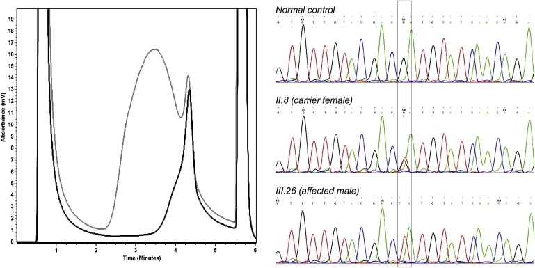
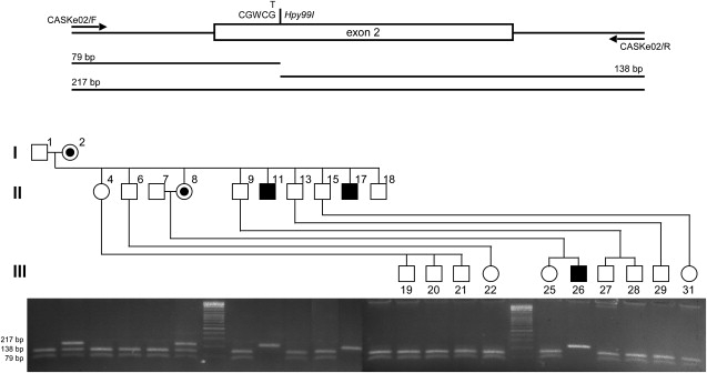
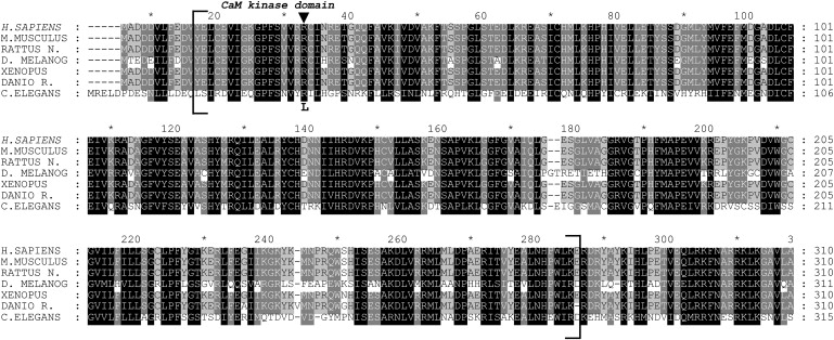
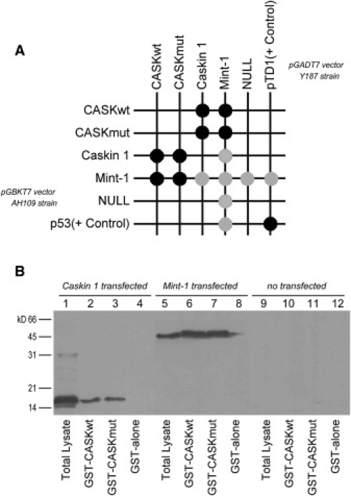
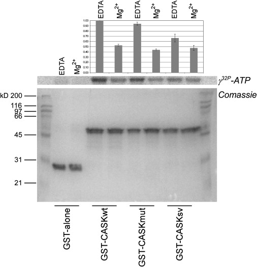
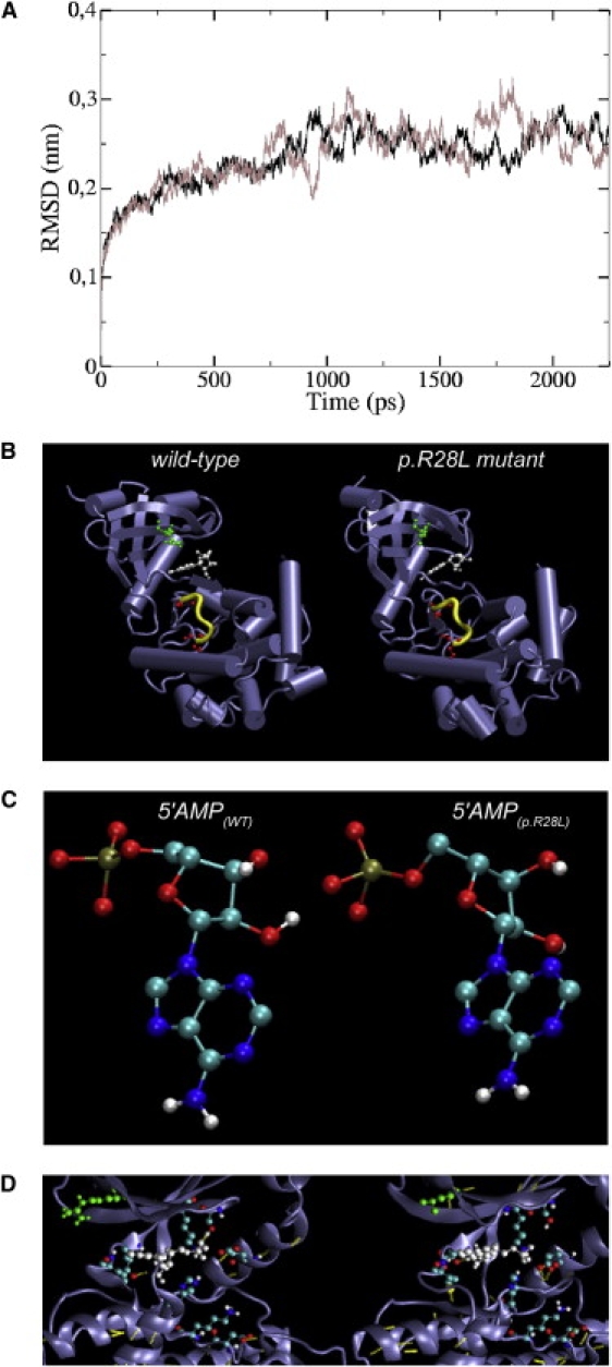
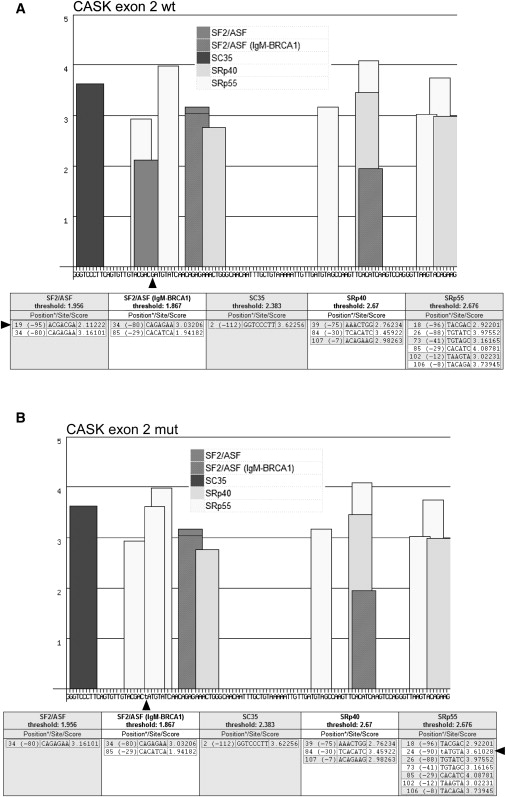
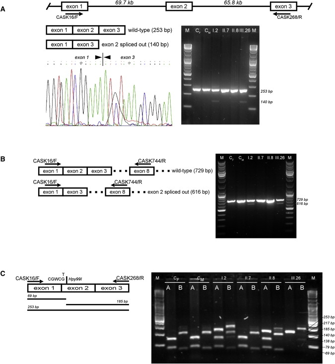
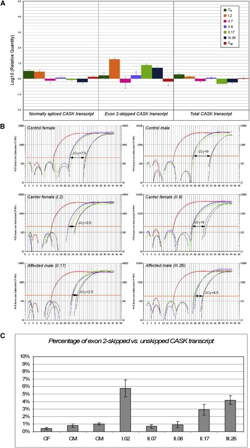
First described in 1974, FG syndrome (FGS) is an X-linked multiple congenital anomaly/mental retardation (MCA/MR) disorder, characterized by high clinical variability and genetic heterogeneity. Five loci (FGS1-5) have so far been linked to this phenotype on the X chromosome, but only one gene, MED12, has been identified to date. Mutations in this gene account for a restricted number of FGS patients with a more distinctive phenotype, referred to as the Opitz-Kaveggia phenotype. We report here that a p.R28L (c.83G-->T) missense mutation in CASK causes FGS phenotype in an Italian family previously mapped to Xp11.4-p11.3 (FGS4). The identified missense mutation cosegregates with the phenotype in this family and is absent in 1000 control X chromosomes of the same ethnic origin. An extensive analysis of CASK protein functions as well as structural and dynamic studies performed by molecular dynamics (MD) simulation did not reveal significant alterations induced by the p.R28L substitution. However, we observed a partial skipping of the exon 2 of CASK, presumably a consequence of improper recognition of exonic splicing enhancers (ESEs) induced by the c.83G-->T transversion. CASK is a multidomain scaffold protein highly expressed in the central nervous system (CNS) with specific localization to the synapses, where it forms large signaling complexes regulating neurotransmission. We suggest that the observed phenotype is most likely a consequence of an altered CASK expression profile during embryogenesis, brain development, and differentiation.


Figures

Figure 1
Figure 1
DHPLC Analysis and Characterization of the c.83G→T Mutation Identified in CASK In the chromatogram on the left, the elution profile of a normal control (black curve) is compared with the mutant profile (gray curve). On the right, electropherograms from a normal control male, a carrier female (II.8), and an affected male (III.26) from pedigree are shown. The nucleotide change is boxed.
Figure 2
Figure 2
The c.83G→T Mutation Fully Cosegregates with the Phenotype in Our FGS Family CASK exon 2 and intronic flanking regions were amplified by PCR in DNA samples from all the individuals in the pedigree and digested by Hpy99I. This restriction site is lost when the c.83G→T substitution is present. Agarose gel electrophoresis of Hpy99I-digested PCR products clearly indicates that the c.83G→T mutation is present only in the affected males (hemizygotes; 217-bp-long band) and carrier females (heterozygotes; 79-, 138-, and 217-bp-long bands) and is absent in all the other individuals from pedigree (79- and 138-bp-long bands).
Figure 3
Figure 3
Alignment of CaM-Kinase Domain from Different Hortologs of CASK The protein sequences of CASK from H. sapiens (NP_003679), M. musculus (NP_033936), R. norvegicus (NP_071520), D. melanogaster (NP_524441), X. tropicalis (NP_001016204), D. rerio (NP_694420), and C. elegans (NP_001024587) were aligned with Clustal W software. Amino acid conservation was highlighted with GeneDoc software. Only the N terminus region is shown and CaM-kinase domain of CASK is enclosed in square brackets. An arrow indicates the position of p.R28L substitution.
Figure 4
Figure 4
In Vivo and In Vitro Binding Assays of CaM-Kinase Domain of CASK with the Interacting Domains of Mint-1 and Caskin 1 (A) By yeast two-hybrid system, the CaM-kinase domain of CASK interacted with these partners and the p.R28L substitution did not modify binding properties. In the grid, strong and weak growth is indicated with black and gray disks, respectively. A mild autoactivation of reporter genes was observed for Mint-1. The p53 plasmid encoding murine p53 fused to GAL4 DNA-binding domain and the pTD1 plasmid encoding SV40 large T-antigen fused to GAL4 activating domain were used as positive control. pGBKT7 and pGADT7 empty plasmids were used as negative control. (B) For GST pull-down assays, the interacting domains of Caskin 1 and Mint-1, N-terminal labeled with c-Myc epitope tag, were transiently expressed in COS-7 cells. Protein extracts from Caskin 1-transfected cells (lanes 1–4), Mint-1-transfected cells (lanes 5–8), and untransfected cells (lanes 9–12) were incubated with GST bead-bound fusion proteins: GST-CASKwt, GST-CASKmut, and GST alone. Western blotting analysis with an anti-Myc monoclonal antibody did not reveal differences in binding properties of CaM-kinase domain of CASK as a result of p.R28L substitution.
Figure 5
Figure 5
In Vitro Autophosphorylation Assay of CASK CaM-Kinase Domain All the experiments were carried out as described. Equimolar quantities of purified GST bead-bound fusion proteins (GST alone, GST-CASKwt, GST-CASKmut, and GST-CASKsv) were incubated with [γ-32P]ATP (specific activity: 2 × 10⁷ cpm) in Tris-KCl buffer under two different conditions: 2 mM EDTA or 4 mM Mg2+. The panel (from bottom) depicts Coomassie blue stain of SDS gel and related autoradiogram. Normalized values of incorporated radioactivity were reported in the upper bar graph where error bars indicate ±SD. When compared with the wild-type (CASKwt) and hypomorphic mutant (CASKsv) of CASK CaM-kinase domain, p.R28L mutation (CASKmut) did not seem to generate significant variations in autophosphorylation of CaM-kinase domain.
Figure 6
Figure 6
Molecular Dynamics Simulation (A) Structural drift of wild-type (black line) and p.R28L mutant CaM-kinase domain of CASK (gray line) is shown as root mean square deviation of all atoms starting from crystallographic coordinates after energy minimization. (B) Comparison between 3D structures of the wild-type (left) and p.R28L mutant (right) CaM-kinase domain of CASK obtained from the 2.5 ns MD simulations at 300 K. The residue at position 28, in which the mutation occurs, was colored in green, the AMPPNP ligand was colored in white, and the serine residues at position 147 and 151, targets of autophosphorylation activity, were colored in red. The residues 51–54 that showed small differences in secondary structure were highlighted in yellow. (C) The conformation adopted by the 5′AMP portion of the AMPPNP ligand is shown for wild-type (left) and p.R28L mutant (right). (D) A detailed view of the nucleotide-binding pocket of CASK CaM-kinase domain in complex with AMPPNP is shown. The color scheme used in (B) is conserved. In addition, side chains of residues involved in AMPPNP binding and H bonds (yellow dots) are shown.
Figure 7
Figure 7
Analysis of the 113-bp-Long Coding Sequence of CASK Exon 2 via ESEfinder Software As suggested by the authors, default threshold values were used. The bar graphs and tables below depict the ESE motifs for different SR proteins (SF2/ASF, SC35, SRp40, and SRp55) found in wild-type (A) and c.83G→T mutant (B) CASK exon 2 sequence. In the tables, both positions () from 5′ end (through 1) and 3′ end (through −1) are given. The arrows indicate the position of nucleotide substitution and ESE motif lost or acquired as a result of the change.
Figure 8
Figure 8
RT-PCR Analysis of CASK Transcripts (A) The CASK cDNA fragment covering exons 1–3 was amplified by RT-PCR. The expected 253-bp-long PCR product was observed in all samples analyzed by agarose gel electrophoresis (right). cDNA samples are indicated with respect to their position on pedigree (Figure 2). CM and CF indicate control male or female, respectively. A 140-bp-long PCR product, corresponding to the CASK exon 2-skipped form, was observed, at low levels, only in the carrier female I.2 and the affected male III.26. The same result was also obtained with the other affected males (II.11 and II.17) from pedigree (data not shown). The band was undetectable in the other carrier female (II.8), mother of III.26. The skipping of CASK exon 2 was confirmed by direct sequencing of 140-bp-long PCR fragment (left). (B) Different primer pairs were used to confirm the CASK exon-2 skipped transcript in the same sample panel. The 616-bp-long PCR product corresponding to the CASK exon-2 skipped transcript form was observed only in the carrier female I.2 and the affected male III.26. (C) Hpy99I digestion of CASK exon 2 and its intronic flanking regions, as previously depicted in Figure 2, is combined with the Hpy99I digestion of CASK cDNA fragment covering exons 1–3. For the same sample panel, agarose gel electrophoresis of Hpy99I-digested genomic (A) and cDNA (B) PCR products are shown. For carrier females (I.2 and II.8), heterozygosity is confirmed (lanes A), but cDNA PCR products (lanes B) present a different digestion pattern in which 253- and 185-bp-long fragments show inverted intensity of the bands. This indicates that the wild-type allele is preferentially used by II.8 in CASK transcription.
Figure 9
Figure 9
Relative Quantification of CASK Expression The analyzed cDNA samples are indicated with respect to their position on pedigree (Figure 2). CM and CF indicate control male or female, respectively. (A) The bar graph shows relative quantification of the expression of different CASK transcripts. The expression of the exon 2-skipped transcript (center), amplified with CASKsp1-2/F and CASK268/R primer pair, is significantly increased in I.2 (carrier female) as well as in II.17 and III.26 (affected males). For II.8 (carrier female), this aberrant transcript is undetectable, even considering the wide interval of error bar ±MSD). Expression levels of CASK (right) detected with the CASK2310/F and CASK2545/R primer pair are reduced in the affected males (II.17 and III.26). The expression of the unskipped CASK transcript (left), specifically amplified with CASKsp1-3/F and CASK398/R primer pair, is increased in carrier female II.8. (B) Amplification plots describing the kinetics of PCR reactions for different targets and samples are presented. Differently colored curves correspond to GAPDH (red), unskipped CASK transcript (blue), CASK exon 2-skipped transcript (black), and total CASK transcript (green). The ΔCT obtained by subtracting the CT values of the unskipped and exon 2-skipped CASK forms is shown. (C) After normalization, expression levels of the unskipped CASK transcript were compared to those of the exon 2-skipped form. For each sample, the bar graph shows the percentage accounting for the CASK exon 2-skipped transcript ranging between 3% and 6% in affected males (II.17 and III.26) and carrier female (I.2).

Similar articles

Cited by

References

    1. Opitz J.M., Kaveggia E.G. Studies of malformation syndromes of man 33: the FG syndrome. An X- linked recessive syndrome of multiple congenital anomalies and mental retardation. Z. Kinderheilkd. 1974;117:1–18. - PubMed
    1. Opitz J.M., Richieri-da Costa A., Aase J.M., Benke P.J. FG syndrome update 1988: note of 5 new patients and bibliography. Am. J. Med. Genet. 1988;30:309–328. - PubMed
    1. Romano C., Baraitser M., Thompson E. A clinical follow-up of British patients with FG syndrome. Clin. Dysmorphol. 1994;3:104–114. - PubMed
    1. Thompson E.M., Baraitser M., Lindenbaum R.H., Zaidi Z.H., Kroll J.S. The FG syndrome: 7 new cases. Clin. Genet. 1985;27:582–594. - PubMed
    1. Neri G., Blumberg B., Miles P.V., Opitz J.M. Sensorineural deafness in the FG syndrome: report on four new cases. Am. J. Med. Genet. 1984;19:369–377. - PubMed

Publication types

LinkOut - more resources