

Mutation in BAG3 causes severe dominant childhood muscular dystrophy
- PMID: 19085932
- PMCID: PMC2639628
- DOI: 10.1002/ana.21553
Mutation in BAG3 causes severe dominant childhood muscular dystrophy
Abstract
Objective: Myofibrillar myopathies (MFMs) are morphologically distinct but genetically heterogeneous muscular dystrophies in which disintegration of Z disks and then of myofibrils is followed by ectopic accumulation of multiple proteins. Cardiomyopathy, neuropathy, and dominant inheritance are frequent associated features. Mutations in alphaB-crystallin, desmin, myotilin, Zasp, or filamin-C can cause MFMs and were detected in 32 of 85 patients of the Mayo MFM cohort. Bag3, another Z-disk-associated protein, has antiapoptotic properties, and its targeted deletion in mice causes fulminant myopathy with early lethality. We therefore searched for mutations in BAG3 in 53 unrelated MFM patients.
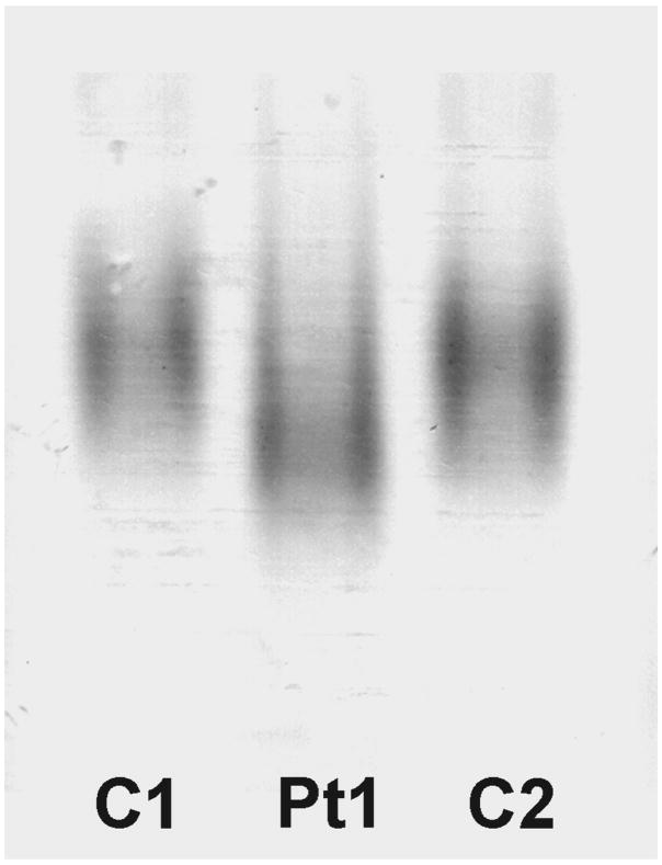
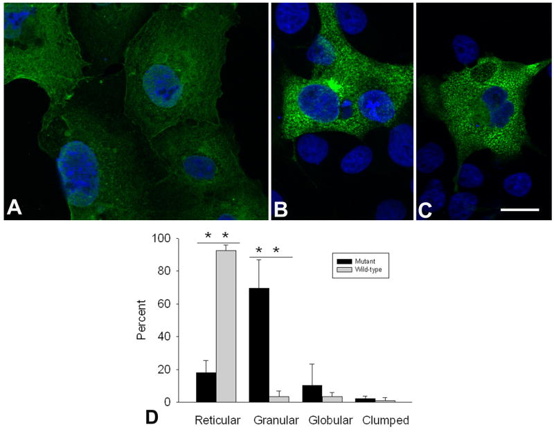
Methods: We searched for mutations in BAG3 by direct sequencing. We analyzed structural changes in muscle by histochemistry, immunocytochemistry, and electron microscopy, examined mobility of the mutant Bag3 by nondenaturing electrophoresis, and searched for abnormal aggregation of the mutant protein in COS-7 (SV-40 transformed monkey kidney fibroblast-7) cells.
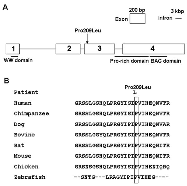
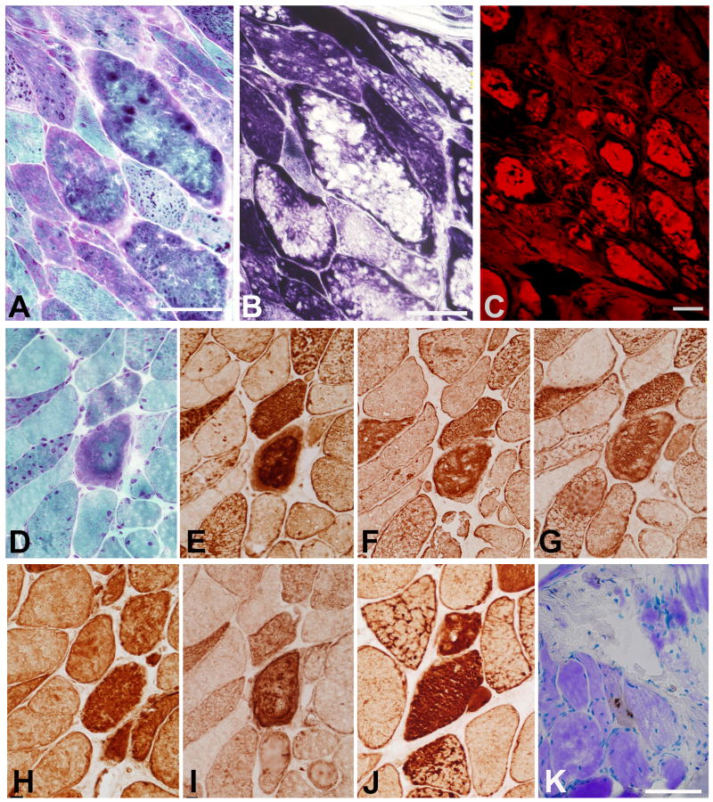
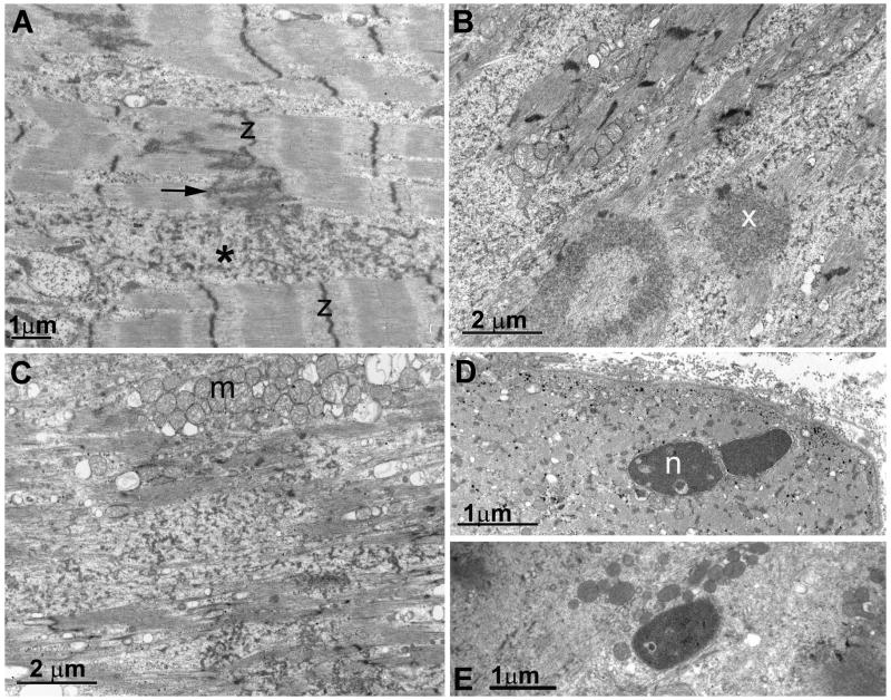
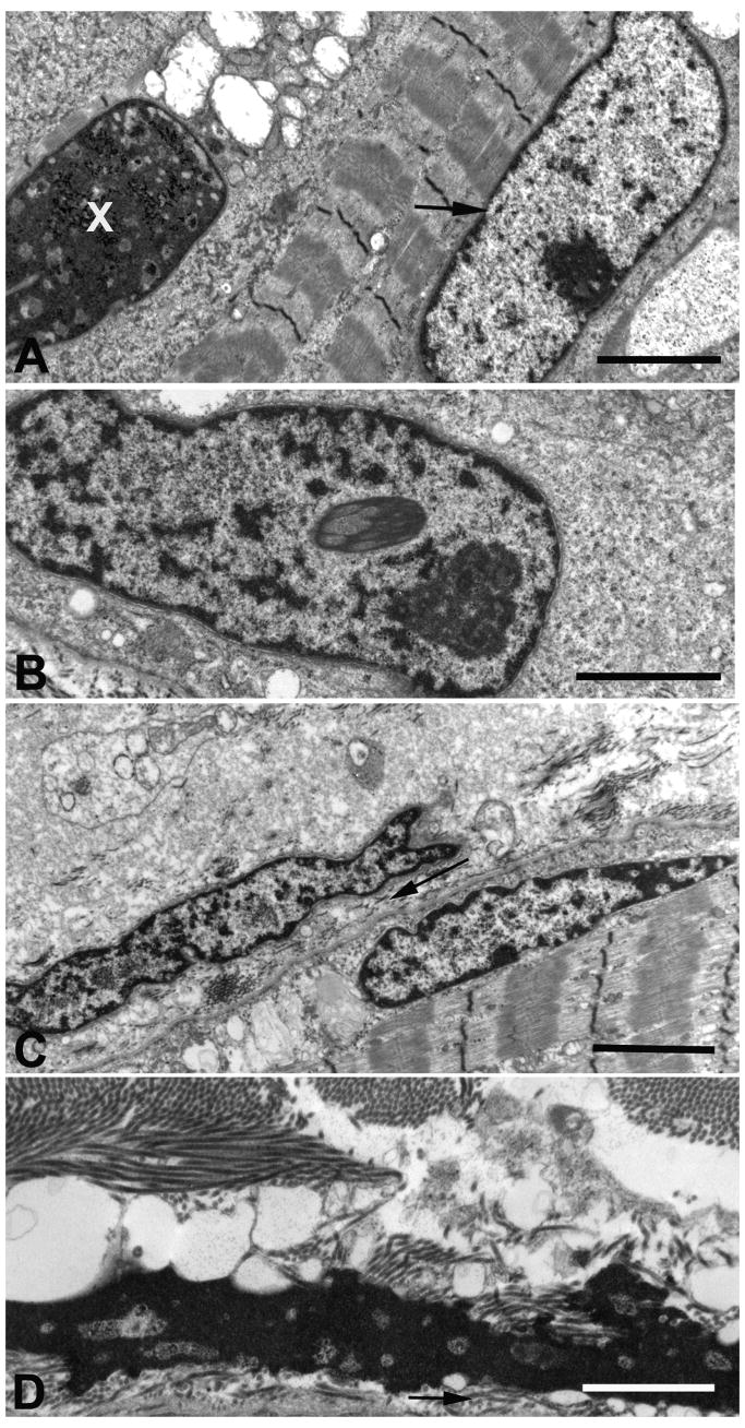
Results: We identified a heterozygous p.Pro209Leu mutation in three patients. All presented in childhood, had progressive limb and axial muscle weakness, and experienced development of cardiomyopathy and severe respiratory insufficiency in their teens; two had rigid spines, and one a peripheral neuropathy. Electron microscopy showed disintegration of Z disks, extensive accumulation of granular debris and larger inclusions, and apoptosis of 8% of the nuclei. On nondenaturing electrophoresis of muscle extracts, the Bag3 complex migrated faster in patient than control extracts, and expression of FLAG-labeled mutant and wild-type Bag3 in COS cells showed abnormal aggregation of the mutant protein.
Interpretation: We conclude mutation in Bag3 defines a novel severe autosomal dominant childhood muscular dystrophy.
Figures
Similar articles
-
Mutations in ZASP define a novel form of muscular dystrophy in humans.Ann Neurol. 2005 Feb;57(2):269-76. doi: 10.1002/ana.20376. Ann Neurol. 2005. PMID: 15668942
-
Myofibrillar myopathies.Curr Opin Neurol. 2010 Oct;23(5):477-81. doi: 10.1097/WCO.0b013e32833d38b0. Curr Opin Neurol. 2010. PMID: 20664348 Review.
-
Clinical, morphological and genetic studies in a cohort of 21 patients with myofibrillar myopathy.Acta Myol. 2011 Oct;30(2):121-6. Acta Myol. 2011. PMID: 22106715 Free PMC article.
-
Reducing bodies and myofibrillar myopathy features in FHL1 muscular dystrophy.Neurology. 2011 Nov 29;77(22):1951-9. doi: 10.1212/WNL.0b013e31823a0ebe. Epub 2011 Nov 16. Neurology. 2011. PMID: 22094483 Free PMC article.
-
Myofibrillar myopathies.Neuromuscul Disord. 2011 Mar;21(3):161-71. doi: 10.1016/j.nmd.2010.12.007. Epub 2011 Jan 20. Neuromuscul Disord. 2011. PMID: 21256014 Free PMC article. Review.
Cited by
-
Two desmin gene mutations associated with myofibrillar myopathies in Polish families.PLoS One. 2014 Dec 26;9(12):e115470. doi: 10.1371/journal.pone.0115470. eCollection 2014. PLoS One. 2014. PMID: 25541946 Free PMC article.
-
The role of BAG3 in health and disease: A "Magic BAG of Tricks".J Cell Biochem. 2022 Jan;123(1):4-21. doi: 10.1002/jcb.29952. Epub 2021 May 14. J Cell Biochem. 2022. PMID: 33987872 Free PMC article. Review.
-
A Crucial Role for the Protein Quality Control System in Motor Neuron Diseases.Front Aging Neurosci. 2020 Jul 21;12:191. doi: 10.3389/fnagi.2020.00191. eCollection 2020. Front Aging Neurosci. 2020. PMID: 32792938 Free PMC article.
-
Exome sequencing reveals DNAJB6 mutations in dominantly-inherited myopathy.Ann Neurol. 2012 Mar;71(3):407-16. doi: 10.1002/ana.22683. Epub 2012 Feb 14. Ann Neurol. 2012. PMID: 22334415 Free PMC article.
-
Evaluating pathogenicity of rare variants from dilated cardiomyopathy in the exome era.Circ Cardiovasc Genet. 2012 Apr 1;5(2):167-74. doi: 10.1161/CIRCGENETICS.111.961805. Epub 2012 Feb 15. Circ Cardiovasc Genet. 2012. PMID: 22337857 Free PMC article.
References
-
- Goldfarb LG, Park KY, Cervenákova L, et al. Missense mutations in desmin associated with familial cardiac and skeletal myopathy. Nat Genet. 1998;19:402–403. - PubMed
-
- Vicart P, Caron A, Guicheney P, et al. A missense mutation in the αB-crystallin chaperone gene causes a desmin-related myopathy. Nat Genet. 1998;20:92–95. - PubMed
-
- Selcen D, Ohno K, Engel AG. Myofibrillar myopathy. Clinical, morphological, and genetic studies in 63 patients. Brain. 2004;127:439–451. - PubMed
-
- Selcen D, Engel AG. Mutations in myotilin cause myofibrillar myopathy. Neurology. 2004;62:1363–1371. - PubMed
-
- Olive M, Goldfarb LG, Shatunov A, et al. Myotilinopathy: refining the clinical and myopathological phenotype. Brain. 2005;128:2315–2326. - PubMed
Publication types
MeSH terms
Substances
Grants and funding
LinkOut - more resources
Full Text Sources
Other Literature Sources
Medical
Molecular Biology Databases
Miscellaneous
