

MR imaging characteristics and neuropathology of the spinal cord in adult-onset autosomal dominant leukodystrophy with autonomic symptoms
- PMID: 18945794
- PMCID: PMC7051393
- DOI: 10.3174/ajnr.A1354
MR imaging characteristics and neuropathology of the spinal cord in adult-onset autosomal dominant leukodystrophy with autonomic symptoms
Abstract
Background and purpose: MR imaging findings in adult-onset autosomal dominant leukodystrophy (ADLD) with autonomic symptoms have been described in the brain, but no descriptions of MR imaging findings in the spinal cord have been published. Here, we describe MR imaging findings in the spinal cord in adult-onset ADLD with autonomic symptoms and histopathologic investigations of the spinal cord.
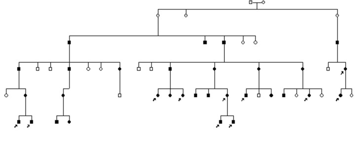
Materials and methods: Twelve subjects from 2 families with adult-onset ADLD with autonomic symptoms identified by clinical investigation underwent MR imaging examination of the spinal cord. Sagittal and transverse sections were obtained. MR imaging examination of the brain was performed in 11 patients. One of the patients underwent postmortem examination, and the spinal cord was subjected to histopathologic analysis.
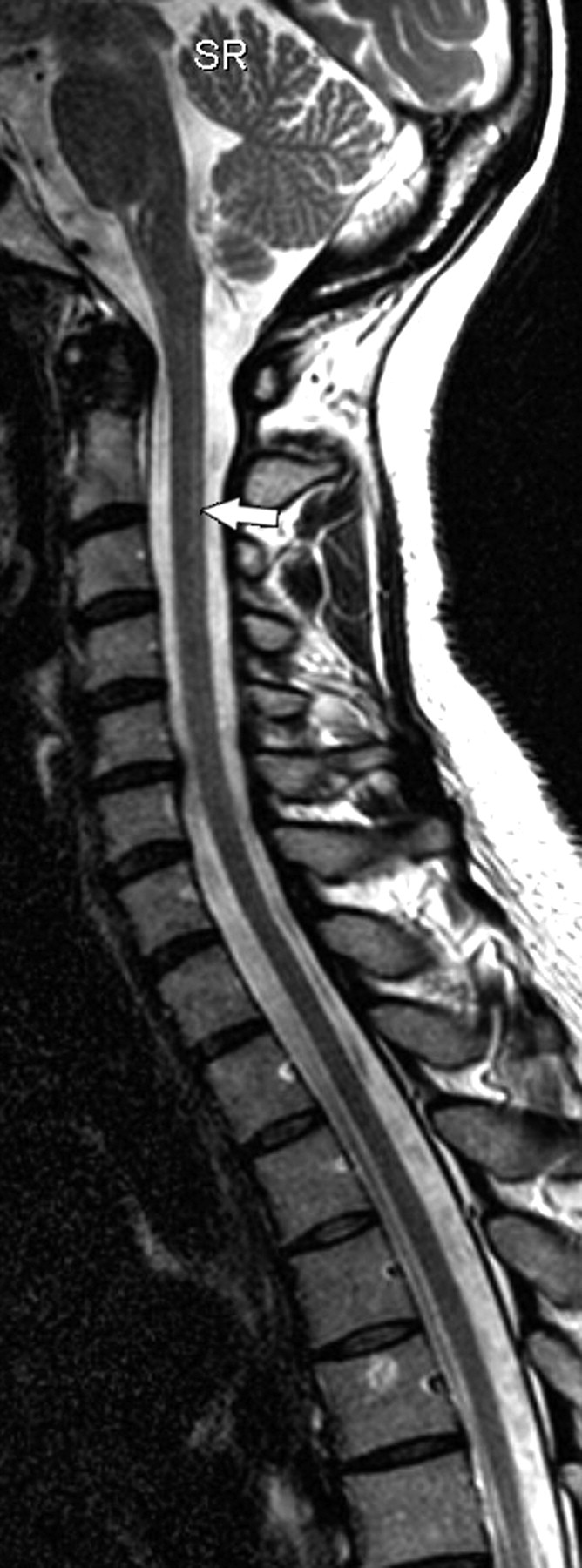
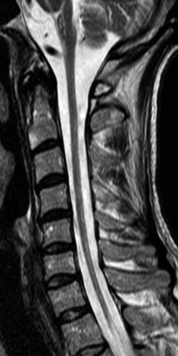
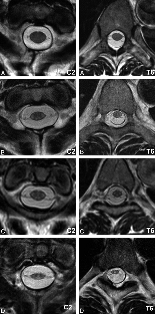
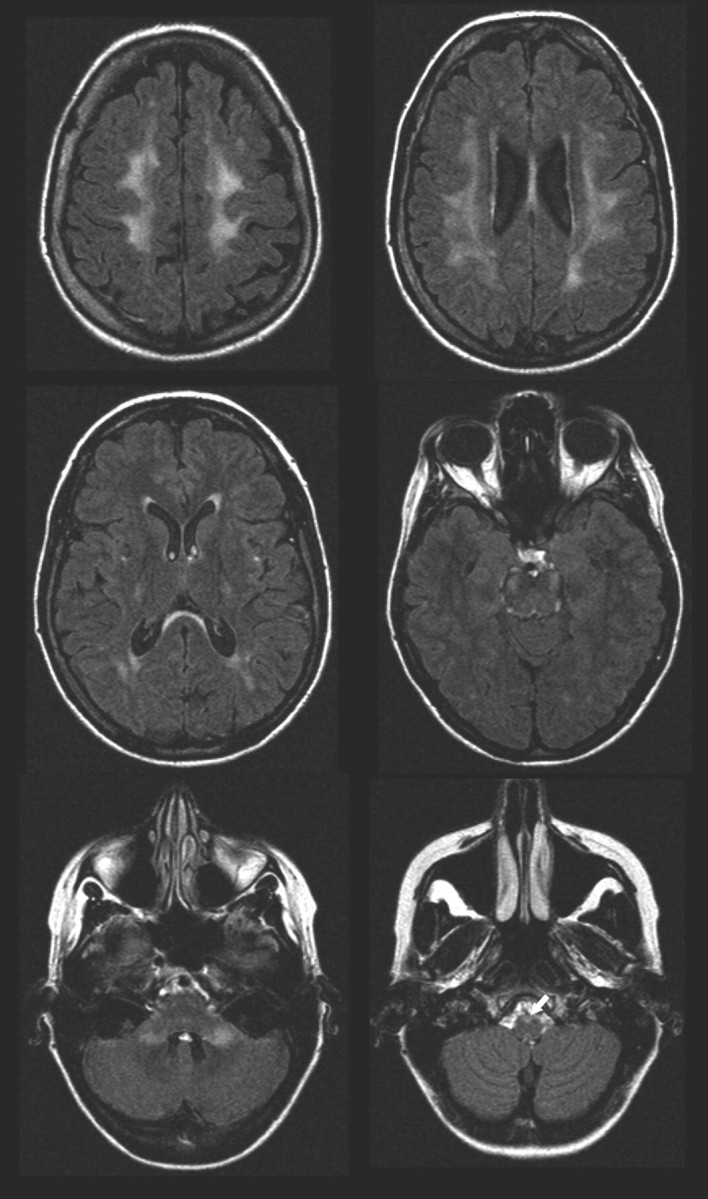
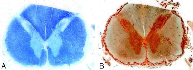
Results: In all family members with adult-onset ADLD with autonomic symptoms, even in the asymptomatic person, the spinal cord was thin. All examined family members also had a slight general white matter signal intensity (SI) increase in the whole spinal cord, mainly visible in T2-weighted transverse images. The pathologic examination revealed a discrete demyelination in the spinal cord. Brain MR imaging also showed increased T2 SI in the white matter.
Conclusions: The spinal cord is affected in adult-onset ADLD with autonomic symptoms. Findings consist of atrophy and a diffuse T2 SI increase in the white matter. Transverse images are needed to assess these findings. The typical SI changes of the spinal cord are also present in subjects without clinical symptoms of the disease and with very limited changes in the brain.
Figures

Similar articles
-
Genomic duplications mediate overexpression of lamin B1 in adult-onset autosomal dominant leukodystrophy (ADLD) with autonomic symptoms.Neurogenetics. 2011 Feb;12(1):65-72. doi: 10.1007/s10048-010-0269-y. Epub 2011 Jan 12. Neurogenetics. 2011. PMID: 21225301
-
Brain magnetic resonance metabolic and microstructural changes in adult-onset autosomal dominant leukodystrophy.Brain Res Bull. 2015 Aug;117:24-31. doi: 10.1016/j.brainresbull.2015.07.002. Epub 2015 Jul 17. Brain Res Bull. 2015. PMID: 26189928
-
Adult-onset autosomal dominant leukodystrophy without early autonomic dysfunctions linked to lamin B1 duplication: a phenotypic variant.J Neurol. 2013 Aug;260(8):2124-9. doi: 10.1007/s00415-013-6958-3. Epub 2013 May 17. J Neurol. 2013. PMID: 23681646
-
[A Japanese family with probably autosomal dominant adult-onset leukodystrophy].Rinsho Shinkeigaku. 1996 Aug;36(8):968-72. Rinsho Shinkeigaku. 1996. PMID: 8958750 Review. Japanese.
-
Magnetic resonance findings in leucodystrophies and MS.Int MS J. 2009 Jun;16(2):47-56. Int MS J. 2009. PMID: 19671368 Review.
Cited by
-
An LMNB1 Duplication Caused Adult-Onset Autosomal Dominant Leukodystrophy in Chinese Family: Clinical Manifestations, Neuroradiology and Genetic Diagnosis.Front Mol Neurosci. 2017 Jul 18;10:215. doi: 10.3389/fnmol.2017.00215. eCollection 2017. Front Mol Neurosci. 2017. PMID: 28769756 Free PMC article.
-
Analysis of LMNB1 duplications in autosomal dominant leukodystrophy provides insights into duplication mechanisms and allele-specific expression.Hum Mutat. 2013 Aug;34(8):1160-71. doi: 10.1002/humu.22348. Epub 2013 May 28. Hum Mutat. 2013. PMID: 23649844 Free PMC article.
-
B-type lamins in health and disease.Semin Cell Dev Biol. 2014 May;29(100):158-63. doi: 10.1016/j.semcdb.2013.12.012. Epub 2013 Dec 28. Semin Cell Dev Biol. 2014. PMID: 24380701 Free PMC article. Review.
-
'Grasshopper sign': the novel imaging of post-COVID-19 myelopathy with delayed longitudinal white matter abnormalities.BMJ Neurol Open. 2024 Jun 12;6(1):e000730. doi: 10.1136/bmjno-2024-000730. eCollection 2024. BMJ Neurol Open. 2024. PMID: 38884066 Free PMC article.
-
Adult-onset autosomal dominant leukodystrophy and neuronal intranuclear inclusion disease: lessons from two new Chinese families.Neurol Sci. 2022 Aug;43(8):1-9. doi: 10.1007/s10072-022-06057-0. Epub 2022 Apr 14. Neurol Sci. 2022. PMID: 35419641
References
-
- Garbern J. Leukodystrophies. In: Lynch DR, ed. Neurogenetics: Scientific and Clinical Advances. New York, NY: Taylor & Francis;2006. :469–554
-
- Eldridge R, Anayiotos CP, Schlesinger S, et al. Hereditary adult-onset leukodystrophy simulating chronic progressive multiple sclerosis. N Engl J Med 1984;311:948–53 - PubMed
-
- Abe K, Ikeda M, Watase K, et al. A kindred of hereditary adult-onset leukodystrophy with sparing of the optic radiations. Neuroradiology 1993;35:281–83 - PubMed
-
- Quattrocolo G, Leombruni S, Vaula G, et al. Autosomal dominant late-onset leukoencephalopathy: clinical report of a new Italian family. Eur Neurol 1997;37:53–61 - PubMed
Publication types
MeSH terms
LinkOut - more resources
Full Text Sources
Medical
Molecular Biology Databases
Research Materials