

In vivo delivery of human acid ceramidase via cord blood transplantation and direct injection of lentivirus as novel treatment approaches for Farber disease
- PMID: 18805722
- PMCID: PMC2614354
- DOI: 10.1016/j.ymgme.2008.08.003
In vivo delivery of human acid ceramidase via cord blood transplantation and direct injection of lentivirus as novel treatment approaches for Farber disease
Abstract
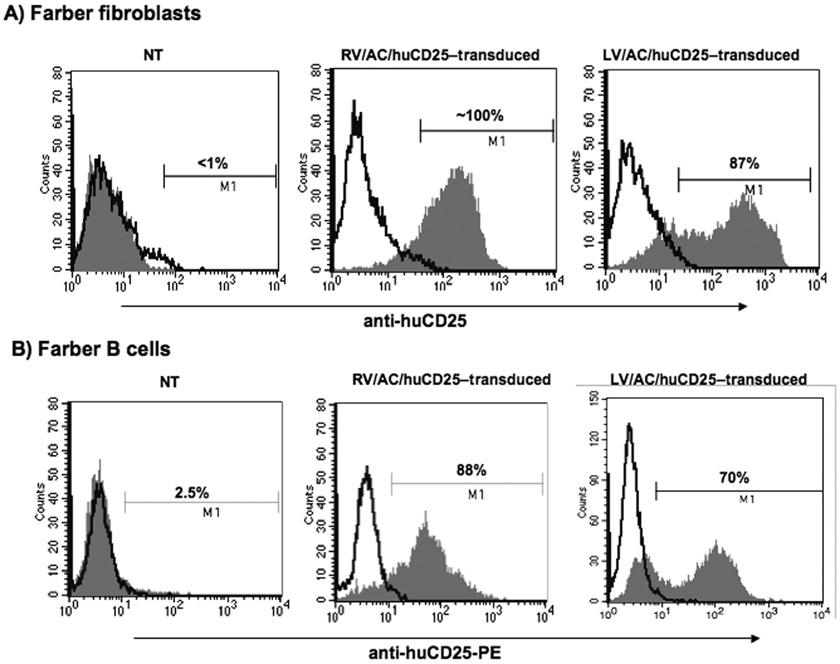
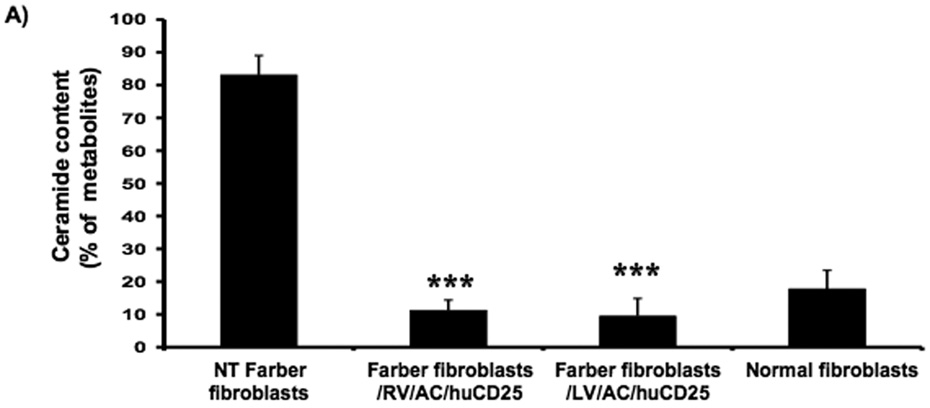
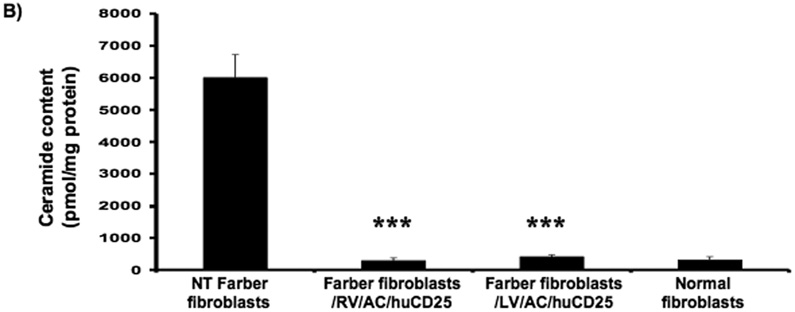
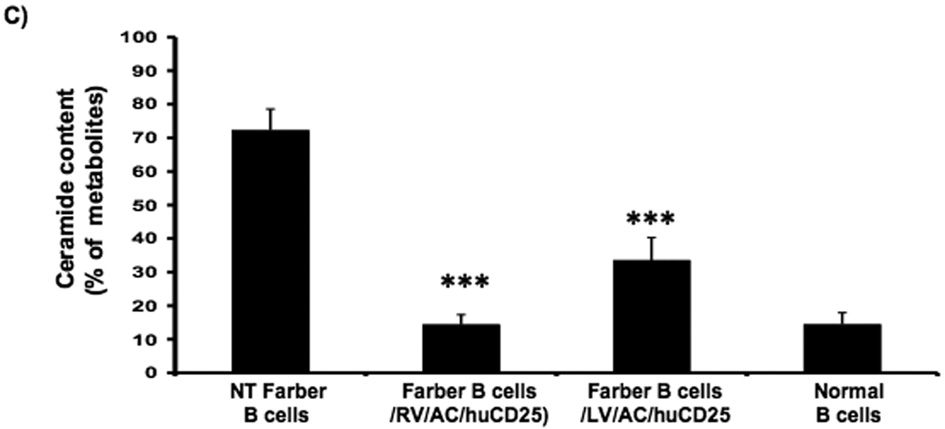
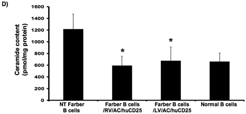
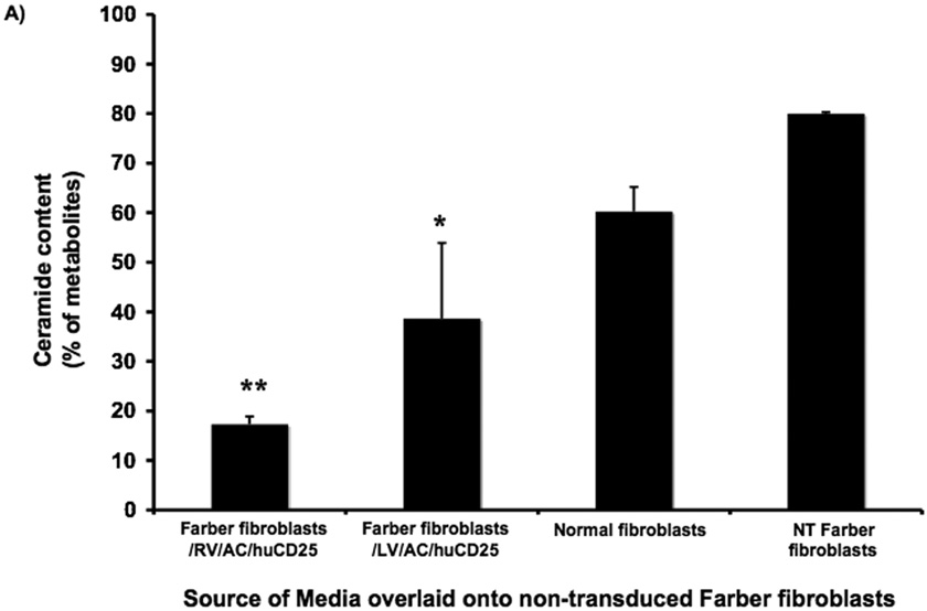
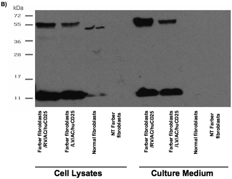
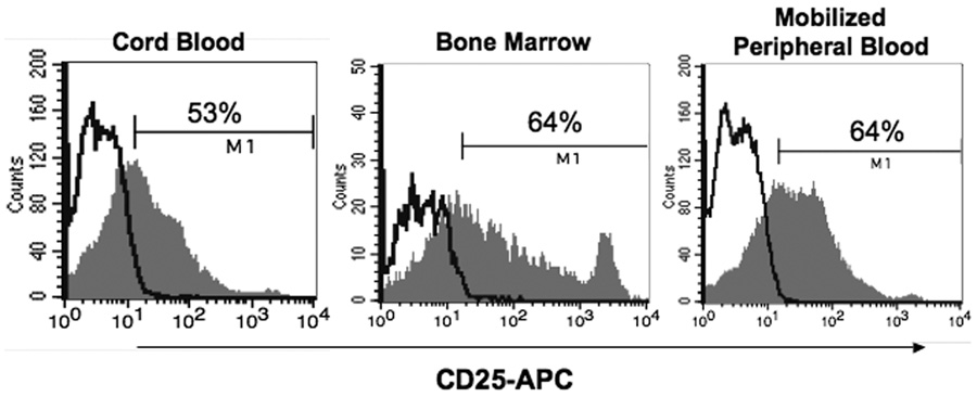
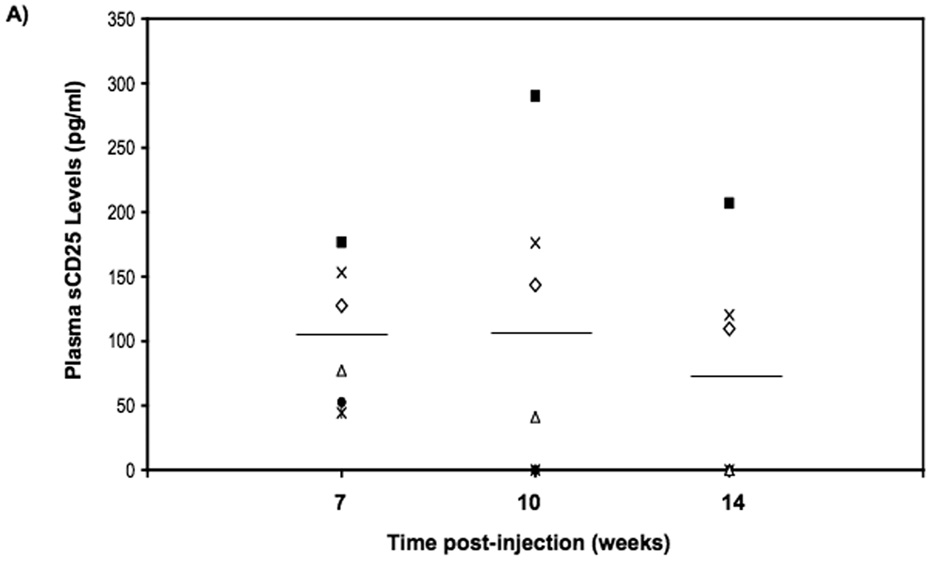
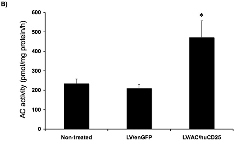
Farber disease is a rare lysosomal storage disorder (LSD) caused by a deficiency of acid ceramidase (AC) activity and subsequent accumulation of ceramide. Currently, there is no treatment for Farber disease beyond palliative care and most patients succumb to the disorder at a very young age. Previously, our group showed that gene therapy using oncoretroviral vectors (RV) could restore enzyme activity in Farber patient cells. The studies described here employ novel RV and lentiviral (LV) vectors that engineer co-expression of AC and a cell surface marking transgene product, human CD25 (huCD25). Transduction of Farber patient fibroblasts and B cells with these vectors resulted in overexpression of AC and led to a 90% and 50% reduction in the accumulation of ceramide, respectively. Vectors were also evaluated in human hematopoietic stem/progenitor cells (HSPCs) and by direct in vivo delivery in mouse models. In a xenotransplantation model using NOD/SCID mice, we found that transduced CD34(+) cells could repopulate irradiated recipient animals, as measured by CD25 expression. When virus was injected intravenously into mice, soluble CD25 was detected in the plasma and increased AC activity was present in the liver up to 14 weeks post-injection. These findings suggest that vector and transgene expression can persist long-term and offer the potential of a lasting cure. To our knowledge, this is the first report of in vivo testing of direct gene therapy strategies for Farber disease.
Figures
Similar articles
-
Autologous transplantation of lentivector/acid ceramidase-transduced hematopoietic cells in nonhuman primates.Hum Gene Ther. 2011 Jun;22(6):679-87. doi: 10.1089/hum.2010.195. Epub 2011 Mar 25. Hum Gene Ther. 2011. PMID: 21280983 Free PMC article.
-
Hematopoietic stem cell transplantation leads to biochemical and functional correction in two mouse models of acid ceramidase deficiency.Mol Ther. 2024 Oct 2;32(10):3402-3421. doi: 10.1016/j.ymthe.2024.08.004. Epub 2024 Aug 5. Mol Ther. 2024. PMID: 39108096 Free PMC article.
-
High levels of transgene expression following transduction of long-term NOD/SCID-repopulating human cells with a modified lentiviral vector.Stem Cells. 2001;19(3):247-59. doi: 10.1634/stemcells.19-3-247. Stem Cells. 2001. PMID: 11359950
-
Acid ceramidase and the treatment of ceramide diseases: The expanding role of enzyme replacement therapy.Biochim Biophys Acta. 2016 Sep;1862(9):1459-71. doi: 10.1016/j.bbadis.2016.05.001. Epub 2016 May 4. Biochim Biophys Acta. 2016. PMID: 27155573 Review.
-
The molecular medicine of acid ceramidase.Biol Chem. 2015 Jun;396(6-7):759-65. doi: 10.1515/hsz-2014-0290. Biol Chem. 2015. PMID: 25938220 Review.
Cited by
-
Autologous transplantation of lentivector/acid ceramidase-transduced hematopoietic cells in nonhuman primates.Hum Gene Ther. 2011 Jun;22(6):679-87. doi: 10.1089/hum.2010.195. Epub 2011 Mar 25. Hum Gene Ther. 2011. PMID: 21280983 Free PMC article.
-
Generation of an induced pluripotent stem cell line (TRNDi030-A) from a patient with Farber disease carrying a homozygous p. Y36C (c. 107 A>G) mutation in ASAH1.Stem Cell Res. 2021 May;53:102387. doi: 10.1016/j.scr.2021.102387. Epub 2021 May 12. Stem Cell Res. 2021. PMID: 34088014 Free PMC article.
-
Acid ceramidase deficiency: Farber disease and SMA-PME.Orphanet J Rare Dis. 2018 Jul 20;13(1):121. doi: 10.1186/s13023-018-0845-z. Orphanet J Rare Dis. 2018. PMID: 30029679 Free PMC article. Review.
-
Lentivector transduction improves outcomes over transplantation of human HSCs alone in NOD/SCID/Fabry mice.Mol Ther. 2012 Jul;20(7):1454-61. doi: 10.1038/mt.2012.64. Epub 2012 Apr 3. Mol Ther. 2012. PMID: 22472949 Free PMC article.
-
Systemic ceramide accumulation leads to severe and varied pathological consequences.EMBO Mol Med. 2013 Jun;5(6):827-42. doi: 10.1002/emmm.201202301. Epub 2013 May 16. EMBO Mol Med. 2013. PMID: 23681708 Free PMC article.
References
-
- Meikle PJ, Hopwood JJ, Clague AE, Carey WF. Prevalence of lysosomal storage disorders. JAMA. 1999;281:249–254. - PubMed
-
- Pinto R, Caseiro C, Lemos M, Lopes L, Fontes A, Ribeiro H, Pinto E, Silva E, Rocha S, Marcao A, Ribeiro I, Lacerda L, Ribeiro G, Amaral O, Sa Miranda MC. Prevalence of lysosomal storage diseases in Portugal. Eur. J. Hum. Genet. 2004;12:87–92. - PubMed
-
- Poorthuis BJ, Wevers RA, Kleijer WJ, Groener JE, de Jong JG, van Weely S, Niezen-Koning KE, van Diggelen OP. The frequency of lysosomal storage diseases in The Netherlands. Hum. Genet. 1999;105:151–156. - PubMed
-
- Moser HW. Acid Ceramidase Deficiency: Farber Lipogranulomatosis. In: Scriver CR, Beaudet AL, Sly WS, Valle D, editors. The Metabolic and Molecular Bases of Inherited Disease. New York: McGraw-Hill; 2001. pp. 3573–3588.
-
- Bar J, Linke T, Ferlinz K, Neumann U, Schuchman EH, Sandhoff K. Molecular analysis of acid ceramidase deficiency in patients with Farber disease. Hum. Mutat. 2001;17:199–209. - PubMed
Publication types
MeSH terms
Substances
Grants and funding
LinkOut - more resources
Full Text Sources
Other Literature Sources
Medical
