

TDP-43 mutations in familial and sporadic amyotrophic lateral sclerosis
- PMID: 18309045
- PMCID: PMC7116650
- DOI: 10.1126/science.1154584
TDP-43 mutations in familial and sporadic amyotrophic lateral sclerosis
Abstract
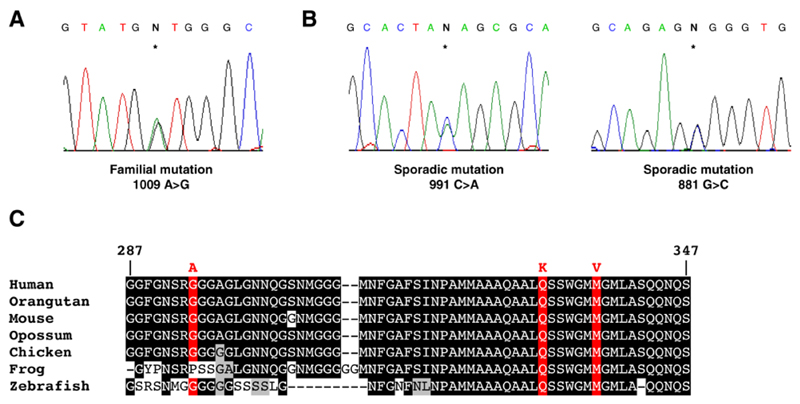
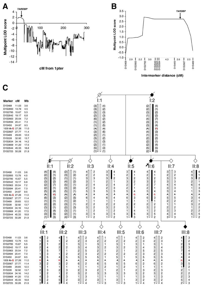
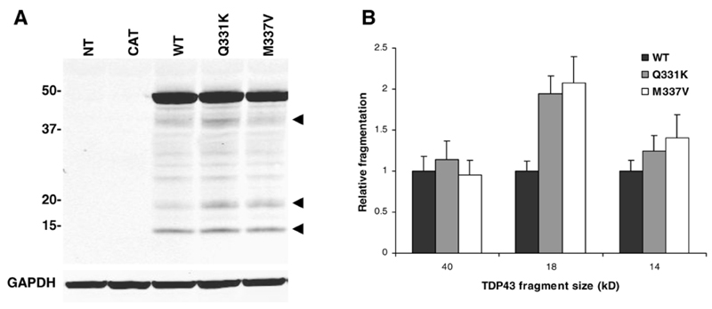
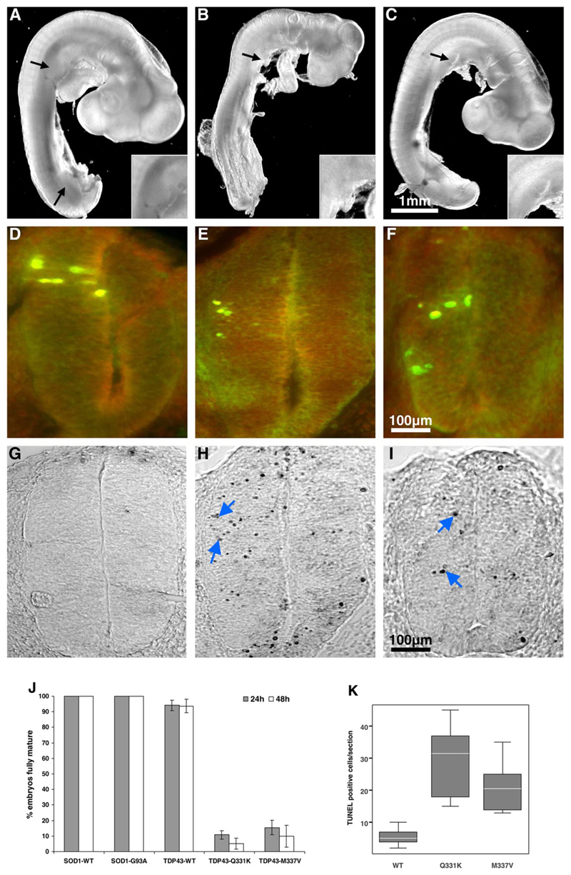
Amyotrophic lateral sclerosis (ALS) is a fatal motor neuron disorder characterized pathologically by ubiquitinated TAR DNA binding protein (TDP-43) inclusions. The function of TDP-43 in the nervous system is uncertain, and a mechanistic role in neurodegeneration remains speculative. We identified neighboring mutations in a highly conserved region of TARDBP in sporadic and familial ALS cases. TARDBPM337V segregated with disease within one kindred and a genome-wide scan confirmed that linkage was restricted to chromosome 1p36, which contains the TARDBP locus. Mutant forms of TDP-43 fragmented in vitro more readily than wild type and, in vivo, caused neural apoptosis and developmental delay in the chick embryo. Our evidence suggests a pathophysiological link between TDP-43 and ALS.
Figures
Similar articles
-
A novel TARDBP mutation in an Australian amyotrophic lateral sclerosis kindred.J Neurol Neurosurg Psychiatry. 2009 Nov;80(11):1286-8. doi: 10.1136/jnnp.2008.163261. J Neurol Neurosurg Psychiatry. 2009. PMID: 19864664
-
Novel mutations in TARDBP (TDP-43) in patients with familial amyotrophic lateral sclerosis.PLoS Genet. 2008 Sep 19;4(9):e1000193. doi: 10.1371/journal.pgen.1000193. PLoS Genet. 2008. PMID: 18802454 Free PMC article.
-
Two German kindreds with familial amyotrophic lateral sclerosis due to TARDBP mutations.Arch Neurol. 2008 Sep;65(9):1185-9. doi: 10.1001/archneur.65.9.1185. Arch Neurol. 2008. PMID: 18779421 Free PMC article.
-
Mutations in TDP-43 link glycine-rich domain functions to amyotrophic lateral sclerosis.Hum Mol Genet. 2009 Oct 15;18(R2):R156-62. doi: 10.1093/hmg/ddp303. Hum Mol Genet. 2009. PMID: 19808791 Free PMC article. Review.
-
[The implications of TDP-43 mutations in pathogenesis of amyotrophic lateral sclerosis].Brain Nerve. 2009 Nov;61(11):1301-7. Brain Nerve. 2009. PMID: 19938687 Review. Japanese.
Cited by
-
Homozygosity analysis in amyotrophic lateral sclerosis.Eur J Hum Genet. 2013 Dec;21(12):1429-35. doi: 10.1038/ejhg.2013.59. Epub 2013 Apr 24. Eur J Hum Genet. 2013. PMID: 23612577 Free PMC article.
-
Limbic-predominant age-related TDP-43 encephalopathy differs from frontotemporal lobar degeneration.Brain. 2020 Sep 1;143(9):2844-2857. doi: 10.1093/brain/awaa219. Brain. 2020. PMID: 32830216 Free PMC article.
-
Controversies and priorities in amyotrophic lateral sclerosis.Lancet Neurol. 2013 Mar;12(3):310-22. doi: 10.1016/S1474-4422(13)70036-X. Lancet Neurol. 2013. PMID: 23415570 Free PMC article. Review.
-
Frontotemporal dementia: implications for understanding Alzheimer disease.Cold Spring Harb Perspect Med. 2012 Feb;2(2):a006254. doi: 10.1101/cshperspect.a006254. Cold Spring Harb Perspect Med. 2012. PMID: 22355793 Free PMC article. Review.
-
Quantification of the Relative Contributions of Loss-of-function and Gain-of-function Mechanisms in TAR DNA-binding Protein 43 (TDP-43) Proteinopathies.J Biol Chem. 2016 Sep 9;291(37):19437-48. doi: 10.1074/jbc.M116.737726. Epub 2016 Jul 21. J Biol Chem. 2016. PMID: 27445339 Free PMC article.
References
Publication types
MeSH terms
Substances
Grants and funding
LinkOut - more resources
Full Text Sources
Other Literature Sources
Medical
Molecular Biology Databases
Miscellaneous
