. 2006 Jul 5;103(27):10438-10443.
doi: 10.1073/pnas.0604066103. Epub 2006 Jun 23.

Aspm specifically maintains symmetric proliferative divisions of neuroepithelial cells

Affiliations

Aspm specifically maintains symmetric proliferative divisions of neuroepithelial cells

Jennifer L Fish et al. Proc Natl Acad Sci U S A. .

Abstract

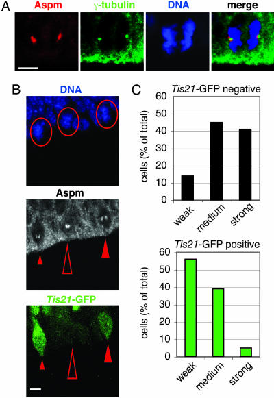
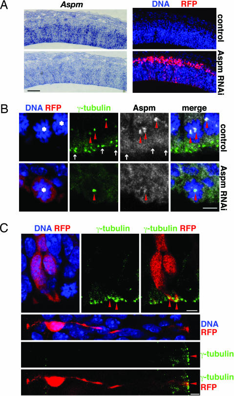
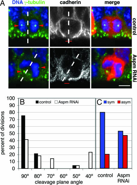
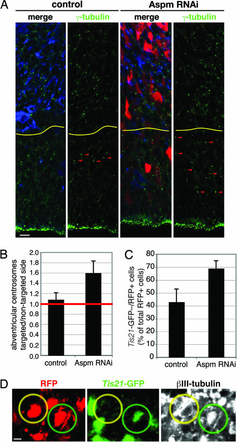
The ASPM (abnormal spindle-like microcephaly-associated) protein has previously been implicated in the determination of human cerebral cortical size, but the cell biological basis of this regulation has not been studied. Here we investigate the role of Aspm in mouse embryonic neuroepithelial (NE) cells, the primary stem and progenitor cells of the mammalian brain. Aspm was found to be concentrated at mitotic spindle poles of NE cells and to be down-regulated with their switch from proliferative to neurogenic divisions. Upon RNA interference in telencephalic NE cells, Aspm mRNA is reduced, mitotic spindle poles lack Aspm protein, and the cleavage plane of NE cells is less frequently oriented perpendicular to the ventricular surface of the neuroepithelium. The alteration in the cleavage plane orientation of NE cells increases the probability that these highly polarized cells undergo asymmetric division, i.e., that apical plasma membrane is inherited by only one of the daughter cells. Concomitant with the resulting increase in abventricular cells in the ventricular zone, a larger proportion of NE cell progeny is found in the neuronal layer, implying a reduction in the number of NE progenitor cells upon Aspm knock-down relative to control. Our results demonstrate that Aspm is crucial for maintaining a cleavage plane orientation that allows symmetric, proliferative divisions of NE cells during brain development. These data provide a cell biological explanation of the primary microcephaly observed in humans with mutations in ASPM, which also has implications for the evolution of mammalian brains.


Conflict of interest statement

Conflict of interest statement: No conflicts declared.

Figures

Fig. 1.
Fig. 1.
Aspm is concentrated at the spindle poles during mitosis and is down-regulated in NE cells undergoing neurogenic divisions. (A) The dorsal telencephalon of E12.5 Tis21–GFP knockin mice was stained with DAPI (DNA, blue) to reveal NE cells in anaphase and immunostained for Aspm (red) and γ-tubulin (green). All cells analyzed were Tis21–GFP-negative (data not shown). Note the concentration of Aspm in the immediate vicinity of the γ-tubulin-stained centrosomes. (Scale bar, 5 μm.) (B) Comparison of Aspm immunoreactivity at spindle poles in metaphase Tis21–GFP-negative NE cells (proliferative divisions, open arrowheads) versus Tis21–GFP-positive NE cells (neurogenic divisions, filled arrowheads) in the dorsal telencephalon of an E14.5 Tis21–GFP knockin mouse. (Top) DNA staining using DAPI (blue); circles indicate the three metaphase cells analyzed. (Middle) Aspm immunoreactivity (white); the small, medium, and large arrowheads indicate weak, medium, and strong Aspm immunoreactivity at spindle poles, respectively. (Bottom) Tis21–GFP fluorescence (green). (Scale bar, 5 μm.) (A and B) The apical surface of the VZ is down. (C) Quantification in the dorsal telencephalon of E14.5 Tis21–GFP knockin mice of prophase or metaphase Tis21–GFP-negative (black bars, 22 cells) and Tis21–GFP-positive (green bars, 18 cells) NE cells showing weak, medium, or strong Aspm immunoreactivity at spindle poles, expressed as a percentage of total (weak plus medium plus strong). Data are from 19 cryosections that originated from at least four brains.
Fig. 2.
Fig. 2.
Knockdown of Aspm results in its loss from centrosomes in mitosis but does not affect the apical localization of centrosomes of interphase NE cells. (A) RNAi of Aspm mRNA. Mouse E10.5 dorsal telencephalon was coelectroporated with Aspm esiRNAs and mRFP plasmid followed by 24 h of whole-embryo culture, and consecutive cryosections were analyzed by in situ hybridization for Aspm mRNA (Left) and RFP fluorescence (Right). Nontargeted (control; Upper) and targeted (Aspm RNAi; Lower) hemispheres are from the same cryosections. The apical surface of the VZ is down. (Scale bar, 100 μm.) (B) Knockdown of Aspm. Mouse E12.5 dorsal telencephalon was coelectroporated with Aspm esiRNAs and mRFP plasmid followed by 24 h of in utero development, and cryosections were analyzed for mitotic NE cells (asterisks) by DNA staining using DAPI (blue), RFP fluorescence (red), and γ-tubulin (green) and Aspm (white) immunofluorescence. Single optical sections are shown. (Upper) Nontargeted hemisphere serving as control. (Lower) Targeted hemisphere subjected to Aspm RNAi. Note the loss of Aspm immunoreactivity from the spindle poles (arrowheads) upon Aspm knockdown. The cell shown is representative of all targeted cells of the electroporated hemisphere. Arrows indicate centrosomes of adjacent NE cells in interphase, which lack Aspm. The apical surface of the VZ is down. (Scale bar, 5 μm.) (C) Interphase centrosomes. Mouse E10.5 dorsal telencephalon was coelectroporated with Aspm esiRNAs and mRFP plasmid followed by 24 h of whole-embryo culture, and cryosections were analyzed for DNA staining with DAPI (blue), RFP fluorescence (red), and γ-tubulin immunofluorescence (green). Note the apical localization of centrosomes (arrowheads) in the targeted NE cells in interphase. (Upper) Two NE cells in G1/G2. The apical surface of the VZ is down. (Scale bar, 5 μm.) (Lower) S-phase NE cell. The apical surface of the VZ is to the right. (Scale bar, 5 μm.)
Fig. 3.
Fig. 3.
Knockdown of Aspm alters the cleavage plane of NE cells, resulting in their asymmetric division. (A) Cleavage plane and cadherin hole analysis. Mouse E12.5 dorsal telencephalon was either electroporated with mRFP plasmid only (Upper) or coelectroporated with Aspm esiRNAs and mRFP plasmid (Lower), followed by 24 h of in utero development, and cryosections were analyzed for NE cells in anaphase or telophase by DNA staining with DAPI (blue), RFP fluorescence (red), and γ-tubulin (green) and cadherin (white) immunofluorescence. The cleavage plane (dashed lines) was deduced from the orientation of the sister chromatids (5, 6). Note the aberrant position of the γ-tubulin-stained centrosomes (arrowheads, stack of optical sections) in the targeted cell and the oblique cleavage plane, which bypasses the cadherin hole (red bar, single optical section), upon Aspm knockdown. The apical surface of the VZ is down. (Scale bar, 5 μm.) (B and C) Quantification of cleavage plane orientation (B) and cadherin hole distribution (C). Dorsal telencephalon of E10.5 Tis21–GFP knockin mice was electroporated with mRFP plasmid only (control) or coelectroporated with mRFP plasmid and Aspm esiRNAs (Aspm RNAi), followed by 24 h of whole-embryo culture. Tis21–GFP-negative, mRFP-expressing NE cells in anaphase or telophase were analyzed for cleavage plane orientation and the position of the cadherin hole as in A. (B) Orientation of the cleavage plane relative to the radial, apical–basal axis of the neuroepithelium (defined as 90°), expressed as a percentage of all divisions for the control (black bars; n = 24) or Aspm RNAi (white bars; n = 22) condition. Groups of cleavage plane angle are ±5°. (C) Cleavage planes bisecting [symmetric (sym), blue bars] or bypassing [asymmetric (asym), red bars] the cadherin hole, expressed as a percentage of all divisions for the control (n = 15) or Aspm RNAi (n = 15) condition.
Fig. 4.
Fig. 4.
Knockdown of Aspm promotes VZ cells to adopt a non-NE fate and increases the appearance of neuron-like NE cell progeny in the neuronal layer. (A and B) Mouse E12.5 dorsal telencephalon was either coelectroporated with Aspm esiRNAs and mRFP plasmid (A and B) or electroporated with mRFP plasmid only (B), followed by 48 h in utero development. (A) Cryosections of the nontargeted (control) and targeted (Aspm RNAi) hemispheres were analyzed for RFP fluorescence (red) and γ-tubulin (green) and βIII-tubulin (blue) immunofluorescence. Note the increase in abventricular centrosomes (arrowheads) upon Aspm knockdown. The yellow lines indicate the boundary between the VZ/sub-VZ and the neuronal layers. The apical surface of the VZ is down. (Scale bar, 10 μm.) (B) Numbers of centrosomes per 10,000 μm2 counted in γ-tubulin-immunostained cryosections are expressed as a ratio of targeted/nontargeted hemispheres for electroporation with mRFP plasmid only (control) and mRFP plasmid plus Aspm esiRNAs (Aspm RNAi). Data are the mean of five cryosections from three different embryos each; error bars indicate SD. (C and D) Dorsal telencephalon of E10.5 Tis21–GFP knockin mice was coelectroporated with Aspm esiRNAs and mRFP plasmid (Aspm RNAi) or electroporated with mRFP plasmid only (control), followed by 24 h of whole-embryo culture. (C) Cryosections were analyzed for the proportion of RFP-positive cells in the neuronal layer that lacked Tis21–GFP fluorescence. Data are the mean of three cryosections from two to three different embryos each (control, 70 cells; Aspm RNAi, 74 cells); error bars indicate SD. (D) Representative example of two neighboring RFP-positive cell bodies (red) in the neuronal layer, with one being positive (green circles) and one being negative (yellow circles) for Tis21–GFP fluorescence (green) but both exhibiting βIII-tubulin immunofluorescence (white). (Scale bar, 5 μm.)

Similar articles

Cited by

References

    1. Rakic P. Trends Neurosci. 1995;18:383–388. - PubMed
    1. Götz M., Huttner W. B. Nat. Rev. Mol. Cell Biol. 2005;6:777–788. - PubMed
    1. Kriegstein A. R., Götz M. Glia. 2003;43:37–43. - PubMed
    1. Chenn A., McConnell S. K. Cell. 1995;82:631–641. - PubMed
    1. Kosodo Y., Röper K., Haubensak W., Marzesco A.-M., Corbeil D., Huttner W. B. EMBO J. 2004;23:2314–2324. - PMC - PubMed

Publication types