

Losartan, an AT1 antagonist, prevents aortic aneurysm in a mouse model of Marfan syndrome
- PMID: 16601194
- PMCID: PMC1482474
- DOI: 10.1126/science.1124287
Losartan, an AT1 antagonist, prevents aortic aneurysm in a mouse model of Marfan syndrome
Abstract
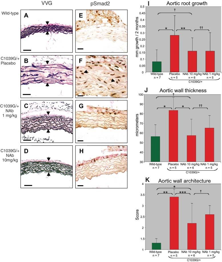
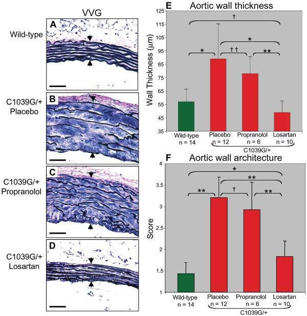
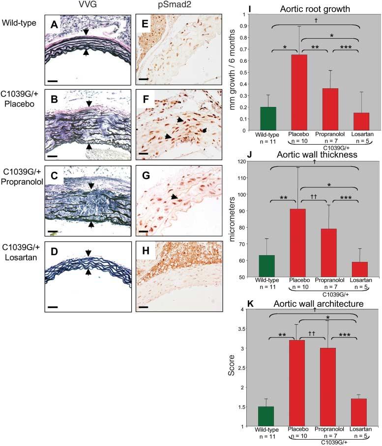
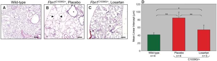
Aortic aneurysm and dissection are manifestations of Marfan syndrome (MFS), a disorder caused by mutations in the gene that encodes fibrillin-1. Selected manifestations of MFS reflect excessive signaling by the transforming growth factor-beta (TGF-beta) family of cytokines. We show that aortic aneurysm in a mouse model of MFS is associated with increased TGF-beta signaling and can be prevented by TGF-beta antagonists such as TGF-beta-neutralizing antibody or the angiotensin II type 1 receptor (AT1) blocker, losartan. AT1 antagonism also partially reversed noncardiovascular manifestations of MFS, including impaired alveolar septation. These data suggest that losartan, a drug already in clinical use for hypertension, merits investigation as a therapeutic strategy for patients with MFS and has the potential to prevent the major life-threatening manifestation of this disorder.
Figures
Comment in
-
Medicine. Old drug, new hope for Marfan syndrome.Science. 2006 Apr 7;312(5770):36-7. doi: 10.1126/science.312.5770.36b. Science. 2006. PMID: 16601163 No abstract available.
Similar articles
-
Dimorphic effects of transforming growth factor-β signaling during aortic aneurysm progression in mice suggest a combinatorial therapy for Marfan syndrome.Arterioscler Thromb Vasc Biol. 2015 Apr;35(4):911-7. doi: 10.1161/ATVBAHA.114.305150. Epub 2015 Jan 22. Arterioscler Thromb Vasc Biol. 2015. PMID: 25614286 Free PMC article.
-
Rationale and design of a trial evaluating the effects of losartan vs. nebivolol vs. the association of both on the progression of aortic root dilation in Marfan syndrome with FBN1 gene mutations.J Cardiovasc Med (Hagerstown). 2009 Apr;10(4):354-62. doi: 10.2459/JCM.0b013e3283232a45. J Cardiovasc Med (Hagerstown). 2009. PMID: 19430350 Clinical Trial.
-
Medicine. Old drug, new hope for Marfan syndrome.Science. 2006 Apr 7;312(5770):36-7. doi: 10.1126/science.312.5770.36b. Science. 2006. PMID: 16601163 No abstract available.
-
Recent advances in understanding Marfan syndrome: should we now treat surgical patients with losartan?J Thorac Cardiovasc Surg. 2008 Feb;135(2):389-94. doi: 10.1016/j.jtcvs.2007.08.047. J Thorac Cardiovasc Surg. 2008. PMID: 18242274 Review.
-
Pathophysiology and Management of Cardiovascular Manifestations in Marfan and Loeys-Dietz Syndromes.Int Heart J. 2016 May 25;57(3):271-7. doi: 10.1536/ihj.16-094. Epub 2016 May 13. Int Heart J. 2016. PMID: 27181042 Review.
Cited by
-
Aortic Dimensions, Biophysical Properties, and Plasma Biomarkers in Children and Adults with Marfan or Loeys-Dietz Syndrome.CJC Open. 2020 Dec 28;3(5):585-594. doi: 10.1016/j.cjco.2020.12.018. eCollection 2021 May. CJC Open. 2020. PMID: 34027363 Free PMC article.
-
Losartan ameliorates dystrophic epidermolysis bullosa and uncovers new disease mechanisms.EMBO Mol Med. 2015 Sep;7(9):1211-28. doi: 10.15252/emmm.201505061. EMBO Mol Med. 2015. PMID: 26194911 Free PMC article.
-
GLUT10 is required for the development of the cardiovascular system and the notochord and connects mitochondrial function to TGFβ signaling.Hum Mol Genet. 2012 Mar 15;21(6):1248-59. doi: 10.1093/hmg/ddr555. Epub 2011 Nov 24. Hum Mol Genet. 2012. PMID: 22116938 Free PMC article.
-
Androgens Accentuate TGF-β Dependent Erk/Smad Activation During Thoracic Aortic Aneurysm Formation in Marfan Syndrome Male Mice.J Am Heart Assoc. 2020 Oct 20;9(20):e015773. doi: 10.1161/JAHA.119.015773. Epub 2020 Oct 16. J Am Heart Assoc. 2020. PMID: 33059492 Free PMC article.
-
Genetic dissection of marfan syndrome and related connective tissue disorders: an update 2012.Mol Syndromol. 2012 Aug;3(2):47-58. doi: 10.1159/000339441. Epub 2012 Jun 12. Mol Syndromol. 2012. PMID: 23326250 Free PMC article.
References
-
- Dietz HC, et al. Nature. 1991;352:337. - PubMed
-
- Sakai LY, Keene DR, Glanville RW, Bachinger HP. J. Biol. Chem. 1991;266:14763. - PubMed
-
- Visconti RP, Barth JL, Keeley FW, Little CD. Matrix Biol. 2003;22:109. - PubMed
-
- Isogai Z, et al. J. Biol. Chem. 2003;278:2750. - PubMed
-
- Neptune ER, et al. Nat. Genet. 2003;33:407. - PubMed
Publication types
MeSH terms
Substances
Grants and funding
LinkOut - more resources
Full Text Sources
Other Literature Sources
Medical
Molecular Biology Databases
Research Materials
