

Fibrillins 1 and 2 perform partially overlapping functions during aortic development
- PMID: 16407178
- PMCID: PMC3052983
- DOI: 10.1074/jbc.M511599200
Fibrillins 1 and 2 perform partially overlapping functions during aortic development
Abstract
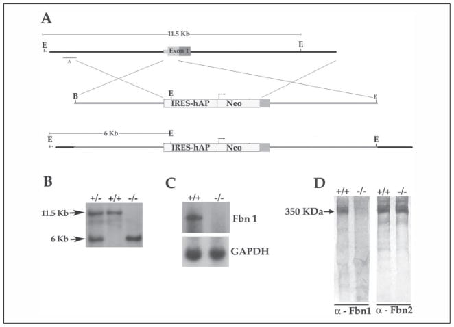
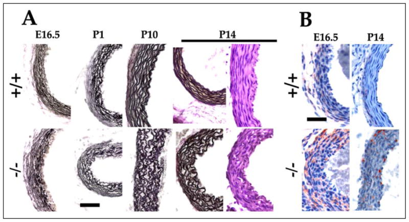
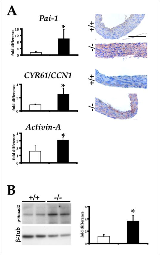
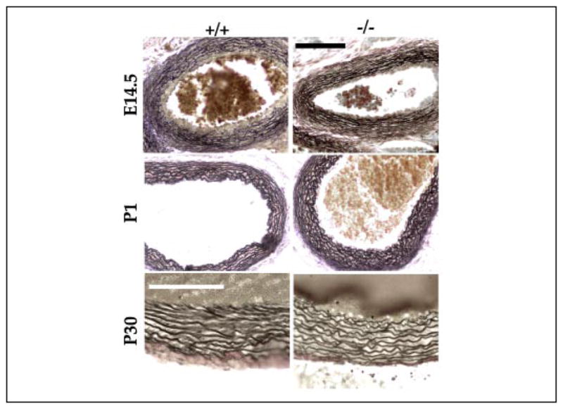
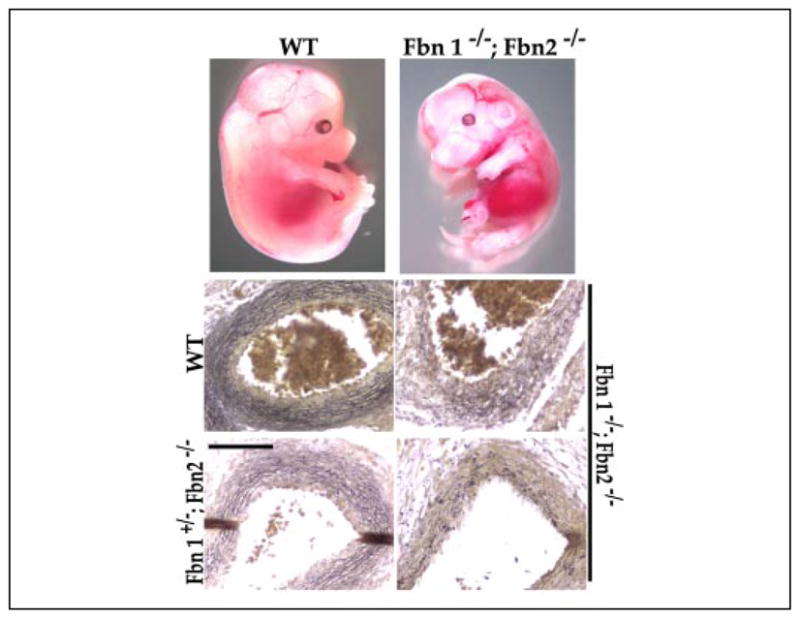
Fibrillin-rich microfibrils are extracellular assemblies that impart structural properties to the connective tissue. To elucidate the contribution of fibrillin-rich microfibrils to organogenesis, we have examined the vascular phenotype of a newly created strain of mice that completely lacks fibrillin-1 and the consequences of combined deficiency of fibrillins 1 and 2 on tissue formation. The results demonstrated that fibrillins 1 and 2 perform partially overlapping functions during aortic development. Fbn1-/- mice died soon after birth from ruptured aortic aneurysm, impaired pulmonary function, and/or diaphragmatic collapse. Analysis of the neonatal Fbn1-/- aorta documented a disorganized and poorly developed medial layer but normal levels of elastin cross-links. Transcriptional profiling revealed that aneurysm progression in Fbn1 null mice is accompanied by unproductive up-regulation of gene products normally involved in tissue repair and vascular integrity, such as plasminogen activator inhibitor-1, activin A, and cysteine-rich angiogenic protein 61. In contrast to Fbn1-/- mice, Fbn2 null mice had a well developed and morphologically normal aortic wall. However, virtually all Fbn1-/-;Fbn2-/- embryos and about half of the Fbn1+/-;Fbn2-/- embryos died in utero and displayed a significantly more severe vascular phenotype than Fbn1-/- mice. Consistent with a specialized function of fibrillin-2, electron microscopy visualized ultrastructurally different microfibrils in Fbn1 null compared with control cell cultures. Collectively, these data demonstrate that involvement of fibrillin-2 in the initial assembly of the aortic matrix overlaps in part with fibrillin-1 and that continued fibrillin-1 deposition is absolutely required for the maturation and function of the vessel during neonatal life.
Figures


Similar articles
-
Nonselective assembly of fibrillin 1 and fibrillin 2 in the rodent ocular zonule and in cultured cells: implications for Marfan syndrome.Invest Ophthalmol Vis Sci. 2013 Dec 23;54(13):8337-44. doi: 10.1167/iovs.13-13121. Invest Ophthalmol Vis Sci. 2013. PMID: 24265020 Free PMC article.
-
Structure and function of the mammalian fibrillin gene family: implications for human connective tissue diseases.Mol Genet Metab. 2012 Dec;107(4):635-47. doi: 10.1016/j.ymgme.2012.07.023. Epub 2012 Aug 3. Mol Genet Metab. 2012. PMID: 22921888 Review.
-
Initial steps in assembly of microfibrils. Formation of disulfide-cross-linked multimers containing fibrillin-1.J Biol Chem. 2000 Jan 21;275(3):2205-10. doi: 10.1074/jbc.275.3.2205. J Biol Chem. 2000. PMID: 10636927
-
New insights into the assembly of extracellular microfibrils from the analysis of the fibrillin 1 mutation in the tight skin mouse.J Cell Biol. 2000 Aug 7;150(3):667-80. doi: 10.1083/jcb.150.3.667. J Cell Biol. 2000. PMID: 10931876 Free PMC article.
-
Fibrillin-rich microfibrils: Structural determinants of morphogenetic and homeostatic events.J Cell Physiol. 2007 Nov;213(2):326-30. doi: 10.1002/jcp.21189. J Cell Physiol. 2007. PMID: 17708531 Review.
Cited by
-
The extracellular matrix glycoprotein fibrillin-1 in health and disease.Front Cell Dev Biol. 2024 Jan 10;11:1302285. doi: 10.3389/fcell.2023.1302285. eCollection 2023. Front Cell Dev Biol. 2024. PMID: 38269088 Free PMC article. Review.
-
In vivo studies of mutant fibrillin-1 microfibrils.J Biol Chem. 2010 Aug 6;285(32):24943-55. doi: 10.1074/jbc.M110.130021. Epub 2010 Jun 7. J Biol Chem. 2010. PMID: 20529844 Free PMC article.
-
Versican facilitates chondrocyte differentiation and regulates joint morphogenesis.J Biol Chem. 2010 Jul 2;285(27):21114-25. doi: 10.1074/jbc.M109.096479. Epub 2010 Apr 19. J Biol Chem. 2010. PMID: 20404343 Free PMC article.
-
Co-expression of FBN1 with mesenchyme-specific genes in mouse cell lines: implications for phenotypic variability in Marfan syndrome.Eur J Hum Genet. 2010 Nov;18(11):1209-15. doi: 10.1038/ejhg.2010.91. Epub 2010 Jun 16. Eur J Hum Genet. 2010. PMID: 20551991 Free PMC article.
-
Elastic fiber ultrastructure and assembly.Matrix Biol. 2019 Nov;84:31-40. doi: 10.1016/j.matbio.2019.10.002. Epub 2019 Oct 24. Matrix Biol. 2019. PMID: 31669522 Free PMC article. Review.
References
-
- Mecham RP, Davis E. In: Extracellular Matrix Assembly and Structure. Yurchenco PD, Birk DE, Mecham RP, editors. Academic Press, Inc; New York: 1994. pp. 281–314.
-
- Handford PA, Downing AK, Reinhardt DP, Sakai LY. Matrix Biol. 2000;19:457–470. - PubMed
-
- Kielty CM, Sherratt MJ, Shuttleworth CA. J Cell Sci. 2002;115:2817–2828. - PubMed
-
- Ramirez F. Curr Opin Genet Dev. 1996;6:309–315. - PubMed
-
- Charbonneau NL, Ono RN, Corson GM, Keene DR, Sakai LY. Birth Defects Res C Embryo Today. 2004;72:37–50. - PubMed
Publication types
MeSH terms
Substances
Grants and funding
LinkOut - more resources
Full Text Sources
Other Literature Sources
Molecular Biology Databases
