. 2004 Oct 1;18(19):2336-41.
doi: 10.1101/gad.1239604. Epub 2004 Sep 15.

Tel/Etv6 is an essential and selective regulator of adult hematopoietic stem cell survival

Affiliations

Tel/Etv6 is an essential and selective regulator of adult hematopoietic stem cell survival

Hanno Hock et al. Genes Dev. .

Abstract

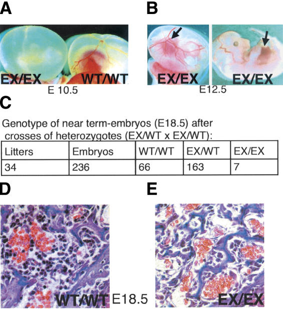
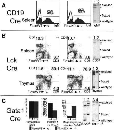
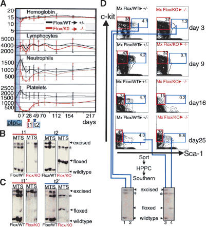
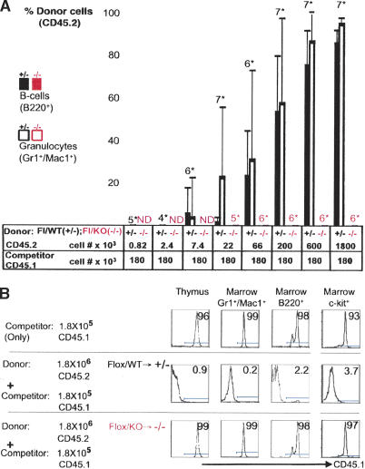
Hematopoietic stem cells (HSCs) sustain blood formation throughout life. Pathways regulating maintenance of adult HSCs are largely unknown. Here we report that the Ets-related transcription factor Tel/Etv6, the product of a locus frequently involved in translocations in leukemia, is a selective regulator of HSC survival. Following inactivation of Tel/Etv6, HSCs are lost in the adult bone marrow but their progeny are unaffected and transiently sustain blood formation. Accordingly, absence of Tel/Etv6 after lineage commitment is ostensibly without consequence except for unexpected impairment of maturation of megakaryocytes. Thus, we establish Tel/Etv6 as a selective and essential regulator of postembryonic HSCs.


Figures

Figure 1.
Figure 1.
Germ-line excision of a floxed Tel/Etv6 allele causes either midembryonic or perinatal lethality with hypocellular bone marrow. (A) Most progeny homozygous for germ-line-excised Tel/Etv6 (EX/EX) die at approximately E10.5 with absent yolk sac angiogenesis; wild-type (WT/WT) yolk sac at the same stage is shown for comparison. (B) A few homozygous excised embryos survive longer; they have normal yolk sac angiogenesis and exhibit red livers indicative of active hematopoiesis (arrows) on E12.5. (C) Homozygous excised embryos are not born alive (not shown) but at E18.5 (C-section), ∼3% of pups that appear normal are homozygous for the excised allele. (D) Pathological examination of E18.5 homozygotes (n = 3) and their wild-type littermates (n = 3) revealed markedly reduced bone marrow hematopoiesis in the mutants (cross-sections through the humeral head, 60×).
Figure 2.
Figure 2.
Tel/Etv6 is dispensable for selected lineage-committed hematopoietic cells. (A) CD19-Cre mediated excision in the B-lymphoid lineage does not affect frequencies of IgM+ B cells in the spleen assessed by FACS analysis (control, left histogram; lineage-specific mutant, right histogram); in each case, germ-line genotype of mice ([WT] wild-type; [Flox] floxed; [KO] conventional knockout) and expected functional status of both alleles in cells excised by Cre (following arrow) are indicated below. Results from representative animals are shown (n = 5). Right panel shows Southern blot analysis of sorted IgM+ B cells from spleen. Note complete excision of the floxed allele in both control (lane 1) and lineage-specific mutant (lane 2). (sstarf;) Band shared by floxed and wild-type allele; see Supplementary Figure 1 for complete explanation of band pattern. (B) Lck-Cre-mediated excision in the T-lymphoid lineage does not affect frequencies of CD4+ and CD8+ T cells in the spleen (upper FACS plots) or distribution of subsets of developing T cells in the thymus (lower FACS plots) (n = 4). Southern blots on the right show complete excision in sorted CD3+ T cells from the spleen (upper) and subtotal excision in the thymus (lower) of both controls (lane 1) and lineage-specific mutants (lane 2). (C) Gata1-Cre-mediated excision in erythroid and megakaryocytic lineages reveals terminal megakaryocyte defect but normal steady-state erythropoiesis. Bar graphs on the left show no difference in hemoglobin levels but ∼50% reduction in peripheral blood platelet counts in the lineage-specific mutants compared with controls. Third bar graph shows approximately fivefold elevation of megakaryocyte colonies from mutant bone marrow. Error bars represent standard deviations. Southern blots show the unexcised allele in sorted B220+ B cells from bone marrow (left; control [lane 1], mutant [lane 2]) but only the excised allele in Ter119+ (erythroid) cells (right; control [lane 3], mutant [lane 4]). (Lane 1) Note that control B220+ cells from Gata1-Cre mice lack detectable excised allele, confirming that excision is intrinsic to the megakaryocyte/erythroid lineage and does not occur earlier; that is, in HSCs.
Figure 3.
Figure 3.
Disruption of Tel/Etv6 in adult mice reveals an essential and selective role for survival of HSCs. (A) Peripheral blood during (shaded blue) and after pIpC injection to induce Cre-mediated disruption of the floxed allele. Note the absence of differences in hemoglobin (g/dL) and lymphocyte counts (cell#/μL), moderate transient drop in neutrophil counts (cell#/μL) and drastic but transient drop in platelet count (cell# × 103/μL) in conditional mutants (genotype: Floxed/conventional knockout [Flox/KO]; n = 6-11) compared with controls (genotype: Floxed/wild-type [Flox/WT]; n = 7-11). (B) Southern blot of bone marrow (M), thymus (T), and spleen (S) 3 wk after the final pIpC injection demonstrates complete excision of the floxed allele in controls (Flox/WT) and conditional mutants (Flox/KO). Left panels (t1) show representative results from analysis of six mice in each group from three independent experiments; complete excision of the floxed allele in bone marrow was confirmed as early as 2 d after the final pIpC injection (n = 6, not shown, see Supplementary Fig. 5A). Right panels (t2, 6 wk after Cre induction) show persistent excision in control mice (Flox/WT) but absence of detectable excised allele associated with re-emergence of the floxed allele in conditional mutants (Flox/KO). Representative results from analysis of six mice in each group from three independent experiments are shown; excision in controls was found to be undiminished as late as 18 mo after Cre-induction (n = 3; not shown). (sstarf;) Band shared by floxed and wild-type allele; see Supplementary Figure 1 for complete explanation of band pattern. (C) Excision analysis in experiment performed as above except that mutant marrow had been transplanted into lethally irradiated wild-type recipients 3 mo prior to Cre induction. Longitudinal development of peripheral blood counts (not shown) and excision status after 3 wk (left panel, t1′) and 6 wk (right panel, t2′) following Cre induction was indistinguishable from that in untransplanted mutants. (D) FACS analysis of HSCs (lineage-, c-Kit+, Sca-1+, marked with blue frames) and myeloid progenitors (lin-, c-Kit-, Sca-1+, marked with red frames) at days 3, 9, 16 and 25 after Cre induction. In controls (Flox/WT), HSC frequency remained stable, but in conditional mutants HSCs were decreased at day 3, hardly detectable on days 9 and 16, but re-emerged on day 24 (results are representative of six mice for each time point in three independent experiments). Southern blot (bottom) shows excision in high proliferative potential colonies (HPPCs) from sorted control (lane 1) and mutant (lane 2) HSC populations at day 3. At day 25, complete excision was still observed in controls (lane 3) but in conditional mutants stem cells that had re-emerged exclusively harbored unexcised Tel/Etv6 (lane 4).
Figure 4.
Figure 4.
Bone marrow transplantation following disruption of Tel/Etv6 confirms absence of HSC activity. (A) Analysis of CD45 isotype variants on blood granulocytes (Gr1+Mac1+) and B cells (B220+) 3 mo after transplantation into lethally irradiated recipients demonstrates absence of competitive repopulation activity in bone marrow after Tel/Etv6 disruption. Bone marrow donors had received pIpC to induce Cre until 3 d prior to transplant. Control marrow (Flox/WT → +/-; CD45.2) competed efficiently with a low constant dose of wild-type marrow (“competitor”, CD45.1) in a dose-dependent fashion, whereas conditional mutant marrow (Flox/KO → -/-; CD45.2) failed to compete. (Bars represent averages on groups of mice; numberssstarf; indicate mice/group; error bars show standard deviation; ND indicates not done; one representative of two experiments is shown). (B) CD45 isotype analysis of thymocytes, bone marrow myeloid cells (Gr1+Mac1+), B cells (B220+), and progenitors (c-Kit+) from representative recipients in the competitive repopulation assay shown in A. Note that CD45.2+ control marrow (middle) efficiently prevents contribution of wild-type “competitor” cells (CD45.1, given alone in top row), whereas conditional mutant marrow (CD45.2, bottom) completely fails to prevent “competitor” engraftment. See Supplementary Figure 5 for excision analysis 3 mo after engraftment.

Similar articles

Cited by

References

    1. Brakebusch C., Fillatreau, S., Potocnik, A.J., Bungartz, G., Wilhelm, P., Svensson, M., Kearney, P., Korner, H., Gray, D., and Fassler, R. 2002. β1 integrin is not essential for hematopoiesis but is necessary for the T cell-dependent IgM antibody response. Immunity 16: 465-477. - PubMed
    1. Brun A.C., Bjornsson, J.M., Magnusson, M., Larsson, N., Leveen, P., Ehinger, M., Nilsson, E., and Karlsson, S. 2004. Hoxb4 deficient mice have normal hematopoietic development but exhibit a mild proliferation defect in hematopoietic stem cells. Blood. 103(11): 4126-4133. - PubMed
    1. Cheng T., Rodrigues, N., Shen, H., Yang, Y., Dombkowski, D., Sykes, M., and Scadden, D.T. 2000. Hematopoietic stem cell quiescence maintained by p21cip1/waf1. Science 287: 1804-1808. - PubMed
    1. Cheshier S.H., Morrison, S.J., Liao, X., and Weissman, I.L. 1999. In vivo proliferation and cell cycle kinetics of long-term self-renewing hematopoietic stem cells. Proc. Natl. Acad. Sci. 96: 3120-3125. - PMC - PubMed
    1. Cobas M., Wilson, A., Ernst, B., Mancini, S.J., MacDonald, H.R., Kemler, R., and Radtke, F. 2004. β-catenin is dispensable for hematopoiesis and lymphopoiesis. J. Exp. Med. 199: 221-229. - PMC - PubMed

Publication types